. 1999;5(1):19-28.
doi: 10.1177/107385849900500104.

Do Antibodies Stimulate Myelin Repair in Multiple Sclerosis?

Affiliations

Do Antibodies Stimulate Myelin Repair in Multiple Sclerosis?

Dorian McGavern et al. Neuroscientist. 1999.

Abstract

One of the major goals in the study of multiple sclerosis (MS) is to identify a beneficial therapeutic intervention that mimics the intrinsic reparative process and results in long-term clinical improvement. As yet, the therapeutic strategies tested in MS have failed to accomplish this task. However, one potential therapy that has shown some promise in rodent models of demyelination involves the administration of antibodies. Studies in various models of demyelination (virus-induced, autoimmune, and toxic) indicate that a subset of autoantibodies with reactivity to CNS antigens promote remyelination. We have identified a prototypic germline IgMk monoclonal antibody, designated SCH 94.03, with reactivity to a surface antigen on oligodendrocytes that promotes CNS remyelination. This antibody has the phenotypic features of polyreactive physiological natural autoantibodies. Additionally, treatment of MS patients with intravenous immunoglobulin, which contains these natural autoantibodies, may be efficacious in a subset of patients. We propose three mechanisms (direct stimulation of oligodendrocytes, immunomodulation, and opsonization of debris) by which polyreactive natural autoantibodies directed against CNS antigen may promote remyelination. Remyelination has the potential to not only improve conduction velocity but also may protect axons from injury and improve neurological function.

Keywords: Autoantibodies; Oligodendrocytes; Remyelination; Theiler’s virus.


Figures
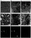
Fig. 1.
Fig. 1.
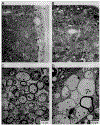
Promotion of remyelination and oligodendrocyte surface reactivity with the natural remyelination-promoting autoantibody designated as SCH 94.03. Spinal cord white matter photographs (A, B, C) and electron micrographs (D, E, F) are shown for uninfected (A, D), chronically infected with Theiler’s murine encephalomyelitis virus (TMEV) and treated with SCH 94.03 (B, E), and chronically-TMEV-infected, untreated (C, F) mice. Normal spinal cord white matter is shown for comparative purposes (A, D). The electron dense material in D corresponds to normal myelin surrounding axons. These dense rings of normal myelin are also apparent in A. In the chronic stages of disease, susceptible SJL/J mice normally show extensive demyelination with little or no remyelination. After treatment twice per week for 4–5 weeks (1 mg total dose) with SCH 94.03, extensive spinal cord remyelination results, as is evident by light (B) and electron (E) microscopy. Note the difference between the thinly remyelinated axons in B and E and the normally myelinated axons in A and D. SCH 94.03 treatment also increases the number of proliferating oligodendrocytes (white O in D and E) in the lesion, which then engage in the process of remyelination (E). Chronically-infected SJL/J mice treated with phosphate-buffered saline (C) or a germline control IgM autoantibody (F) continue to have extensive demyelination with little or no remyelination. (Original magnifications for A-F are 900×, 900×, 1200×, 4000×, 6000×, and 7500×, respectively.) SCH 94.03 shows punctate labeling on the surface of oligodendrocytes in culture (G). Approximately 50% of the SCH 94.03 positive cells in culture are positive for myelin basic protein (a marker for a mature oligodendrocyte) (H). G and H are the same field, double-labeled with SCH 94.03 and an antibody to MBP. Note the cell (white arrow) labeled with both markers, whereas the MBP-positive cell (double white arrows) at the bottom of the figure does not label for the SCH 94.03 antigen. The germline control IgM that does not promote remyelination (F) shows no reactivity on the surface of oligodendrocytes in culture (I). (Original magnification for G, H, and I is 500×.)
Fig. 2.
Fig. 2.
The schematic describes three potential mechanisms by which remyelination-promoting autoantibodies could function. In the direct hypothesis, it is proposed that autoantibodies reactive with the surface of oligodendrocytes could stimulate a receptor to induce the proliferation and differentiation of progenitors. The indirect hypotheses include modulation of the immune response or the opsonization of debris in the lesion. Autoantibodies could act by interfering with the effector function of a macrophage or T lymphocyte. Alternatively, polyreactive autoantibodies directed to the CNS may aid in the clearance of cellular debris, permitting the normal process of remyelination to proceed. This is referred to as the “scavenger” hypothesis and explains the broad antigen specificity of remyelination-promoting autoantibodies toward both surface and intracellular proteins.
Fig. 3.
Fig. 3.
Proliferation of oligodendrocytes in remyelinating lesions after treatment with purified IgG directed against SCH. SJL/J mice chronically infected with Theiler’s murine encephalomyelitis virus (TMEV) were treated twice weekly for 4–5 weeks (1 mg total dose) with purified IgG directed against SCH (SCH/IgG) (A, C, D) or IgG obtained from animals injected with phosphate-buffered saline (PBS) as a control (PBS/IgG) (B). Before sacrifice, an intraperitoneal injection of [3H]thymidine was given to aid in the localization of proliferating cells. After SCH/IgG treatment, extensive spinal cord remyelination and proliferating cells (black arrows) were observed by light microscopy (A), whereas extensive demyelination with little or no remyelination was observed in control mice treated with PBS/IgG (B). Remyelination in A is evidenced by many thinly myelinated axons in the field. (Original magnifications for A and B are 1,300× and 1,000×, respectively.) To identify the proliferating cells in SCH/IgG treated mice, electron microscopy (C) and immunohistochemistry (D) were used. C shows an electron micrograph of a presumed oligodendrocyte (black O) that incorporated the [3H]thymidine label (white arrow) and is extending a process to myelinate a nearby axon (black arrow). (Original magnification for panel C is 14,000×). D shows three cells that have incorporated [3H]thymidine and are positive for the oligodendrocyte marker, galactocerebroside (GC) (single black arrows). However, there are cells in the lesion that have incorporated [3H]thymidine, but do not stain positively for GC (white arrows). Additionally, there are GC-positive cells that do not incorporate [3H]thymidine (double black arrows). Some of the unidentified, proliferating cells in the lesion may be of the oligodendrocyte lineage and may represent progenitors. (Original magnification for panel D is 1,400×.)
Fig. 4.
Fig. 4.
Promotion of remyelination by immunoglobulins reactive with MBP. Spinal cord white matter photographs (A, B) and electron micrographs (C, D) are shown for mice chronically infected with Theiler’s murine encephalomyelitis virus (TMEV) and treated with affinity-purified immunoglobulin against myelin basic protein (MBP) (MBP/Ig) (A, C) or affinity-purified immunoglobulin against incomplete Freund’s adjuvant (IFA/Ig) (B, D) as a control. Chronically-infected SJL/J mice from both groups were injected twice a week for 4–5 weeks (total dose of 240 mg). After treatment with MBP/Ig, extensive remyelination was evident by both light (A) and electron (C) microscopy. Note the presence of the three oligodendrocytes (white O) in the remyelinated region (C). In contrast, control mice treated with IFA/Ig showed extensive demyelination with minimal repair (B, D). This is evident by both light (B) and electron (D) microscopy. (Original magnifications for panels A-D are 1000×, 1000×, 2500×, and 8000×, respectively.)

References

    1. Lucchinetti CF, Rodriguez M. The controversy surrounding the pathogenesis of the multiple sclerosis lesion. Mayo Clin Proc 1997;72:665–678. - PubMed
    1. Gilbert JJ, Sadler M. Unsuspected multiple sclerosis. Arch Neurol 1983;40:533–536. - PubMed
    1. Mackay RP, Hirano A. Forms of benign multiple sclerosis: Report of two “clinically silent” cases discovered at autopsy. Arch Neurol 1967;17:588–600. - PubMed
    1. Lucchinetti CF, Bruck W, Rodriguez M, Lassman H. Distinct patterns of multiple sclerosis pathology indicates heterogeneity in pathogenesis. Brain Pathol 1996;6:259–274.. - PMC - PubMed
    1. Rodriguez M, Scheithauer B. Ultrastructure of multiple sclerosis. Ultrastruct Pathol 1994;18:3–13. - PubMed