Review
. 2019 May 2:6:54.
doi: 10.3389/fcvm.2019.00054. eCollection 2019.

Autoimmune Calcium Channelopathies and Cardiac Electrical Abnormalities

Affiliations
Review

Autoimmune Calcium Channelopathies and Cardiac Electrical Abnormalities

Yongxia Sarah Qu et al. Front Cardiovasc Med. .

Abstract

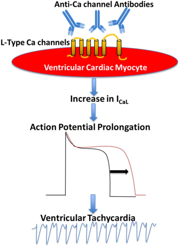
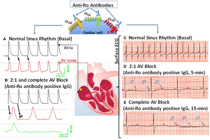
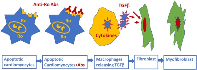
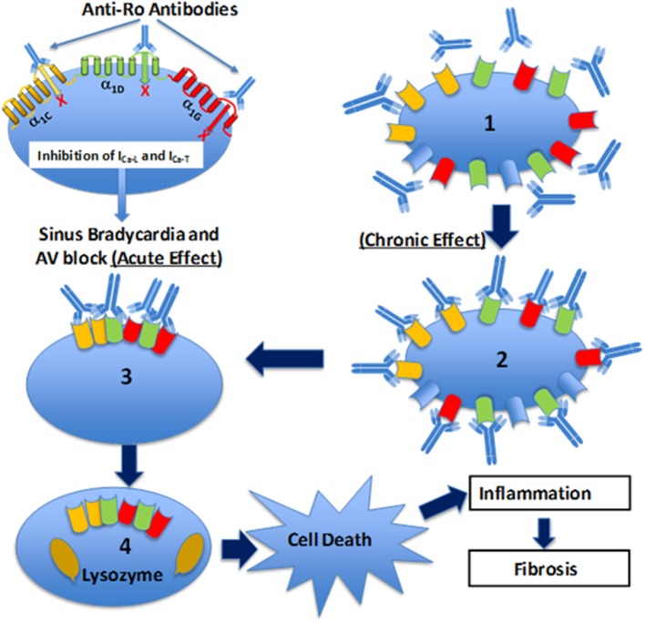
Patients with autoimmune diseases are at increased risk for developing cardiovascular diseases, and abnormal electrocardiographic findings are common. Voltage-gated calcium channels play a major role in the cardiovascular system and regulate cardiac excitability and contractility. Particularly, by virtue of their localization and expression in the heart, calcium channels modulate pace making at the sinus node, conduction at the atrioventricular node and cardiac repolarization in the working myocardium. Consequently, emerging evidence suggests that calcium channels are targets to autoantibodies in autoimmune diseases. Autoimmune-associated cardiac calcium channelopathies have been recognized in both sinus node dysfunction atrioventricular block in patients positive for anti-Ro/La antibodies, and ventricular arrhythmias in patients with dilated cardiomyopathy. In this review, we discuss mechanisms of autoimmune-associated calcium channelopathies and their relationship with the development of cardiac electrical abnormalities.

Keywords: autoantibodies; autoimmune; calcium channel; cardiac electrical abnormalities; channelopathy.


Figures

Figure 1
Figure 1
Proposed mechanism of the pathogenic role of anti-Ca channels autoantibodies in Dilated Cardiomyopathy. Anti-Ca channels autoantibodies target L-type Ca channels in the ventricular myocyte resulting in an increase in L-type Ca current (ICaL) which in turn leads to action potential prolongation and ventricular arrhythmias.
Figure 2
Figure 2
Effects of anti-Ro antibodies from mothers of children with congenital heart block on an isolated multicellular AV nodal preparation (left) and Langendorff perfused whole heart (Right). (A) Simultaneous control action potentials from the crista terminalis (black tracing) and the AV node area (red tracing). (B) Superfusion of the preparation with positive IgG (800 μg/mL) for 10 min resulted in 2:1 AV block (indicated by the arrows) which progressed to near complete inhibition of the AV node action potential by 15 min (B), (green tracing). (C) ECG was recorded by the conventional ECG machine in lead I, except for the use of silver wires at the recording end of the leads. One lead was inserted in the atrium, the second in the left ventricle near the apex, and the third in Tyrode's solution (ground). “P” indicates, the P wave and on the ECG. Regular sinus rhythm (horizontal scale, 50 mm/s and vertical scale: 5 mm/mV) at 300 beats/min in Tyrode's solution. (D) After 5 min of perfusion with positive IgG (800 μg/mL), there was bradycardia associated with a 2:1 second degree AV block that degenerated into complete AV block by 15 min of IgG perfusion (E). The sectioned heart in the middle panel illustrates the location of the microelectrode recordings.
Figure 3
Figure 3
Schematic representation of alternative mechanism of linking anti-Ro antibodies to the development of atrioventricular block: fetal cardiomyocytes undergoing “physiological” apoptosis cause the surface translocation of the intracellular located Ro antigens. Circulating maternal anti-Ro antibodies which can cross the placenta, subsequently bind to the translocated Ro antigens at the cell surface; provoke the secretion of proinflammatory cytokines such as TGFβ from macrophages. Excessive TGFβ secretion activates fibroblasts leading to scars promoting myofibroblasts in the Atrioventricular node, resulting in atrioventricular block.
Figure 4
Figure 4
Schematic illustration of the Ca channel hypothesis. Maternal anti-Ro antibodies cross react and bind to α1C (yellow), α1D (green), and α1G (red) Ca channels in the fetal human heart, inhibit all three Ca currents leading to sinus bradycardia and atrioventricular (AV) block (acute effect). Furthermore, fetal heart Ca channels are exposed chronically (chronic effect) (1) to maternal anti-Ro antibodies during pregnancy. Binding of anti-Ro antibodies to Ca channels (2), can cause cross-linking of the adjacent ion channels by the two Fab arms of IgG (3) to increase the internalization of the channel/antibody complex and thereby decrease of the channel density on the cell membrane. Internalized Ca channels are lysed by lysosomes (4). If the number of Ca channels on cell surface decreased to a critical level, then cell death will occur. Cell death, per se, could trigger inflammation subsequent to leukocytic influx resulting in damage of the surrounding healthy myocytes such as in sinoatrial node and AV node which can cause permanent sinus bradycardia and AV block.

References

    1. Rougier JS, Abriel H. Cardiac voltage-gated calcium channel macromolecular complexes. Biochim Biophys Acta. (2016) 1863:1806–12. 10.1016/j.bbamcr.2015.12.014 - DOI - PubMed
    1. Betzenhauser MJ, Pitt GS, Antzelevitch C. Calcium channel mutations in cardiac arrhythmia syndromes. Curr Mol Pharmacol. (2015) 8:133–42. 10.2174/1874467208666150518114857 - DOI - PMC - PubMed
    1. Dolphin AC. Calcium channel diversity: multiple roles of calcium channel subunits. Curr Opin Neurobiol. (2009) 19:237–44. 10.1016/j.conb.2009.06.006 - DOI - PubMed
    1. Catterall WA. Signaling complexes of voltage-gated sodium and calcium channels. Neurosci Lett. (2010) 486:107–16. 10.1016/j.neulet.2010.08.085 - DOI - PMC - PubMed
    1. Mesirca P, Torrente AG, Mangoni ME. Functional role of voltage gated Ca(2+) channels in heart automaticity. Front Physiol. (2015) 6:19. 10.3389/fphys.2015.00019 - DOI - PMC - PubMed