

Temporal Metabolite, Ion, and Enzyme Activity Profiling Using Fluorescence Microscopy and Genetically Encoded Biosensors
- PMID: 31119673
- PMCID: PMC6901385
- DOI: 10.1007/978-1-4939-9236-2_21
Temporal Metabolite, Ion, and Enzyme Activity Profiling Using Fluorescence Microscopy and Genetically Encoded Biosensors
Abstract
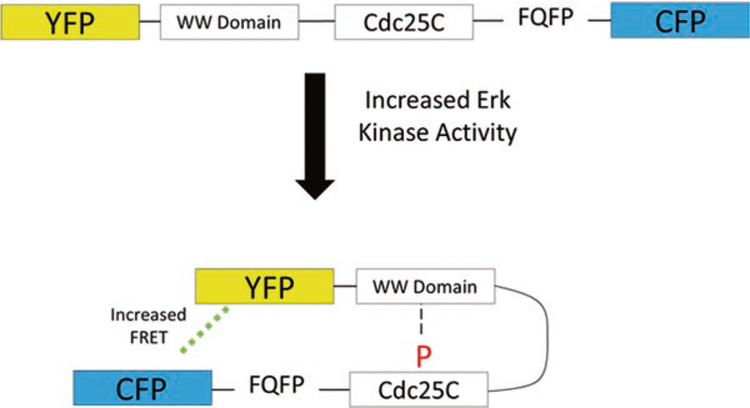
Living cells employ complex and highly dynamic signaling networks and transcriptional circuits to maintain homeostasis and respond appropriately to constantly changing environments. These networks enable cells to maintain tight control on intracellular concentrations of ions, metabolites, proteins, and other biomolecules and ensure a careful balance between a cell's energetic needs and catabolic processes required for growth. Establishing molecular mechanisms of genetic and pharmacological perturbations remains challenging, due to the interconnected nature of these networks and the extreme sensitivity of cellular systems to their external environment. Live cell imaging with genetically encoded fluorescent biosensors provides a powerful new modality for nondestructive spatiotemporal tracking of ions, small molecules, enzymatic activities, and molecular interactions in living systems, from cells, tissues, and even living organisms. By deploying large panels of cell lines, each with distinct biosensors, many critical biochemical pathways can be monitored in a highly parallel and high-throughput fashion to identify pharmacological vulnerabilities and combination therapies unique to a given cell type or genetic background. Here we describe the experimental and analytical methods required to conduct multiplexed parallel fluorescence microscopy experiments on live cells expressing stable transgenic synthetic protein biosensors.
Keywords: FRET; Fluorescence microscopy; Fluorescent biosensor; Mechanism of action; Profiling.
Figures

Similar articles
-
Multiplexed visualization of dynamic signaling networks using genetically encoded fluorescent protein-based biosensors.Pflugers Arch. 2013 Mar;465(3):373-81. doi: 10.1007/s00424-012-1175-y. Epub 2012 Nov 9. Pflugers Arch. 2013. PMID: 23138230 Free PMC article. Review.
-
Genetically encoded FRET-based biosensors for multiparameter fluorescence imaging.Curr Opin Biotechnol. 2009 Feb;20(1):19-27. doi: 10.1016/j.copbio.2009.01.003. Epub 2009 Feb 14. Curr Opin Biotechnol. 2009. PMID: 19223167
-
FRET microscopy for real-time monitoring of signaling events in live cells using unimolecular biosensors.J Vis Exp. 2012 Aug 20;(66):e4081. doi: 10.3791/4081. J Vis Exp. 2012. PMID: 22929080 Free PMC article.
-
FRET Microscopy for Real-Time Visualization of Second Messengers in Living Cells.Methods Mol Biol. 2017;1563:85-90. doi: 10.1007/978-1-4939-6810-7_6. Methods Mol Biol. 2017. PMID: 28324603
-
Imaging and tracing of intracellular metabolites utilizing genetically encoded fluorescent biosensors.Biotechnol J. 2013 Nov;8(11):1280-91. doi: 10.1002/biot.201300001. Epub 2013 Sep 25. Biotechnol J. 2013. PMID: 24591186 Review.
Cited by
-
Deciphering cell signaling networks with massively multiplexed biosensor barcoding.Cell. 2021 Dec 9;184(25):6193-6206.e14. doi: 10.1016/j.cell.2021.11.005. Epub 2021 Nov 26. Cell. 2021. PMID: 34838160 Free PMC article.
References
-
- Terai T, Nagano T (2013) Small-molecule fluorophores and fluorescent probes for bioimaging. Pflugers Arch 465:347–359 - PubMed
-
- Mohsin M, Ahmad A, Iqbal M (2015) FRET-based genetically-encoded sensors for quantitative monitoring of metabolites. Biotechnol Lett 37:1919–1928 - PubMed
-
- Greer LF 3rd, Szalay AA (2002) Imaging of light emission from the expression of luciferases in living cells and organisms: a review. Luminescence 17:43–74 - PubMed
-
- Padilla-Parra S, Tramier M (2012) FRET microscopy in the living cell: different approaches, strengths and weaknesses. BioEssays 34:369–376 - PubMed
-
- Sanford L, Palmer A (2017) Recent advances in development of genetically encoded fluorescent sensors. Methods Enzymol 589:1–49 - PubMed
Publication types
MeSH terms
Substances
Grants and funding
LinkOut - more resources
Full Text Sources
