. 2019 Aug;23(8):5063-5075.
doi: 10.1111/jcmm.14367. Epub 2019 May 23.

Sulodexide attenuates endoplasmic reticulum stress induced by myocardial ischaemia/reperfusion by activating the PI3K/Akt pathway

Affiliations

Sulodexide attenuates endoplasmic reticulum stress induced by myocardial ischaemia/reperfusion by activating the PI3K/Akt pathway

Danping Shen et al. J Cell Mol Med. 2019 Aug.

Abstract

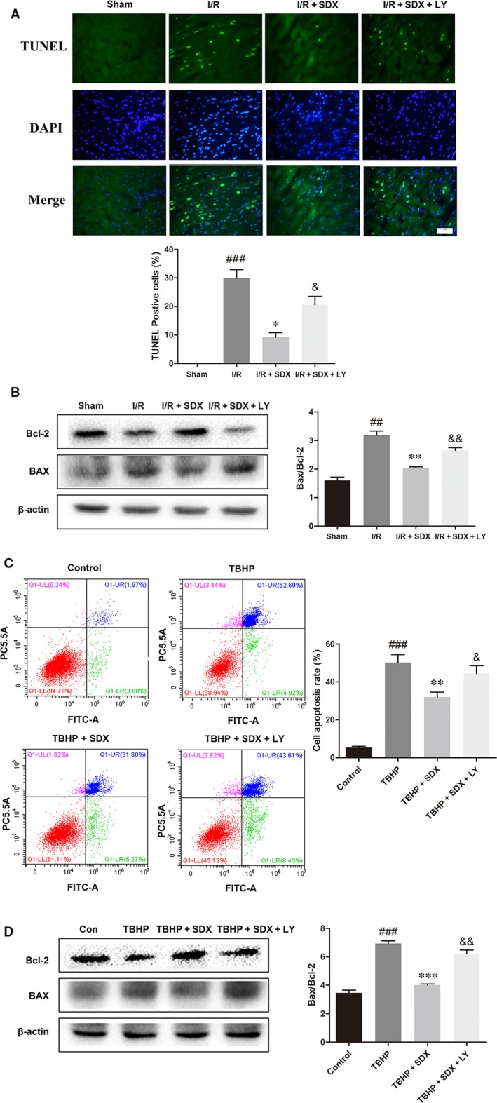
Acute myocardial ischaemia/reperfusion (MI/R) injury causes severe arrhythmias with a high rate of lethality. Extensive research focus on endoplasmic reticulum (ER) stress and its dysfunction which leads to cardiac injury in MI/R Our study evaluated the effects of sulodexide (SDX) on MI/R by establishing MI/R mice models and in vitro oxidative stress models in H9C2 cells. We found that SDX decreases cardiac injury during ischaemia reperfusion and decreased myocardial apoptosis and infarct area, which was paralleled by increased superoxide dismutase and reduced malondialdehyde in mice plasm, increased Bcl-2 expression, decreased BAX expression in a mouse model of MI/R. In vitro, SDX exerted a protective effect by the suppression of the ER stress which induced by tert-butyl hydroperoxide (TBHP) treatment. Both of the in vivo and in vitro effects were involved in the phosphatidylinositol 3-kinase (PI3K)/Akt signalling pathway. Inhibition of PI3K/Akt pathway by specific inhibitor, LY294002, partially reduced the protective effect of SDX. In short, our results suggested that the cardioprotective role of SDX was related to the suppression of ER stress in mice MI/R models and TBHP-induced H9C2 cell injury which was through the PI3K/Akt signalling pathway.

Keywords: PI3K/Akt; endoplasmic reticulum stress; myocardial ischaemia / reperfusion; sulodexide.


Conflict of interest statement

The authors confirm that the content of this article has no conflicts of interest.

Figures

Figure 1
Figure 1
Sulodexide (SDX) pre‐treatment reduces myocardial ischaemia/reperfusion injury. A, Representative images and quantitative analysis of the infarct area (INF: white), area at risk (AAR: red and white) and normal area (blue). B, Superoxide dismutase activities and malondialdehyde leaves in mice serum. Data are shown as means ± SEM; # P < 0.05, ### P < 0.001, I/R (pre‐treated with normal saline) control group vs Sham group; *P < 0.05, **P < 0.01, I/R + SDX (pre‐treated with SDX 60 µg) group vs I/R control group; n = 3 per group
Figure 2
Figure 2
Sulodexide (SDX) reduces myocardial apoptosis and the BAX/Bcl‐2 proteins level in the hearts of mice after myocardial ischaemia/reperfusion. A, Representative images and quantitative analysis of terminal deoxynucleotidyl transferase‐mediated dUTP nick end labelling (TUNEL) immunofluorescence from the ischaemic area in the hearts of mice. B, The detection and optical density analysis of BAX/Bcl‐2 apoptosis proteins in the hearts. Data are shown as means ± SEM; ### P < 0.001, I/R group vs Sham group, *P < 0.05, **P < 0.01, I/R + SDX group vs I/R group; n = 3 per group
Figure 3
Figure 3
The effect of sulodexide (SDX) on endoplasmic reticulum stress‐related proteins in the hearts of mice after myocardial ischaemia/reperfusion. A, The protein expression levels and optical density analysis of C/EBP homologous protein (CHOP), glucose‐regulated protein 78 (GRP78) and activating transcription factor 6 (ATF‐6) in the hearts. B, The protein expression levels and optical density analysis of p‐PI3K, PI3K, p‐AKT and AKT in the hearts. Data are shown as means ± SEM; # P < 0.05, ## P < 0.01, ### P < 0.001, I/R group vs Sham group, *P < 0.05, **P < 0.01, I/R + SDX group vs I/R group; n = 3 per group
Figure 4
Figure 4
Sulodexide (SDX) inhibits apoptosis induced by tert‐butyl hydroperoxide (TBHP) in H9C2 cells. A, H9C2 cells were treated with different concentrations of TBHP for 12 h and then cell viability was assessed by the MTS assay. H9C2 cells were pre‐treated with different concentrations of SDX for 30 min, 100 µmol/L TBHP was added for an additional 12 h and then cell viability was assessed by the MTS assay. **P < 0.01, ***P < 0.001, different groups vs control group. B, H9C2 cells were pre‐treated with 0.25 µg/mL SDX for 30 min and then 100 µmol/L TBHP was added for an additional 12 h. Cells were then stained with Annexin V‐fluorescein isothiocyanate (FITC)/propidium iodide and detected by flow cytometry. The apoptosis ratio was quantified by BD FACS software. C, The detection and optical density analysis of BAX/Bcl‐2 apoptosis proteins in the H9C2 cells. Data are shown as means ± SEM; ### P < 0.001, TBHP group vs control group; *P < 0.05, **P < 0.01, TBHP + SDX group vs TBHP group; n = 3 per group
Figure 5
Figure 5
Sulodexide (SDX) attenuates endoplasmic reticulum stress‐related proteins induced by tert‐butyl hydroperoxide (TBHP) in H9C2 cells. A, The protein expression levels and optical density analysis of C/EBP homologous protein (CHOP), glucose‐regulated protein 78 (GRP78) and activating transcription factor 6 (ATF‐6) in H9C2 cells. B, The protein expression levels and optical density analysis of p‐PI3K, PI3K, p‐AKT and AKT in H9C2 cells. Data are shown as means ± SEM; # P < 0.05, ## P < 0.01, TBHP group vs control group; *P < 0.05, TBHP + SDX group vs TBHP group; n = 3 per group
Figure 6
Figure 6
Inhibition of the PI3K/Akt pathway partially attenuates sulodexide (SDX)‐mediated reduction of myocardial cell apoptosis in vivo and in vitro. A, Representative images and quantitative analysis of terminal deoxynucleotidyl transferase‐mediated dUTP nick end labelling (TUNEL) immunofluorescence from the ischaemic area in the hearts of mice. B, The detection and optical density analysis of BAX/Bcl‐2 apoptosis proteins in the hearts. C, H9C2 cells were pre‐treated with 0.25 µg/mL SDX with or without the specific inhibitors LY294002 (20 µmol/L) for 30 min and then 100 µmol/L tert‐butyl hydroperoxide (TBHP) was added for an additional 12 h. Cells were collected and stained with Annexin V‐FITC/propidium iodide and detected by flow cytometry. The apoptosis ratio was quantified by BD FACS software. D, The detection and optical density analysis of BAX/Bcl‐2 apoptosis proteins in the H9C2 cells. Data are shown as means ± SEM; ## P < 0.01, ### P < 0.001, I/R group vs Sham group, TBHP group vs control group; *P < 0.05, **P < 0.01, ***P < 0.001, I/R + SDX group vs I/R group, TBHP + SDX group vs TBHP group; & P < 0.05, && P < 0.01, I/R + SDX + LY294002 (LY) group vs I/R + SDX group, TBHP + SDX + LY vs TBHP + SDX; n = 3 per group
Figure 7
Figure 7
Inhibition of the PI3K/Akt pathway partially attenuates sulodexide (SDX)‐mediated reduction in the endoplasmic reticulum stress in mice. A, The protein expression levels and optical density analysis of C/EBP homologous protein (CHOP), glucose‐regulated protein 78 (GRP78) and activating transcription factor 6 (ATF‐6) in the hearts. B, The protein expression levels and optical density analysis of p‐PI3K, PI3K, p‐AKT and AKT in the hearts. Data are shown as means ± SEM; ## P < 0.01, ### P < 0.001, I/R group vs Sham group; *P < 0.05, **P < 0.01, ***P < 0.001, I/R + SDX group vs I/R group; & P < 0.05, && P < 0.01, &&& P < 0.001, I/R + SDX + LY294002 (LY) group vs I/R + SDX group; n = 3 per group
Figure 8
Figure 8
Inhibition of the PI3K/Akt pathway partially attenuates sulodexide (SDX)‐mediated reduction in the endoplasmic reticulum stress in H9C2 cells. A, The protein expression levels and optical density analysis of C/EBP homologous protein (CHOP), glucose‐regulated protein 78 (GRP78) and activating transcription factor 6 (ATF‐6) in H9C2 cells. B, The protein expression levels and optical density analysis of p‐PI3K, PI3K, p‐AKT and AKT in H9C2 cells. Data are shown as means ± SEM; # P < 0.05, ## P < 0.01, ### P < 0.001, tert‐butyl hydroperoxide (TBHP) group vs control group; *P < 0.05, **P < 0.01, ***P < 0.001, TBHP + SDX group vs TBHP group; & P < 0.05, && P < 0.01, &&& P < 0.001, TBHP + SDX + LY294002 (LY) vs TBHP + SDX, n = 3 per group

Similar articles

Cited by

References

    1. Yang CF. Clinical manifestations and basic mechanisms of myocardial ischemia/reperfusion injury. Ci Ji Yi Xue Za Zhi. 2018;30:209‐215. - PMC - PubMed
    1. Forouzanfar MH, Moran AE, Flaxman AD, et al. Assessing the global burden of ischemic heart disease, part 2: analytic methods and estimates of the global epidemiology of ischemic heart disease in 2010. Glob Heart. 2012;7:331‐342. - PMC - PubMed
    1. Monassier JP. Reperfusion injury in acute myocardial infarction. From bench to cath lab. Part I: basic considerations. Arch Cardiovasc Dis. 2008;101:491‐500. - PubMed
    1. Penna C, Perrelli MG, Tullio F, et al. Diazoxide postconditioning induces mitochondrial protein S‐nitrosylation and a redox‐sensitive mitochondrial phosphorylation/translocation of RISK elements: no role for SAFE. Basic Res Cardiol. 2013;108:371. - PubMed
    1. Zhang L, Cao S, Deng S, Yao G, Yu T. Ischemic postconditioning and pinacidil suppress calcium overload in anoxia‐reoxygenation cardiomyocytes via down‐regulation of the calcium‐sensing receptor. PeerJ. 2016;4:e2612. - PMC - PubMed

Publication types

MeSH terms

Substances