

Mechanically Biomimetic Gelatin-Gellan Gum Hydrogels for 3D Culture of Beating Human Cardiomyocytes
- PMID: 31120238
- PMCID: PMC6750838
- DOI: 10.1021/acsami.8b22343
Mechanically Biomimetic Gelatin-Gellan Gum Hydrogels for 3D Culture of Beating Human Cardiomyocytes
Abstract
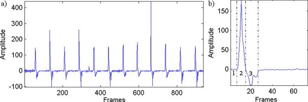
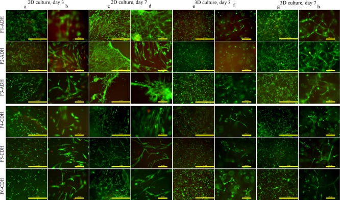
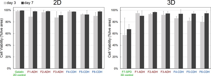
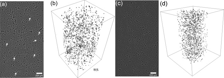
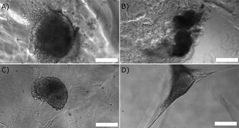
To promote the transition of cell cultures from 2D to 3D, hydrogels are needed to biomimic the extracellular matrix (ECM). One potential material for this purpose is gellan gum (GG), a biocompatible and mechanically tunable hydrogel. However, GG alone does not provide attachment sites for cells to thrive in 3D. One option for biofunctionalization is the introduction of gelatin, a derivative of the abundant ECM protein collagen. Unfortunately, gelatin lacks cross-linking moieties, making the production of self-standing hydrogels difficult under physiological conditions. Here, we explore the functionalization of GG with gelatin at biologically relevant concentrations using semiorthogonal, cytocompatible, and facile chemistry based on hydrazone reaction. These hydrogels exhibit mechanical behavior, especially elasticity, which resembles the cardiac tissue. The use of optical projection tomography for 3D cell microscopy demonstrates good cytocompatibility and elongation of human fibroblasts (WI-38). In addition, human-induced pluripotent stem cell-derived cardiomyocytes attach to the hydrogels and recover their spontaneous beating in 24 h culture. Beating is studied using in-house-built phase contrast video analysis software, and it is comparable with the beating of control cardiomyocytes under regular culture conditions. These hydrogels provide a promising platform to transition cardiac tissue engineering and disease modeling from 2D to 3D.
Keywords: 3D hydrogel; compression testing; gelatin; gellan gum; hiPSC-derived cardiomyocytes.
Conflict of interest statement
The authors declare no competing financial interest.
Figures

Similar articles
-
Contractile force generation by 3D hiPSC-derived cardiac tissues is enhanced by rapid establishment of cellular interconnection in matrix with muscle-mimicking stiffness.Biomaterials. 2017 Jul;131:111-120. doi: 10.1016/j.biomaterials.2017.03.039. Epub 2017 Mar 30. Biomaterials. 2017. PMID: 28384492 Free PMC article.
-
Bioamine-crosslinked gellan gum hydrogel for neural tissue engineering.Biomed Mater. 2017 Mar 24;12(2):025014. doi: 10.1088/1748-605X/aa62b0. Biomed Mater. 2017. PMID: 28233757
-
Optical projection tomography as a quantitative tool for analysis of cell morphology and density in 3D hydrogels.Sci Rep. 2021 Mar 22;11(1):6538. doi: 10.1038/s41598-021-85996-8. Sci Rep. 2021. PMID: 33753803 Free PMC article.
-
Advances in tissue engineering of gellan gum-based hydrogels.Carbohydr Polym. 2024 Jan 15;324:121484. doi: 10.1016/j.carbpol.2023.121484. Epub 2023 Oct 13. Carbohydr Polym. 2024. PMID: 37985043 Review.
-
Biological Role of Gellan Gum in Improving Scaffold Drug Delivery, Cell Adhesion Properties for Tissue Engineering Applications.Molecules. 2019 Dec 10;24(24):4514. doi: 10.3390/molecules24244514. Molecules. 2019. PMID: 31835526 Free PMC article. Review.
Cited by
-
Gellan gum-gelatin based cardiac models support formation of cellular networks and functional cardiomyocytes.Cytotechnology. 2024 Aug;76(4):483-502. doi: 10.1007/s10616-024-00630-5. Epub 2024 May 2. Cytotechnology. 2024. PMID: 38933872 Free PMC article.
-
Effect of hydroxychloroquine sulfate on the gelation behavior, water mobility and structure of gelatin.Colloids Surf A Physicochem Eng Asp. 2022 Jan 20;633:127849. doi: 10.1016/j.colsurfa.2021.127849. Epub 2021 Nov 3. Colloids Surf A Physicochem Eng Asp. 2022. PMID: 34744314 Free PMC article.
-
Building blocks of microphysiological system to model physiology and pathophysiology of human heart.Front Physiol. 2023 Jul 6;14:1213959. doi: 10.3389/fphys.2023.1213959. eCollection 2023. Front Physiol. 2023. PMID: 37485060 Free PMC article. Review.
-
Cardiac Tissue Engineering for the Treatment of Myocardial Infarction.J Cardiovasc Dev Dis. 2021 Nov 8;8(11):153. doi: 10.3390/jcdd8110153. J Cardiovasc Dev Dis. 2021. PMID: 34821706 Free PMC article. Review.
-
Advances in Engineering Human Tissue Models.Front Bioeng Biotechnol. 2021 Jan 28;8:620962. doi: 10.3389/fbioe.2020.620962. eCollection 2020. Front Bioeng Biotechnol. 2021. PMID: 33585419 Free PMC article.
References
-
- Langley G. R.; Adcock I. M.; Busquet F.; Crofton K. M.; Csernok E.; Giese C.; Heinonen T.; Herrmann K.; Hofmann-Apitius M.; Landesmann B.; Marshall L. J.; McIvor E.; Muotri A. R.; Noor F.; Schutte K.; Seidle T.; van de Stolpe A.; Van Esch H.; Willett C.; Woszczek G. Towards a 21st-century roadmap for biomedical research and drug discovery: consensus report and recommendations. Drug Discov. Today 2017, 22, 327–339. 10.1016/j.drudis.2016.10.011. - DOI - PubMed
-
- Akopian V.; Andrews P. W.; Beil S.; Benvenisty N.; Brehm J.; Christie M.; Ford A.; Fox V.; Gokhale P. J.; Healy L.; Holm F.; Hovatta O.; Knowles B. B.; Ludwig T. E.; McKay R. D. G.; Miyazaki T.; Nakatsuji N.; Oh S. K. W.; Pera M. F.; Rossant J.; Stacey G. N.; Suemori H. Comparison of defined culture systems for feeder cell free propagation of human embryonic stem cells. In Vitro Cell. Dev. Biol.: Anim. 2010, 46, 247–258. 10.1007/s11626-010-9297-z. - DOI - PMC - PubMed
-
- Lu H. R.; Whittaker R.; Price J. H.; Vega R.; Pfeiffer E. R.; Cerignoli F.; Towart R.; Gallacher D. J. High Throughput Measurement of Ca++Dynamics in Human Stem Cell-Derived Cardiomyocytes by Kinetic Image Cytometery: A Cardiac Risk Assessment Characterization Using a Large Panel of Cardioactive and Inactive Compounds. Toxicol. Sci. 2015, 148, 503–516. 10.1093/toxsci/kfv201. - DOI - PubMed
MeSH terms
Substances
LinkOut - more resources
Full Text Sources
Other Literature Sources
