. 2019 May 23;13(5):e0007270.
doi: 10.1371/journal.pntd.0007270. eCollection 2019 May.

Revisiting the taxonomy and evolution of pathogenicity of the genus Leptospira through the prism of genomics

Affiliations

Revisiting the taxonomy and evolution of pathogenicity of the genus Leptospira through the prism of genomics

Antony T Vincent et al. PLoS Negl Trop Dis. .

Abstract

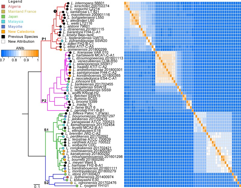
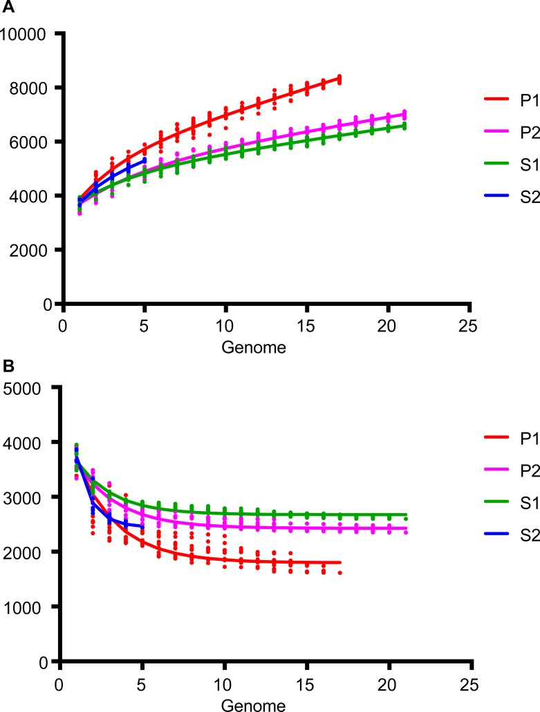
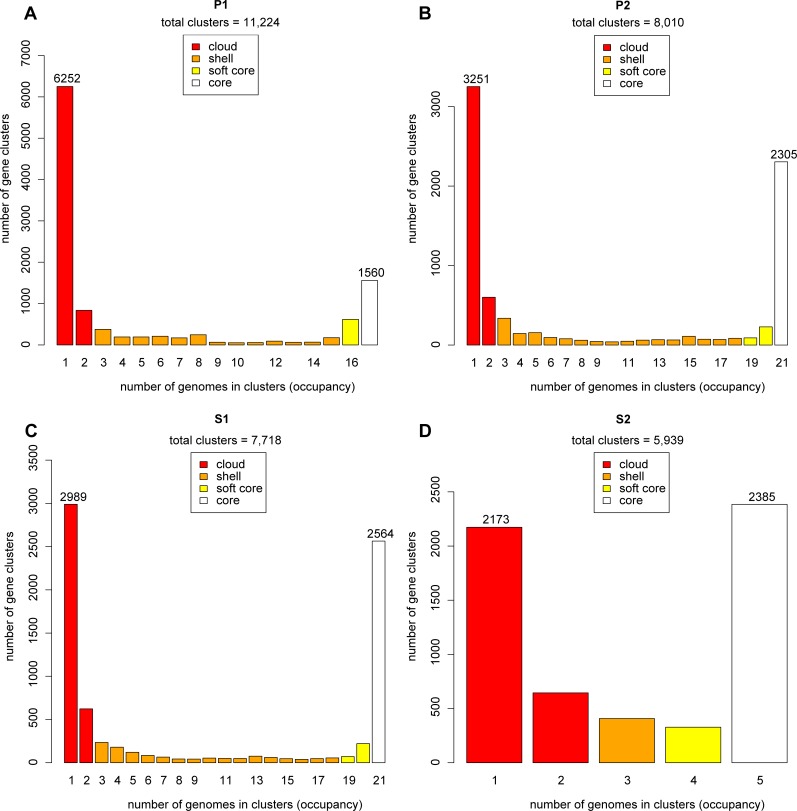
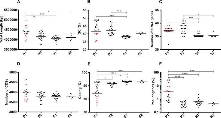
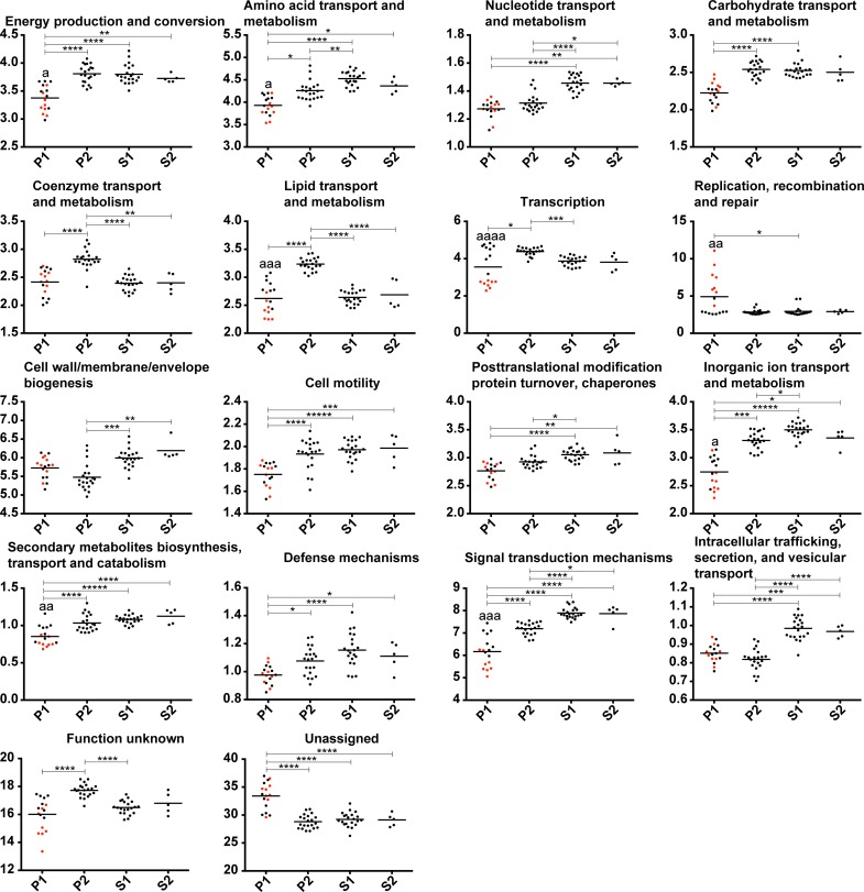
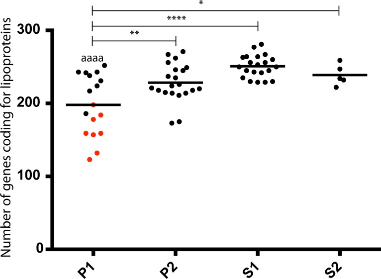
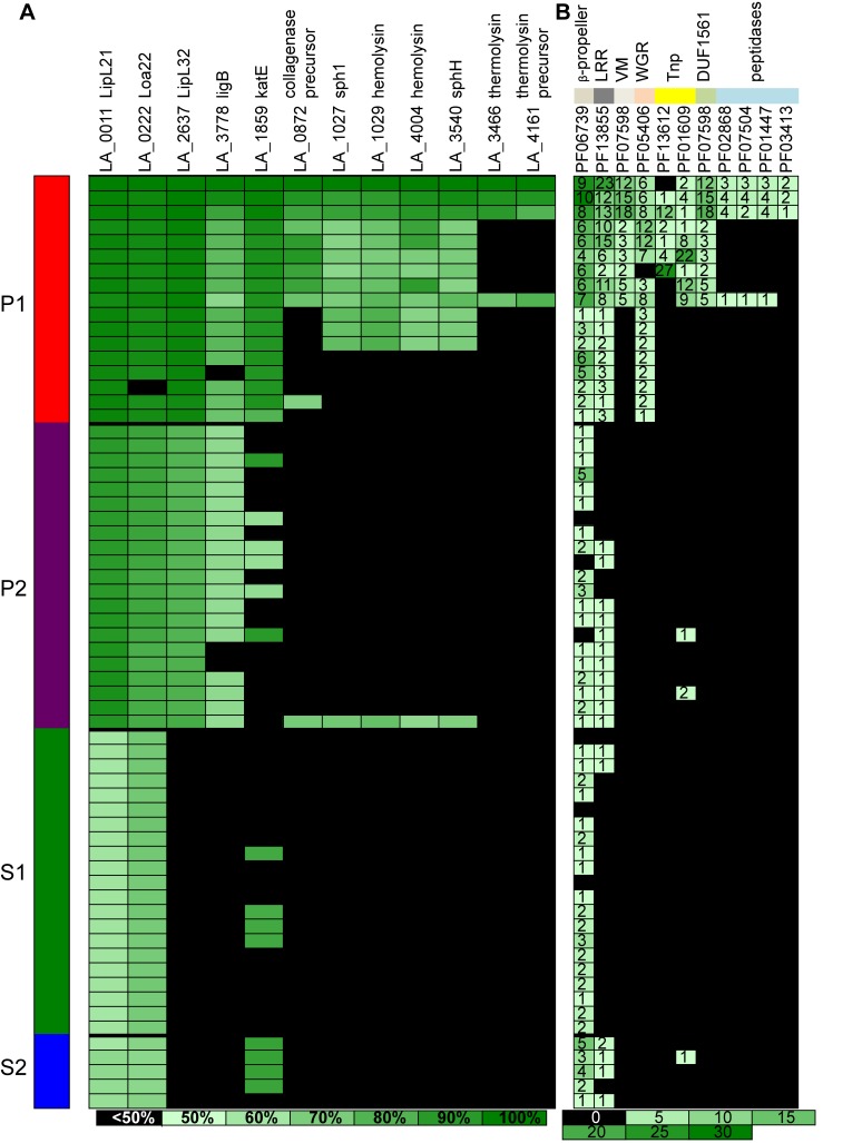
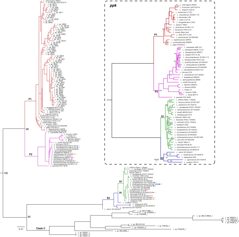
The causative agents of leptospirosis are responsible for an emerging zoonotic disease worldwide. One of the major routes of transmission for leptospirosis is the natural environment contaminated with the urine of a wide range of reservoir animals. Soils and surface waters also host a high diversity of non-pathogenic Leptospira and species for which the virulence status is not clearly established. The genus Leptospira is currently divided into 35 species classified into three phylogenetic clusters, which supposedly correlate with the virulence of the bacteria. In this study, a total of 90 Leptospira strains isolated from different environments worldwide including Japan, Malaysia, New Caledonia, Algeria, mainland France, and the island of Mayotte in the Indian Ocean were sequenced. A comparison of average nucleotide identity (ANI) values of genomes of the 90 isolates and representative genomes of known species revealed 30 new Leptospira species. These data also supported the existence of two clades and 4 subclades. To avoid classification that strongly implies assumption on the virulence status of the lineages, we called them P1, P2, S1, S2. One of these subclades has not yet been described and is composed of Leptospira idonii and 4 novel species that are phylogenetically related to the saprophytes. We then investigated genome diversity and evolutionary relationships among members of the genus Leptospira by studying the pangenome and core gene sets. Our data enable the identification of genome features, genes and domains that are important for each subclade, thereby laying the foundation for refining the classification of this complex bacterial genus. We also shed light on atypical genomic features of a group of species that includes the species often associated with human infection, suggesting a specific and ongoing evolution of this group of species that will require more attention. In conclusion, we have uncovered a massive species diversity and revealed a novel subclade in environmental samples collected worldwide and we have redefined the classification of species in the genus. The implication of several new potentially infectious Leptospira species for human and animal health remains to be determined but our data also provide new insights into the emergence of virulence in the pathogenic species.


Conflict of interest statement

The authors have declared that no competing interests exist.

Figures

Fig 1
Fig 1. Phylogenetic tree based on the sequences of 1371 genes inferred as orthologous.
The matrix represents the calculated ANIb values for all the genomic sequences. The branches are colored according to their belonging to the four main subclades: P1 (red), P2 (purple), S1 (green) and S2 (blue). The bootstrap value is indicated for a single node (that corresponding to the separation between L. biflexa strain Patoc1 and L. bouyouniensis strain 201601297) since all the others have the maximum value of 100. A circle of color, according to the legend, represents the geographical origin of each of the new species described by this study. Node 1 indicates the node from which descent pathogenic species most frequently involved in human disease.
Fig 2
Fig 2
Evolution of (A) pan-genome and (B) core-genome for the genomes of the four leptospiral subclades. Analyses done with GET_HOMOLOGUES (using 17 genomes for P1, 21 for P2, 21 for S1 and 5 for S2) highlighting that the P1 group has a more open pan-genome.
Fig 3
Fig 3
Pan-genome distribution in four categories (cloud, shell, soft core and core) for species from subclades (A) P1, (B) P2, (C) S1 and (D) S2. Analyses done with GET_HOMOLOGUES (using 17 genomes for P1, 21 for P2, 21 for S1 and 5 for S2) showing the U-shaped distribution of pan-genome from the four groups. However, strains of the P1 group show asymmetry by having four times more single species than core genes.
Fig 4
Fig 4
Distribution of (A) total length, (B) GC %, (C) number of tRNA genes, (D) number of CDSs, (E) coding % and (F) pseudogenes % (values in log) in the four major subclades. The points representing the genome-specific values of the species that diverged after node 1 in Fig 1 (L. interrogans, L. kirschneri, L. noguchii, L. santarosai, L. mayottensis, L. borgpetersenii, L. alexanderi and L. weilii) are in red. The "*" represent the level of significance between the different groups: * P ≤ 0.05, ** P ≤ 0.01, *** P ≤ 0.001, and **** P ≤ 0.0001. The level of significance between the two P1 groups separated by node 1 is represented by the same code, but for the sake of clarity the symbol is "a".
Fig 5
Fig 5. Distribution in functional categories of the predicted CDSs (%).
The points representing the genome-specific values of the species that diverged after node 1 in Fig 1 (L. interrogans, L. kirschneri, L. noguchii, L. santarosai, L. mayottensis, L. borgpetersenii, L. alexanderi and L. weilii) are in red. The "*" represent the level of significance between the different groups: * P ≤ 0.05, ** P ≤ 0.01, *** P ≤ 0.001, and **** P ≤ 0.0001. The level of significance between the two pathogenic groups is represented by the same code, but for the sake of clarity the symbol is "a". Only functional categories showing significant difference are shown.
Fig 6
Fig 6. Distribution of genes encoding lipoproteins.
The "*" represent the level of significance between the different groups: * P ≤ 0.05, ** P ≤ 0.01, *** P ≤ 0.001, and **** P ≤ 0.0001. The level of significance between the two pathogenic P1 groups (before and after node 1) is represented by the same code, but for the sake of clarity the symbol is "a". The points representing the genome-specific values of the species that diverged after node 1 in Fig 1 (L. interrogans, L. kirschneri, L. noguchii, L. santarosai, L. mayottensis, L. borgpetersenii, L. alexanderi and L. weilii) are in red.
Fig 7
Fig 7
Distribution of genes involved in virulence (A) and PFAM motifs (B). The gradient represents for (A) the percentage of similarity according to the homologous proteins sequences in L. interrogans strain 56601 and for (B) the number of genes having the different PFAM motifs.
Fig 8
Fig 8. Phylogenetic tree based on the 16S rRNA and ppk sequences to evaluate the diversity within the Leptospira genus.
In addition to the 16S rRNA sequences from the 64 genomes investigated in the present study, those from uncultured strains from the Peruvian Amazon (Clade C) [26] and from insectivorous bats from eastern China [27] were added. The branches are colored according to their belonging to the four main subclades: P1 (red), P2 (purple), S1 (green) and S2 (blue), while the strains of the “clade C” are in black. For the sake of clarity, the bootstrap values are only indicated for the nodes that correspond to the major splits. A tree constructed with the ppk gene sequences is included in the dashed box for comparison. In this case, all bootstrap values less than 100 are indicated at the different nodes.

References

    1. Costa F, Hagan JE, Calcagno J, Kane M, Torgerson P, Martinez-Silveira MS, et al. Global Morbidity and Mortality of Leptospirosis: A Systematic Review. PLoS Negl Trop Dis 2015;9:e0003898 10.1371/journal.pntd.0003898 - DOI - PMC - PubMed
    1. Ellis WA. Animal Leptospirosis In: Adler B, editor. Leptospira and Leptospirosis: Springer; 2014. p. 99–137.
    1. Faine SB, Adler B, Bolin C, Perolat P. Leptospira and leptospirosis. 2nd ed Melbourne, Australia: MediSci; 1999.
    1. Ko AI, Goarant C, Picardeau M. Leptospira: the dawn of the molecular genetics era for an emerging zoonotic pathogen. Nat Rev Microbiol 2009;7:736–47. 10.1038/nrmicro2208 - DOI - PMC - PubMed
    1. Stimson AM. Note on an organism found in yellow-fever tissue. Public Health Reports (Washington). 1907;22:541.

Publication types

LinkOut - more resources