

Transient Receptor Potential Vanilloid 4 Is Required for Foreign Body Response and Giant Cell Formation
- PMID: 31121133
- PMCID: PMC6717909
- DOI: 10.1016/j.ajpath.2019.04.016
Transient Receptor Potential Vanilloid 4 Is Required for Foreign Body Response and Giant Cell Formation
Abstract
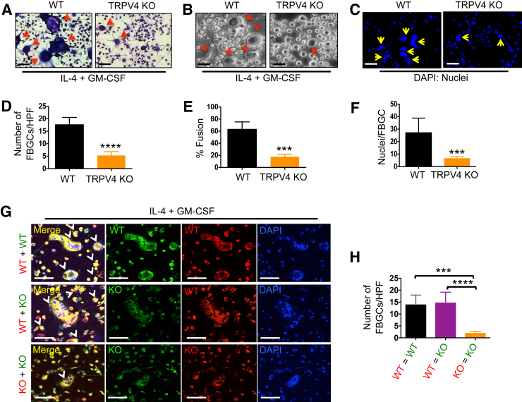
The presence of biomaterials and devices implanted into soft tissue is associated with development of a foreign body response (FBR), a chronic inflammatory condition that can ultimately lead to implant failure, which may cause harm to or death of the patient. Development of FBR includes activation of macrophages at the tissue-implant interface, generation of destructive foreign body giant cells (FBGCs), and generation of fibrous tissue that encapsulates the implant. However, the mechanisms underlying the FBR remain poorly understood, as neither the materials composing the implants nor their chemical properties can explain triggering of the FBR. Herein, we report that genetic ablation of transient receptor potential vanilloid 4 (TRPV4), a Ca2+-permeable mechanosensitive cation channel in the transient receptor potential vanilloid family, protects TRPV4 knockout mice from FBR-related events. The mice showed diminished collagen deposition along with reduced macrophage accumulation and FBGC formation compared with wild-type mice in a s.c. implantation model. Analysis of macrophage markers in spleen tissues and peritoneal cavity showed that the TRPV4 deficiency did not impair basal macrophage maturation. Furthermore, genetic deficiency or pharmacologic antagonism of TRPV4 blocked cytokine-induced FBGC formation, which was restored by lentivirus-mediated TRPV4 reintroduction. Taken together, these results suggest an important, previously unknown, role for TRPV4 in FBR.
Copyright © 2019 American Society for Investigative Pathology. Published by Elsevier Inc. All rights reserved.
Figures


Similar articles
-
Mechanosensing by TRPV4 mediates stiffness-induced foreign body response and giant cell formation.Sci Signal. 2021 Nov 2;14(707):eabd4077. doi: 10.1126/scisignal.abd4077. Epub 2021 Nov 2. Sci Signal. 2021. PMID: 34726952 Free PMC article.
-
Mechanotransduction via a TRPV4-Rac1 signaling axis plays a role in multinucleated giant cell formation.J Biol Chem. 2021 Jan-Jun;296:100129. doi: 10.1074/jbc.RA120.014597. Epub 2020 Dec 4. J Biol Chem. 2021. PMID: 33262217 Free PMC article.
-
The Implant-Induced Foreign Body Response Is Limited by CD13-Dependent Regulation of Ubiquitination of Fusogenic Proteins.J Immunol. 2024 Feb 15;212(4):663-676. doi: 10.4049/jimmunol.2300688. J Immunol. 2024. PMID: 38149920 Free PMC article.
-
Formation and biological activities of foreign body giant cells in response to biomaterials.Acta Biomater. 2024 Oct 15;188:1-26. doi: 10.1016/j.actbio.2024.08.034. Epub 2024 Sep 7. Acta Biomater. 2024. PMID: 39245307 Review.
-
A call for standardization: Evaluating different methodologies to induce in vitro foreign body giant cell formation for biomaterials research and design.Acta Biomater. 2025 Mar 1;194:20-37. doi: 10.1016/j.actbio.2025.01.026. Epub 2025 Jan 16. Acta Biomater. 2025. PMID: 39826854 Review.
Cited by
-
Mechanosensing by TRPV4 mediates stiffness-induced foreign body response and giant cell formation.Sci Signal. 2021 Nov 2;14(707):eabd4077. doi: 10.1126/scisignal.abd4077. Epub 2021 Nov 2. Sci Signal. 2021. PMID: 34726952 Free PMC article.
-
A Case for Material Stiffness as a Design Parameter in Encapsulated Islet Transplantation.Tissue Eng Part B Rev. 2023 Aug;29(4):334-346. doi: 10.1089/ten.TEB.2022.0157. Epub 2023 Feb 1. Tissue Eng Part B Rev. 2023. PMID: 36475851 Free PMC article. Review.
-
Resident cardiac macrophages mediate adaptive myocardial remodeling.Immunity. 2021 Sep 14;54(9):2072-2088.e7. doi: 10.1016/j.immuni.2021.07.003. Epub 2021 Jul 27. Immunity. 2021. PMID: 34320366 Free PMC article.
-
Role of mechanosensitive channels/receptors in atherosclerosis.Am J Physiol Cell Physiol. 2022 May 1;322(5):C927-C938. doi: 10.1152/ajpcell.00396.2021. Epub 2022 Mar 30. Am J Physiol Cell Physiol. 2022. PMID: 35353635 Free PMC article. Review.
-
TRPV4 Plays a Role in Matrix Stiffness-Induced Macrophage Polarization.Front Immunol. 2020 Dec 14;11:570195. doi: 10.3389/fimmu.2020.570195. eCollection 2020. Front Immunol. 2020. PMID: 33381111 Free PMC article.
References
-
- Major M.R., Wong V.W., Nelson E.R., Longaker M.T., Gurtner G.C. The foreign body response: at the interface of surgery and bioengineering. Plast Reconstr Surg. 2015;135:1489–1498. - PubMed
Publication types
MeSH terms
Substances
Grants and funding
LinkOut - more resources
Full Text Sources
Molecular Biology Databases
Miscellaneous
