. 2019 Aug;189(8):1664-1679.
doi: 10.1016/j.ajpath.2019.04.014. Epub 2019 May 20.

Endothelial Hypoxia-Inducible Factor-1α Is Required for Vascular Repair and Resolution of Inflammatory Lung Injury through Forkhead Box Protein M1

Affiliations

Endothelial Hypoxia-Inducible Factor-1α Is Required for Vascular Repair and Resolution of Inflammatory Lung Injury through Forkhead Box Protein M1

Xiaojia Huang et al. Am J Pathol. 2019 Aug.

Abstract

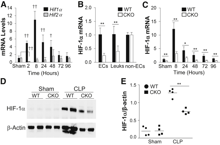
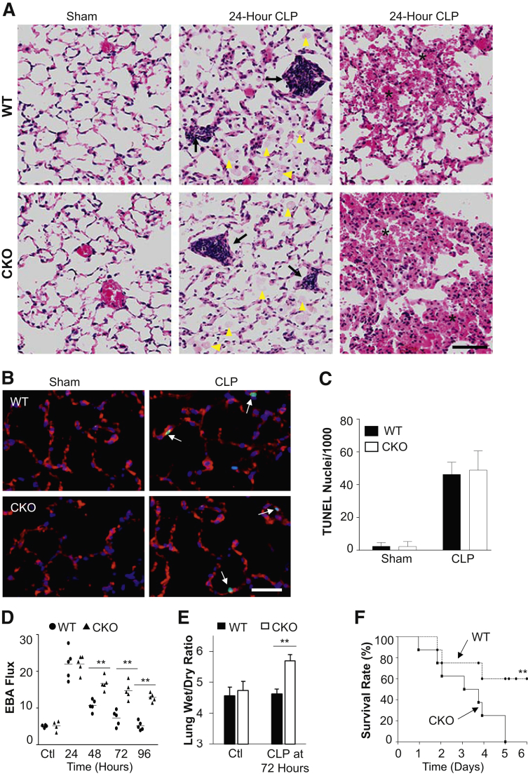
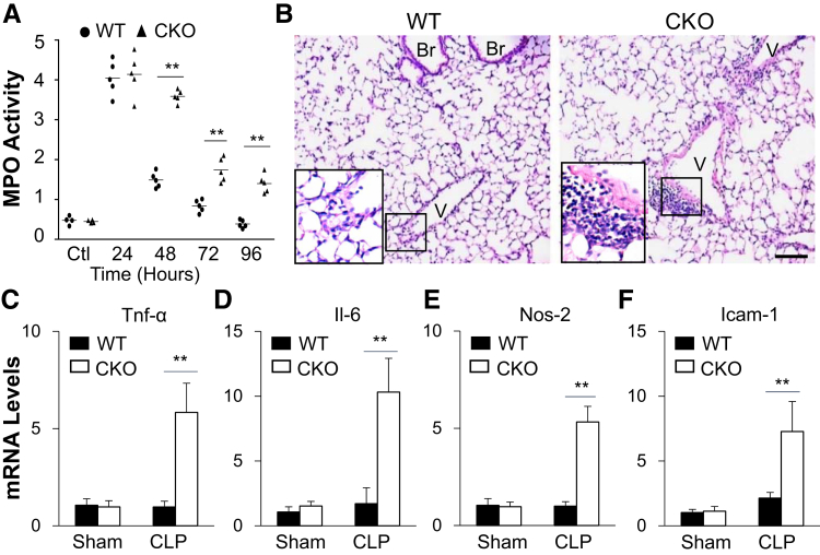
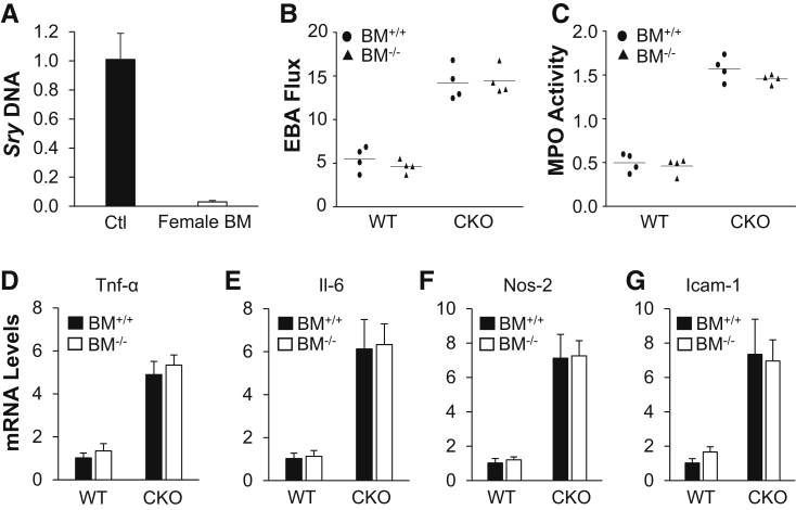
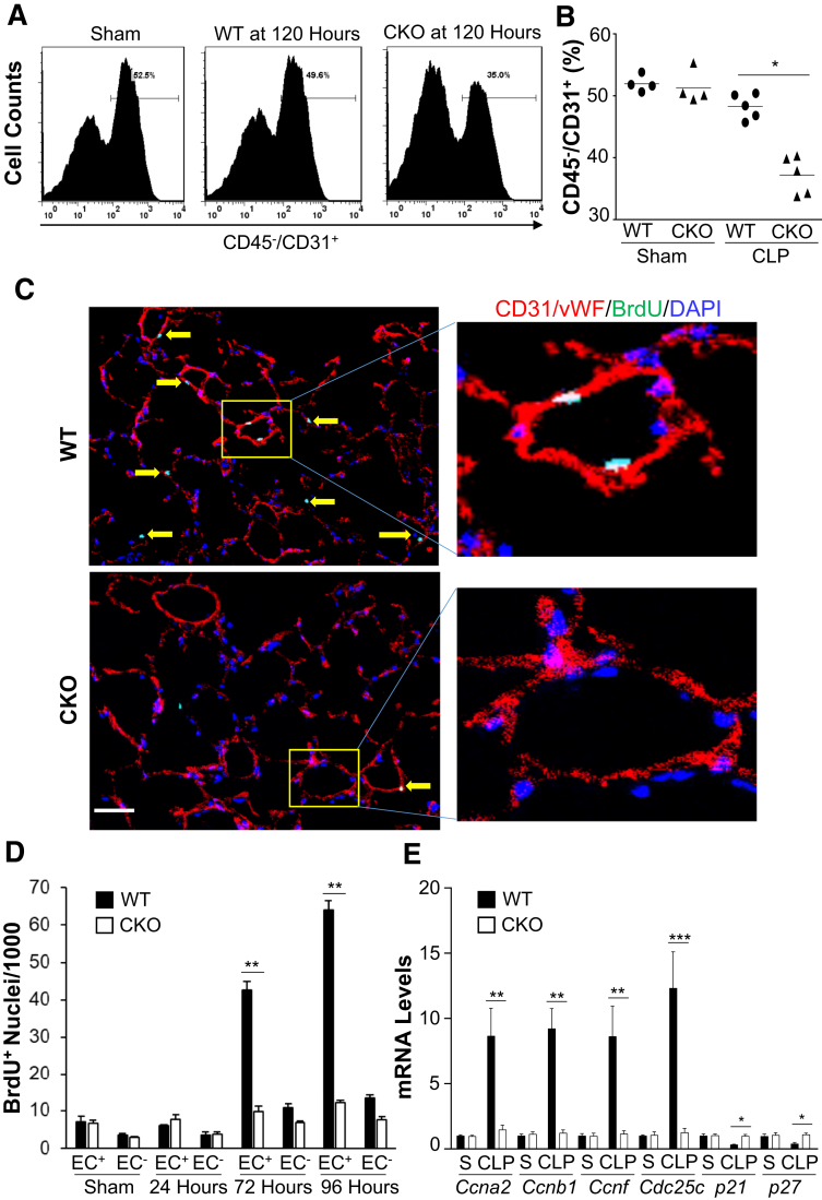
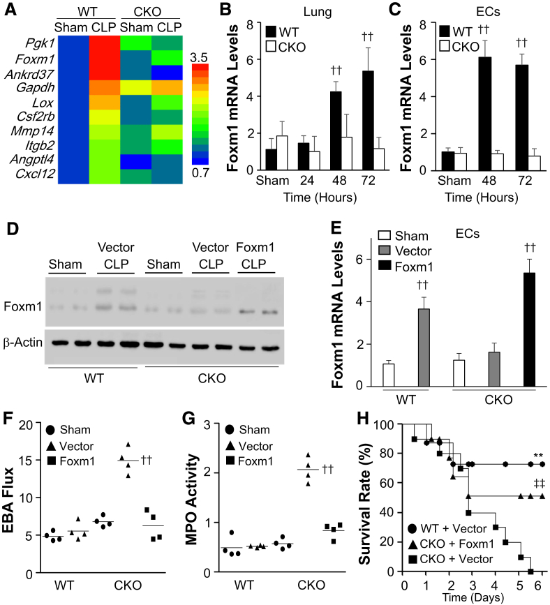
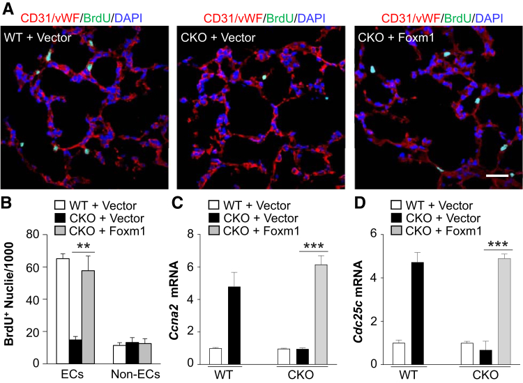
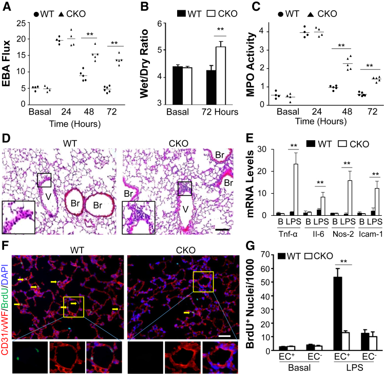
Endothelial barrier dysfunction is a central factor in the pathogenesis of persistent lung inflammation and protein-rich edema formation, the hallmarks of acute respiratory distress syndrome. However, little is known about the molecular mechanisms that are responsible for vascular repair and resolution of inflammatory injury after sepsis challenge. Herein, we show that hypoxia-inducible factor-1α (HIF-1α), expressed in endothelial cells (ECs), is the critical transcriptional factor mediating vascular repair and resolution of inflammatory lung injury. After sepsis challenge, HIF-1α but not HIF-2α expression was rapidly induced in lung vascular ECs, and mice with EC-restricted disruption of Hif1α (Hif1af/f/Tie2Cre+) exhibited defective vascular repair, persistent inflammation, and increased mortality in contrast with the wild-type littermates after polymicrobial sepsis or endotoxemia challenge. Hif1af/f/Tie2Cre+ lungs exhibited marked decrease of EC proliferation during recovery after sepsis challenge, which was associated with inhibited expression of forkhead box protein M1 (Foxm1), a reparative transcription factor. Therapeutic restoration of endothelial Foxm1 expression, via liposomal delivery of Foxm1 plasmid DNA to Hif1af/f/Tie2Cre+ mice, resulted in reactivation of the vascular repair program and improved survival. Together, our studies, for the first time, delineate the essential role of endothelial HIF-1α in driving the vascular repair program. Thus, therapeutic activation of HIF-1α-dependent vascular repair may represent a novel and effective therapy to treat inflammatory vascular diseases, such as sepsis and acute respiratory distress syndrome.


Figures

Figure 1
Figure 1
HIF-1α rapidly induces mouse lung endothelial cells (ECs) after cecal ligation and puncture (CLP) challenge. A: Real-time quantitative RT-PCR (RT-qPCR) analysis demonstrating rapid induction of HIF-1α, but not HIF-2α, expression in lungs of wild-type (WT) mice after CLP. HIF-2α expression is initially decreased at 2 hours after CLP and then returns to basal levels, whereas HIF-1α expression is markedly induced and peaks at 8 hours after CLP. B: HIF-1α mRNA expression in isolated lung ECs (CD45/CD31+), leukocytes (Leuks; CD45+/CD31+), and non-ECs, including epithelial cells and fibroblasts (CD45/CD31), from WT and Hif1af/f/Tie2Cre+ (CKO) mouse lungs by fluorescence-activated cell sorting. C: RT-qPCR analysis demonstrating inhibited HIF-1α induction in Hif1af/f/Tie2Cre+ lungs after CLP challenge. D: Representative Western blot analysis demonstrating marked inhibition of HIF-1α protein expression in CKO mouse lungs at 8 hours after CLP compared with WT lungs. β-Actin was used as a loading control. E: Quantification of Western blot analysis band intensity using ImageJ software version 1.51a (NIH, Bethesda, MD; http://imagej.nih.gov/ij). Data are expressed as means ± SD (AC) and means (E). n = 5 mice per group (A); n = 4 mice (B, demonstrating Tie2Cre-mediated Hif1a deletion in ECs and leukocytes, and C, per group). P < 0.05, ∗∗P < 0.01 (t-test); P < 0.05, ††P < 0.01 compared with WT-sham (one-way analysis of variance with a Tukey's post hoc analysis for multiple-group comparisons and t-test for two-group comparison).
Figure 2
Figure 2
Impaired lung vascular repair and increased mortality in Hif1af/f/Tie2Cre+ (CKO) mice after cecal ligation and puncture (CLP) challenge. A: Representative hematoxylin and eosin staining showing characteristics of the pathology of acute lung injury, including protein leakage (arrowheads), alveolar septum thickening, and inflammatory cell infiltration (arrows), and hemorrhaging (asterisks) at 24 hours after CLP in both wild-type (WT) and Hif1af/f/Tie2Cre+ mice. B: Representative micrographs of terminal deoxynucleotidyl transferase-mediated dUTP nick-end labeling (TUNEL) staining showing apoptosis (green nuclei) at 24 hours after CLP in WT and Hif1af/f/Tie2Cre+ mouse lungs. Endothelial cells (ECs) were immunostained with anti-CD31 and anti–von Willebrand factor (red). Apoptotic nuclei were stained with TUNEL (green). Nuclei were counterstained with DAPI (blue). Arrows indicate apoptotic ECs. C: Quantification of TUNEL-positive ECs. D: Pulmonary transvascular Evans Blue Dye–conjugated albumin (EBA) flux demonstrating defective vascular repair in Hif1af/f/Tie2Cre+ mouse lungs. E: Lung wet/dry weight ratio analysis revealing lung edema in Hif1af/f/Tie2Cre+ mouse at 72 hours after CLP challenge. F:Hif1af/f/Tie2Cre+ mice exhibited greater mortality rate after CLP challenge. Mortality rate was monitored for 6 days after CLP. Data are expressed as means ± SD (C and E) or means (D). n = 5 mice per group (C and E); n = 10 mice per group (F). ∗∗P < 0.01 [one-way analysis of variance with Tukey's post-hoc analysis (D), t-test (E), and Mantel-Cox test (F)]. Scale bar = 100 μm (A and B). Ctl, control.
Figure 3
Figure 3
Impaired resolution of inflammation in Hif1af/f/Tie2Cre+ (CKO) lungs after cecal ligation and puncture (CLP) challenge. A: Time course of myeloperoxidase (MPO) activity in mouse lungs after CLP challenge. B: Representative micrographs of hematoxylin and eosin staining of lung sections showing perivascular leukocyte infiltration in Hif1af/f/Tie2Cre+ mouse lungs at 72 hours after CLP. The boxed areas are shown at higher magnification in the insets in the lower left corners. CF: Marked increases of expression of proinflammatory mediators in Hif1af/f/Tie2Cre+ lungs evaluated by real-time quantitative RT-PCR analysis. At 72 hours after CLP, mouse lungs were collected for analysis. Data are expressed as means (A) or means ± SD (CF). n = 4 mice per group (CF). ∗∗P < 0.01 [one-way analysis of variance with Tukey's post-hoc analysis (A) or t-test (CF)]. Scale bar = 50 μm (B). Br, bronchiole; Icam-1, intercellular adhesion molecule 1; Nos-2, inducible nitric oxide synthase; Tnf-α, tumor necrosis factor–α V, vessel; WT, wild type.
Figure 4
Figure 4
Impaired vascular repair in Hif1af/f/Tie2Cre+ (CKO) mouse lungs was not ascribed to HIF-1α deficiency in bone marrow (BM) cells. A: Quantitative PCR analysis demonstrating >95% efficiency of bone marrow reconstitution. Bone marrow cells from wild-type (WT) female mice were transplanted to lethally irradiated Hif1af/f/Tie2Cre+ male mice. Six weeks after transplantation, bone marrow samples from these chimeric male mice and WT male mice [positive control (Ctl)] were isolated for genomic DNA isolation and PCR analysis of Y-chromosome–specific gene Sry. B: Defective vascular repair in lungs of Hif1af/f/Tie2Cre+ mice transplanted with bone marrow cells from either Hif1α WT (BM+/+) or CKO (BM−/−) mice. At 6 weeks after bone marrow cell transplantation, the mice were challenged with cecal ligation and puncture (CLP). At 72 hours after CLP, lung tissues were collected for transvascular Evans Blue Dye–conjugated albumin (EBA) flux measurement. C: Lung myeloperoxidase (MPO) activity measurement (72 hours after CLP). DG: Real-time quantitative RT-PCR analysis demonstrating increased expression of proinflammatory mediators in Hif1af/f/Tie2Cre+ mouse lungs reconstituted with either WT or CKO bone marrow cells at 72 hours after CLP challenge. Data are expressed as means ± SD (A and D–G) or means (B and C). n = 4 mice per group (A and DG). Icam-1, intercellular adhesion molecule 1; Nos-2, inducible nitric oxide synthase; Tnf-α, tumor necrosis factor–α.
Figure 5
Figure 5
Defective endothelial regeneration in Hif1af/f/Tie2Cre+ (CKO) mouse lungs after cecal ligation and puncture (CLP) challenge. A and B: Flow cytometry quantification of endothelial cells (ECs; CD45/CD31+) in mouse lungs at 120 hours after CLP or sham (S). At 120 hours after CLP or sham, lung tissues were collected for digestion and dissociation. Freshly prepared lung cells were immunostained with anti-CD45 and anti-CD31 antibodies for fluorescence-activated cell sorting analysis by gating CD45- cells. A: Representative histograms showing percentage of ECs in mouse lungs. B: Quantification of ECs (% CD45CD31+ in total CD45 cells) in mouse lungs. C: Representative micrographs showing EC proliferation. Cryosections of lungs (5 μm thick) collected at 96 hours after CLP were immunostained with anti–5-bromo-2-deoxyuridine (BrdU) antibody to identify proliferating cells (green) and with anti-CD31 and anti–von Willebrand factor (vWF) antibodies to identify ECs (red). Nuclei were counterstained with DAPI (blue). The boxed areas in the left panels are shown at higher magnification in the right panels. Arrows point to proliferating ECs. D: Quantification of cell proliferation in mouse lungs. Three consecutive cryosections from each mouse lung were examined; the average number of BrdU-positive nuclei was used. BrdU was administered intraperitoneally to mice at 5 hours before tissue collection. E: Expression of genes regulating cell cycle progression determined by real-time quantitative RT-PCR (RT-qPCR) analysis. At 96 hours after CLP, lung ECs were isolated for RNA extraction and RT-qPCR analysis. Data are expressed as means (B) or means ± SD (D and E). n = 4 (D and E). P < 0.05, ∗∗P < 0.01, and ∗∗∗P < 0.001 (t-test). Scale bar = 50 μm (C). WT, wild type.
Figure 6
Figure 6
Restoration of endothelial forkhead box protein M1 (Foxm1) expression in Hif1af/f/Tie2Cre+ (CKO) mice rescues the defective vascular repair phenotype. A: Heat map of RNA-sequencing (RNA-seq) analysis of gene expression in wild-type (WT) and Hif1af/f/Tie2Cre+ mouse lungs at 72 hours after cecal ligation and puncture (CLP) compared with sham. RNA samples from three mouse lungs were combined for RNA-seq analysis in each group. B and C: Real-time quantitative RT-PCR (RT-qPCR) analysis of Foxm1 expression in WT and Hif1af/f/Tie2Cre+ mouse lung tissues at various times and freshly isolated lung endothelial cells (ECs) after CLP. D: Representative Western blot analysis showing restored Foxm1 protein expression in Foxm1 plasmid DNA-transduced Hif1af/f/Tie2Cre+ mouse lungs. At 12 hours after CLP challenge, a mixture of liposome/plasmid DNA expressing Foxm1 under the control of the CDH5 promoter (Foxm1) or empty vector (Vector) was administered intravenously to Hif1af/f/Tie2Cre+ mice. WT mice were also administered with Vector as controls. At 72 hours after CLP, lung tissues were collected for Western blot analysis with anti-Foxm1 and anti–β-actin (loading control). E: RT-qPCR analysis of Foxm1 expression in ECs freshly isolated from lungs of mice treated as described in D. F: Pulmonary transvascular Evans Blue Dye–conjugated albumin (EBA) flux measurement demonstrating normalized vascular repair of Hif1af/f/Tie2Cre+ mice transduced with Foxm1 plasmid DNA at 72 hours after CLP. G: Myeloperoxidase (MPO) assay demonstrating normalized resolution of lung inflammation in Hif1af/f/Tie2Cre+ mice transduced with Foxm1 plasmid DNA at 72 hours after CLP. H: Restored Foxm1 expression in ECs of Hif1af/f/Tie2Cre+ mouse lungs promotes survival. At 12 hours after CLP challenge, a mixture of liposome/plasmid DNA was administered to WT or Hif1af/f/Tie2Cre+ mice. Survival rate was monitored for 6 days. Data are expressed as means ± SD (B, C, and E) or means (F and G). n = 5 per group (B); n = 4 per group (C and E). ∗∗P < 0.01 versus CKO + Vector; ††P < 0.01 versus WT-sham (one-way analysis of variance); ‡‡P < 0.01 versus CKO + Vector (Mantel-Cox test).
Figure 7
Figure 7
Restoration of endothelial forkhead box protein M1 (Foxm1) expression in Hif1af/f/Tie2Cre+ (CKO) mice normalizes endothelial cell (EC) proliferation. A: Representative micrographs of immunostaining of lung sections. At 12 hours after cecal ligation and puncture (CLP), Hif1af/f/Tie2Cre+ mice were administered intravenously with liposome/Foxm1 plasmid DNA (CKO + Foxm1) or vector (CKO + Vector). Wild-type (WT) mice were also administered with vector. At 72 hours after CLP, mouse lungs were collected. Cryosections were immunostained with anti–5-bromo-2-deoxyuridine (BrdU) (green) and anti-CD31/anti–von Willebrand factor (vWF; red) antibodies. Nuclei were counterstained with DAPI (blue). B: Quantification of BrdU-positive ECs and non-ECs. C and D: Real-time quantitative RT-PCR analysis of expression of genes regulating cell cycle progression. Data are expressed as means ± SD (BD). ∗∗P < 0.01, ∗∗∗P < 0.001 (t-test). Scale bar = 50 μm (A).
Figure 8
Figure 8
Endothelial regeneration and vascular repair after endotoxemia-induced injury is also HIF-1α dependent. A: Pulmonary transvascular Evans Blue Dye–conjugated albumin (EBA) flux assay demonstrating defective vascular repair in Hif1af/f/Tie2Cre+ (CKO) mouse lungs after lipopolysaccharide (LPS) challenge (2.5 mg/kg, intraperitoneally). B: Lung wet/dry weight ratio analysis revealing lung edema in Hif1af/f/Tie2Cre+ mice at 72 hours after LPS.C: Time course of lung myeloperoxidase (MPO) activity after LPS challenge. D: Representative micrographs of hematoxylin and eosin staining of lung sections showing perivascular leukocyte sequestration in Hif1af/f/Tie2Cre+ mouse lungs at 72 hours after LPS. The boxed areas are shown at higher magnification in the insets in the left lower corners. E: Real-time quantitative RT-PCR analysis showing increased expression of proinflammatory mediators in Hif1af/f/Tie2Cre+ mouse lungs at 72 hours after LPS. F: Representative micrographs showing endothelial cell (EC) proliferation. Cryosections of lungs (5 μm thick) collected at 72 hours after LPS were immunostained with anti–5-bromo-2-deoxyuridine (BrdU) antibody to identify proliferating cells (green) and with anti-CD31/von Willebrand factor (vWF) antibodies to identify ECs (red). Arrows indicate proliferating Ecs. The boxed areas in the top panels are shown at higher magnification in the three bottom panels. G: Quantification of cell proliferation in mouse lungs. Three consecutive cryosections from each mouse lung were examined, and the average number of BrdU+ nuclei was used for each mouse. Data are expressed as means (A and C) or means ± SD (B, E, and G). n = 5 mice per group (B); n = 4 (E and G). ∗∗P < 0.01 (t-test). Scale bars = 50 μm (D and F). B, basal; Br, bronchiole; Icam-1, intercellular adhesion molecule 1; Nos-2, inducible nitric oxide synthase; Tnf-α, tumor necrosis factor–α; V, vessel; WT, wild type.

Similar articles

Cited by

References

    1. Cines D.B., Pollak E.S., Buck C.A., Loscalzo J., Zimmerman G.A., McEver R.P., Pober J.S., Wick T.M., Konkle B.A., Schwartz B.S., Barnathan E.S., McCrae K.R., Hug B.A., Schmidt A.M., Stern D.M. Endothelial cells in physiology and in the pathophysiology of vascular disorders. Blood. 1998;91:3527–3561. - PubMed
    1. Deanfield J.E., Halcox J.P., Rabelink T.J. Endothelial function and dysfunction: testing and clinical relevance. Circulation. 2007;115:1285–1295. - PubMed
    1. Aird W.C. Phenotypic heterogeneity of the endothelium, I: structure, function, and mechanisms. Circ Res. 2007;100:158–173. - PubMed
    1. Libby P., Ridker P.M., Maseri A. Inflammation and atherosclerosis. Circulation. 2002;105:1135–1143. - PubMed
    1. Austin G.E., Ratliff N.B., Hollman J., Tabei S., Phillips D.F. Intimal proliferation of smooth muscle cells as an explanation for recurrent coronary artery stenosis after percutaneous transluminal coronary angioplasty. J Am Coll Cardiol. 1985;6:369–375. - PubMed

Publication types

MeSH terms

Substances