Review
. 2019 Oct;1863(10):1480-1497.
doi: 10.1016/j.bbagen.2019.05.012. Epub 2019 May 20.

Exploitation of glycosylation in enveloped virus pathobiology

Affiliations
Review

Exploitation of glycosylation in enveloped virus pathobiology

Yasunori Watanabe et al. Biochim Biophys Acta Gen Subj. 2019 Oct.

Abstract

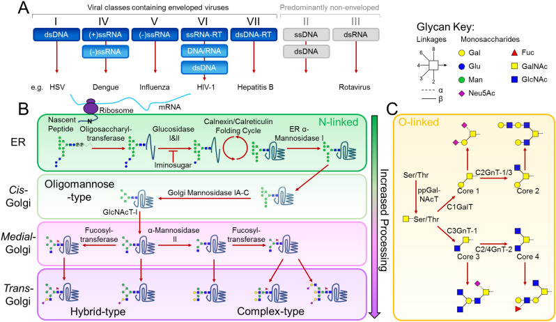
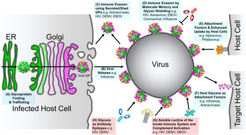
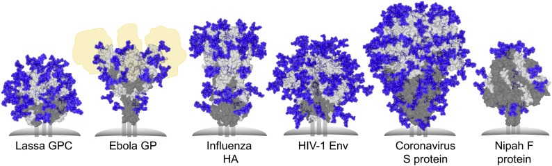
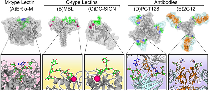
Glycosylation is a ubiquitous post-translational modification responsible for a multitude of crucial biological roles. As obligate parasites, viruses exploit host-cell machinery to glycosylate their own proteins during replication. Viral envelope proteins from a variety of human pathogens including HIV-1, influenza virus, Lassa virus, SARS, Zika virus, dengue virus, and Ebola virus have evolved to be extensively glycosylated. These host-cell derived glycans facilitate diverse structural and functional roles during the viral life-cycle, ranging from immune evasion by glycan shielding to enhancement of immune cell infection. In this review, we highlight the imperative and auxiliary roles glycans play, and how specific oligosaccharide structures facilitate these functions during viral pathogenesis. We discuss the growing efforts to exploit viral glycobiology in the development of anti-viral vaccines and therapies.

Keywords: Glycan shielding; Glycoprotein; Glycosylation; Structure; Virus; Virus-host interactions.


Figures

Fig. 1
Fig. 1
Glycosylation of a viral glycoprotein in the classical mammalian N-linked and mucin-type O-linked pathways. (A) Viral classes containing predominantly enveloped and non-enveloped viruses are coloured in blue and grey, respectively. Although not enveloped, viruses such as rotaviruses can also exploit the host-glycosylation pathways to modify their proteins. (B) Following mRNA synthesis, the mature tri-glucosylated N-linked glycan precursor, dolichol-P-P-glycan, is co-translationally transferred en bloc by oligosaccharyltransferase to the asparagine residue of an Asn-X-Ser/Thr sequon on a nascent polypeptide chain. Following transfer of the precursor glycan to the protein, glucosidases in the ER remove the three glucose residues while the protein folds in the Calnexin/Calreticulin cycle. A series of ER and Golgi mannosidases subsequently cleave mannose residues down to the Man5GlcNAc2 glycan. The action of GlcNAc transferase-I (GlcNAcT-I) initiates the first branch of the N-glycan. Once α-Mannosidase II removes the two remaining outer mannose residues, other glycan processing enzymes such as galactosyl-, fucosyl- and sialyl-transferases, can act to construct a huge assortment of complex-type glycans. (C) The mucin-type O-linked glycosylation pathway is initiated by a family of ppGalNAc transferases that covalently link a N-acetylgalactosamine (GalNAc) monosaccharide to any serine, threonine and tyrosine residue. Following this conjugation, a series of glycosyltransferases can act upon the primary GalNAc residue to generate the four common O-linked glycan cores. Each of these cores can be extended and processed further to generate numerous mucin-type O-linked glycans. Glycans are presented using Consortium for Functional Glycomics symbolic nomenclature and Oxford system linkages [31], as per the key.
Fig. 2
Fig. 2
Roles of Glycosylation in Viral Pathogenesis. The roles contributing to viral pathogenesis and host-cell strategies used to respond to viral infection are coloured blue and red, respectively. Green and pink indicate oligomannose and complex-type N-linked glycan processing states, respectively. (A) Glycoprotein Folding and Trafficking. As with all glycoproteins, glycans on viral glycoproteins aid in folding and trafficking through the host secretory pathway. (B) Glycosylation in Viral Release. Glycosylation of infected host cell proteins can influence viral spread. (C) Immune Evasion Using Secreted/Shed Glycoproteins. Viruses can shed or secrete glycoproteins to act as immune decoys. (D) Immune Evasion by Molecular Mimicry and Glycan Shielding. Extensively glycosylated viral proteins shield themselves from the host immune response by occluding the immunogenic proteinous surface with a dense coat of host-derived glycans. (E) Glycans as Attachment Factors and Enhanced Uptake by Immune Cells. Some virus envelope-displayed glycoproteins contain under-processed oligomannose-type glycans that function as host-cell attachment factors to augment or facilitate infection of immune cells. (F) Host Glycans as Attachment Factors. Viruses can recognise glycans presented on host cell-surface proteins to facilitate host cell attachment. (G) Soluble Lectins of the Innate Immune System and Complement Activation. As under-processed glycans are rarely presented on mature host cell glycoproteins [67,68], the innate immune system is able to recognise these glycans as pathogen-associated molecular patterns (PAMPs) using soluble lectins. (H) Glycans as Antibody Epitopes. Where glycan shielding is conserved on viral glycoproteins, it is possible for the humoral immune response, in rare cases, to elicit neutralising antibodies that target sugars as part of their epitopes.
Fig. 3
Fig. 3
Glycan Shielding of Viral Class I Fusion Proteins. Left to right: Glycan shield models of Lassa virus GPC (PDB ID: 5VK2) [117,140], Ebola GP (PDB ID: 5JQ3) [141], A/H3N2/361/Victoria/2011 H3N2 Influenza virus hemagglutinin (PDB ID: 4O5N) [142,143], BG505 SOSIP.664 HIV-1 Env (PDB ID: 4ZMJ) [3,144], human coronavirus-NL63 (HCoV-NL63) S protein (PDB ID: 5SZS) [8], Nipah F protein (PDB ID: 5EVM) [145]. Glycans and proteins are shown in blue and grey, respectively. The fusion protein subunit is shown in dark grey. The positions of mucin-like domains of Ebola GP are shown in yellow. Most predominant sugar compositions were modelled onto each N-linked glycan site, using pre-existing GlcNAc residues if possible, with Man5GlcNAc2 modelled on if compositional information was lacking.
Fig. 4
Fig. 4
Diverse modes of oligomannose-type glycan recognition. (A) The glycan processing enzyme, ER α-mannosidase. Notice the deep binding pocket of the protein that nearly encapsulates the entire Man9GlcNAc2 glycan (PDB ID: 5KIJ) [242]. (B) The collagen-containing C-type lectin, mannose binding lectin (MBL), that recognises oligomannose-type glycans via a carbohydrate recognition domain containing a calcium ion (pink), which is responsible for mediating the primary interactions to bind the sugar (PDB ID: 1KX1) [243]. (C) The membrane bound C-type lectin receptor, DC-SIGN recognises oligomannose-type glycans via a Ca2+ ion and initiates phagocytosis of the pathogen (PDB ID: 1SL4) [216]. (D) An anti-HIV-1 broadly neutralising antibody, PGT128, that recognises conserved oligomannose-type glycans on Env using an extended complementarity-determining region (CDR) loop (orange) that penetrates the glycan shield (PDB ID: 5ACO) [244]. (E) The unique anti-oligomannose antibody, 2G12, featuring its unique “domain-exchanged” architecture where the heavy chains (orange) of the opposite Fab domain are swapped over (PDB ID: 6N2X) [245,246].

References

    1. Jones K.E., Patel N.G., Levy M.A., Storeygard A., Balk D., Gittleman J.L., Daszak P. Global trends in emerging infectious diseases. Nature. 2008;451:990–993. doi: 10.1038/nature06536. - DOI - PMC - PubMed
    1. Cao L., Pauthner M., Andrabi R., Rantalainen K., Berndsen Z., Diedrich J.K., Menis S., Sok D., Bastidas R., Park S.-K.R., Delahunty C.M., He L., Guenaga J., Wyatt R.T., Schief W.R., Ward A.B., Yates J.R., Burton D.R., Paulson J.C. Differential processing of HIV envelope glycans on the virus and soluble recombinant trimer. Nat. Commun. 2018;9:3693. doi: 10.1038/s41467-018-06121-4. - DOI - PMC - PubMed
    1. Struwe W.B., Chertova E., Allen J.D., Seabright G.E., Watanabe Y., Harvey D.J., Medina-Ramirez M., Roser J.D., Smith R., Westcott D., Keele B.F., Bess J.W., Sanders R.W., Lifson J.D., Moore J.P., Crispin M. Site-specific glycosylation of virion-derived HIV-1 Env Is mimicked by a soluble trimeric immunogen. Cell Rep. 2018;24:1958–1966.e5. doi: 10.1016/j.celrep.2018.07.080. - DOI - PMC - PubMed
    1. Panico M., Bouché L., Binet D., O'Connor M.-J., Rahman D., Pang P.-C., Canis K., North S.J., Desrosiers R.C., Chertova E., Keele B.F., Bess J.W., Lifson J.D., Haslam S.M., Dell A., Morris H.R. Mapping the complete glycoproteome of virion-derived HIV-1 gp120 provides insights into broadly neutralizing antibody binding. Sci. Rep. 2016;6:32956. doi: 10.1038/srep32956. - DOI - PMC - PubMed
    1. Kobayashi Y., Suzuki Y. Evidence for N-glycan shielding of antigenic sites during evolution of human influenza A virus hemagglutinin. J. Virol. 2012;86:3446–3451. doi: 10.1128/JVI.06147-11. - DOI - PMC - PubMed

Publication types