

Amyloid Beta and Phosphorylated Tau-Induced Defective Autophagy and Mitophagy in Alzheimer's Disease
- PMID: 31121890
- PMCID: PMC6562604
- DOI: 10.3390/cells8050488
Amyloid Beta and Phosphorylated Tau-Induced Defective Autophagy and Mitophagy in Alzheimer's Disease
Abstract
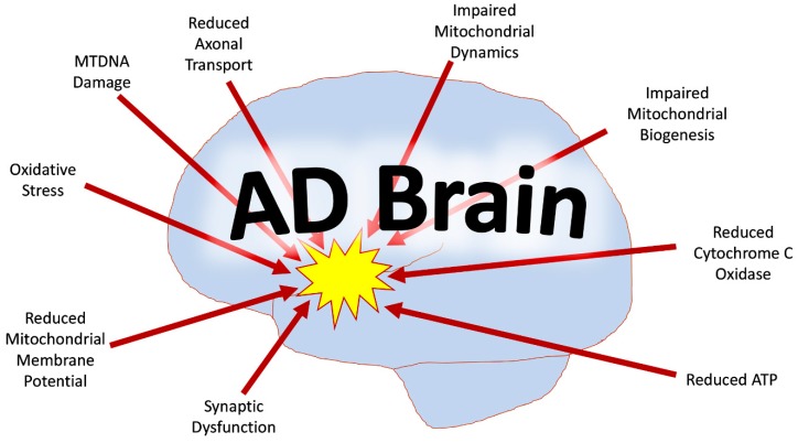
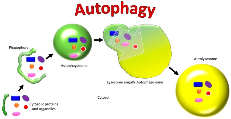
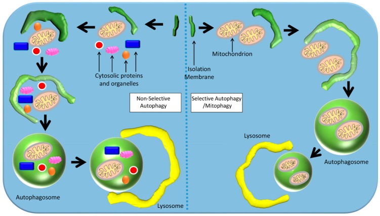
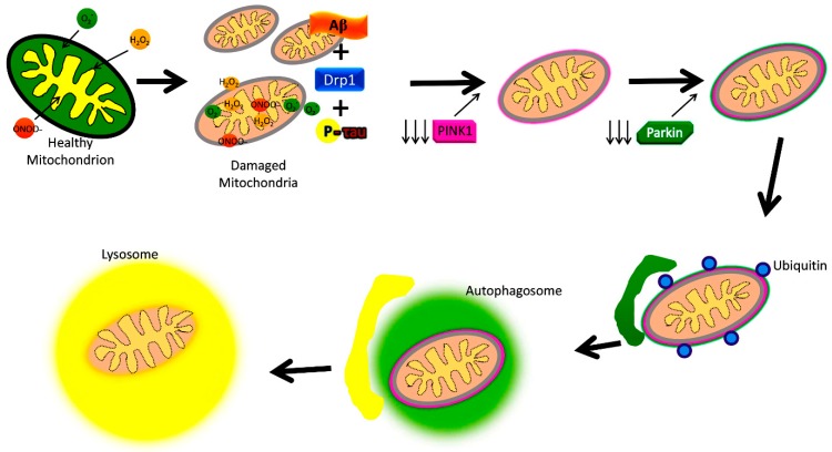
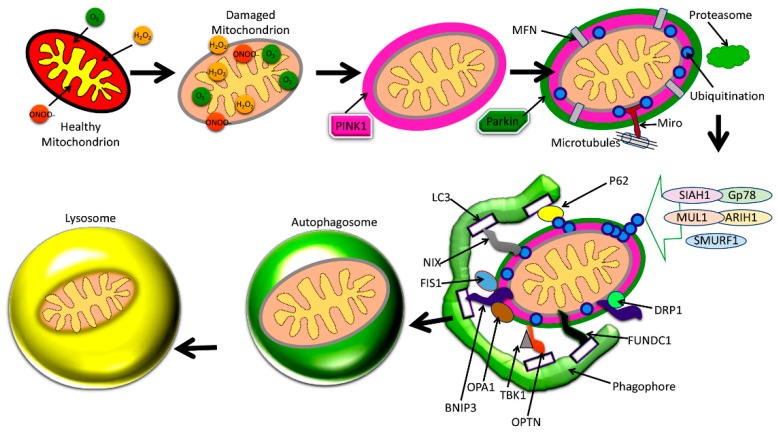
Alzheimer's disease (AD) is a progressive neurodegenerative disease characterized by memory loss and multiple cognitive impairments. Several decades of intense research have revealed that multiple cellular changes are implicated in the development and progression of AD, including mitochondrial damage, synaptic dysfunction, amyloid beta (Aβ) formation and accumulation, hyperphosphorylated tau (P-Tau) formation and accumulation, deregulated microRNAs, synaptic damage, and neuronal loss in patients with AD. Among these, mitochondrial dysfunction and synaptic damage are early events in the disease process. Recent research also revealed that Aβ and P-Tau-induced defective autophagy and mitophagy are prominent events in AD pathogenesis. Age-dependent increased levels of Aβ and P-Tau reduced levels of several autophagy and mitophagy proteins. In addition, abnormal interactions between (1) Aβ and mitochondrial fission protein Drp1; (2) P-Tau and Drp1; and (3) Aβ and PINK1/parkin lead to an inability to clear damaged mitochondria and other cellular debris from neurons. These events occur selectively in affected AD neurons. The purpose of our article is to highlight recent developments of a Aβ and P-Tau-induced defective autophagy and mitophagy in AD. This article also summarizes several aspects of mitochondrial dysfunction, including abnormal mitochondrial dynamics (increased fission and reduced fusion), defective mitochondrial biogenesis, reduced ATP, increased free radicals and lipid peroxidation, and decreased cytochrome c oxidase (COX) activity and calcium dyshomeostasis in AD pathogenesis. Our article also discusses how reduced levels of Drp1, Aβ, and P-Tau can enhance the clearance of damaged mitochondria and other cellular debris by autophagy and mitophagy mechanisms.
Keywords: Alzheimer’s disease; amyloid beta; mitochondria and reactive oxygen species; phosphorylated tau.
Conflict of interest statement
None declared.
Figures
References
-
- Hyman B.T., Phelps C.H., Beach T.G., Bigio E.H., Cairns N.J., Carrillo M.C., Dickson D.W., Duyckaerts C., Frosch M.P., Masliah E., et al. National Institute on Aging-Alzheimer’s Association guidelines for the neuropathologic assessment of Alzheimer’s disease. Alzheimers Dement. 2012;8:1–13. doi: 10.1016/j.jalz.2011.10.007. - DOI - PMC - PubMed
Publication types
MeSH terms
Substances
Grants and funding
LinkOut - more resources
Full Text Sources
Medical
Miscellaneous
