. 2019 May 22;8(5):729.
doi: 10.3390/jcm8050729.

Nuclear Phospho-SOD1 Protects DNA from Oxidative Stress Damage in Amyotrophic Lateral Sclerosis

Affiliations

Nuclear Phospho-SOD1 Protects DNA from Oxidative Stress Damage in Amyotrophic Lateral Sclerosis

Matteo Bordoni et al. J Clin Med. .

Abstract

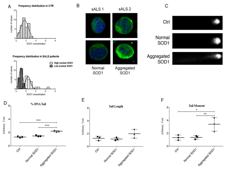
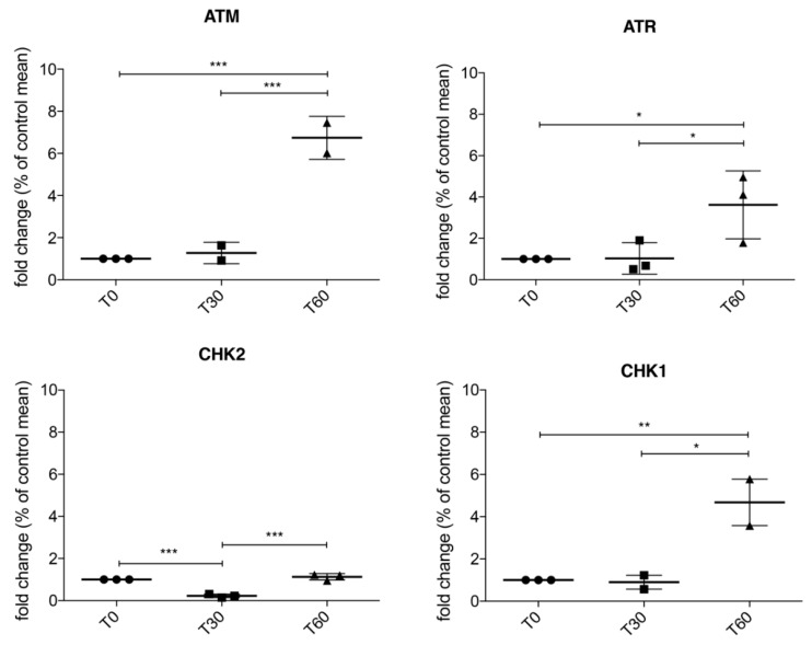
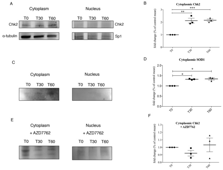
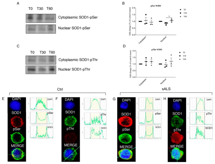
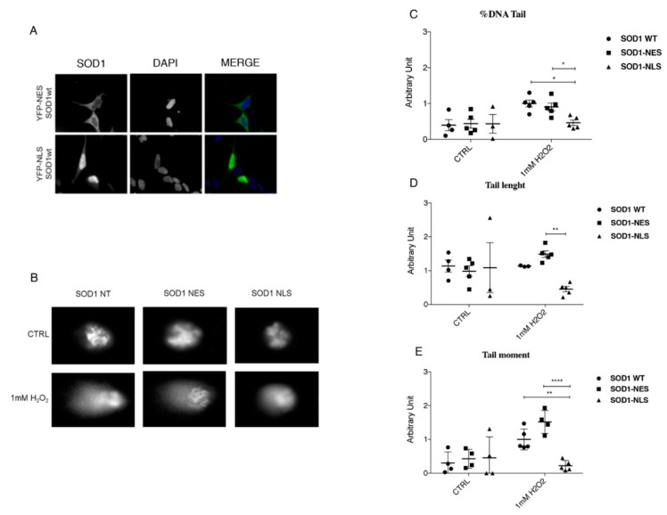
We already demonstrated that in peripheral blood mononuclear cells (PBMCs) of sporadic amyotrophic lateral sclerosis (sALS) patients, superoxide dismutase 1 (SOD1) was present in an aggregated form in the cytoplasmic compartment. Here, we investigated the possible effect of soluble SOD1 decrease and its consequent aggregation. We found an increase in DNA damage in patients PBMCs characterized by a high level of aggregated SOD1, while we found no DNA damage in PBMCs with normal soluble SOD1. We found an activation of ataxia-telangiectasia-mutated (ATM)/Chk2 and ATM and Rad3-related (ATR)/Chk1 DNA damage response pathways, which lead to phosphorylation of SOD1. Moreover, data showed that phosphorylation allows SOD1 to shift from the cytoplasm to the nucleus, protecting DNA from oxidative damage. Such pathway was finally confirmed in our cellular model. Our data lead us to suppose that in a sub-group of patients this physiologic pathway is non-functional, leading to an accumulation of DNA damage that causes the death of particularly susceptible cells, like motor neurons. In conclusion, during oxidative stress SOD1 is phosphorylated by Chk2 leading to its translocation in the nuclear compartment, in which SOD1 protects DNA from oxidative damage. This pathway, inefficient in sALS patients, could represent an innovative therapeutic target.

Keywords: ALS; DNA damage; SOD1; oxidative stress; peripheral blood mononuclear cells.


Conflict of interest statement

The authors declare no conflict of interest.

Figures

Figure 1
Figure 1
(A) Frequency Distribution of SOD1 in healthy controls (CTRL) (n = 40) and sporadic Amyotrophic Lateral Sclerosis (sALS) patients (n = 39). We found a bell-shaped distribution of the normalized values corresponding to SOD1 concentration for healthy controls, while a bimodal distribution was described for sALS patients confirming the presence of two sub-group. (B) Aggregation of cytoplasmic SOD1 in a subgroup of sALS patients observed by immunofluorescence. (C) Analysis of DNA damage in Peripheral Blood Mononuclear Cells (PBMCs) of control and patients with normal SOD1 and aggregated SOD1. Patients with normal SOD1 showed low damaged DNA (very weak Comet tail) similar to that observed in healthy CTRL; on the other hand, a very bright tail was observed in sALS patients characterized by cytoplasmic SOD1 aggregates. (DF) Comet assay quantification by three different parameters: Tail length, % tail DNA and tail moment. Data were analyzed by ANOVA (n = 3), followed by Newman-Keuls Multiple Comparison Test; * p < 0.05; ** p < 0.01 and *** p < 0.001.
Figure 2
Figure 2
RT-qPCR in SH-SY5Y treated with 1 mM H2O2 for 30 and 60 min showed that ATM/Chk2 and ATR/Chk1 are actively transcribed after 60 min of oxidative stress, suggesting that SOD1 localization in the nuclear compartment is involved in DNA damage response. Data were analyzed by ANOVA (n = 3), followed by Newman-Keuls Multiple Comparison Test; * p < 0.05; ** p < 0.01 and *** p < 0.001.
Figure 3
Figure 3
(A,B) Representative immunoblotting of cytoplasmic and nuclear Chk2 and quantification of Chk2 levels in the cytoplasmic compartment in SH-SY5Y at T0, T30 and T60. (C,D) Immunoprecipitation of Chk2 and quantification of cytoplasmic and nuclear SOD1 after oxidative stress. (E,F) The binding between Chk2 and SOD1 was further confirmed by immunoprecipitation also using AZD7762, a Chk1 and Chk2 inhibitor, to SH-SY5Y cells. After AZD7762 treatment no differences were observed in both nuclear and cytoplasm compartment. Data were analyzed by ANOVA (n = 3), followed by Newman-Keuls Multiple Comparison Test; * p < 0.05; ** p < 0.01 and *** p < 0.001.
Figure 4
Figure 4
(A,B) 1 mM H2O2 treatment determines significant phosphorylation at Ser residue at T60 in the nuclear compartment. SOD1 was immunoprecipitated and representative immunoblotting for pSer was reported for the nucleus. (C,D) 1 mM H2O2 treatment induces significant phosphorylation also at Thr residue at T60 in the nuclear compartment. SOD1 was immunoprecipitated and representative immunoblotting for pThr was reported for the nucleus. Data were analyzed by ANOVA (n = 3), followed by Newman-Keuls Multiple Comparison Test; * p < 0.05. PBMCs from healthy controls and from a subgroup of sALS patients were analyzed for pThr, pSer and SOD1 by immunofluorescence followed by confocal microscopy analysis. (E,F) In healthy controls both pThr and pSer were not observed in the nuclear fraction; (G,H) in PBMCs of sALS patients we observed a bright signal of SOD1 and pThr in the nuclear compartment, while SOD1 and pSer co-localization showed a slight signal. Nuclei were visualized using the fluorescent nuclear dye DAPI (blue).
Figure 5
Figure 5
(A) Cytoplasmic and nuclear localization of SOD1; chimeric fluorescent-tagged proteins bearing either a nuclear export signal (NES; YFP-NES-wtSOD1) or a nuclear localization signal (NLS NLS; YFP-NLS-wtSOD1) in SH-SY5Y cells were used. (B) Protective role of nuclear SOD1 against DNA damage in SH-SY5Y cells; 60 min of 1 mM H2O2 Treatment induced marked DNA damage in both NT SOD1 and SOD1-NES in SH-SY5Y cells. In SOD1-NLS cells, instead, no comets were observed, indicating that minor or no DNA fragmentation occurred. (CE) Comet assay quantification by means tail length, % tail DNA and tail moment were carried out. Data were analyzed by ANOVA (n = 3), followed by Newman-Keuls Multiple Comparison Test; * p < 0.05; ** p < 0.01 and **** p < 0.0001.

References

    1. Riancho J., Gonzalo I., Ruiz-Soto M., Berciano J. Why do motor neurons degenerate? Actualization in the pathogenesis of amyotrophic lateral sclerosis. Neurologia. 2019;34:27–37. doi: 10.1016/j.nrl.2015.12.001. - DOI - PubMed
    1. Zufiría M., Gil-Bea F.J., Fernández-Torrón R., Poza J.J., Muñoz-Blanco J.L., Rojas-García R., Riancho J., López de Munain A. Als: A bucket of genes, environment, metabolism and unknown ingredients. Prog. Neurobiol. 2016;142:104–129. doi: 10.1016/j.pneurobio.2016.05.004. - DOI - PubMed
    1. Migliore L., Coppedè F. Genetics, environmental factors and the emerging role of epigenetics in neurodegenerative diseases. Mutat. Res. 2009;667:82–97. doi: 10.1016/j.mrfmmm.2008.10.011. - DOI - PubMed
    1. Cirulli E.T., Lasseigne B.N., Petrovski S., Sapp P.C., Dion P.A., Leblond C.S., Couthouis J., Lu Y.F., Wang Q., Krueger B.J., et al. Exome sequencing in amyotrophic lateral sclerosis identifies risk genes and pathways. Science. 2015;347:1436–1441. doi: 10.1126/science.aaa3650. - DOI - PMC - PubMed
    1. Taylor J.P., Brown R.H., Cleveland D.W. Decoding als: From genes to mechanism. Nature. 2016;539:197–206. doi: 10.1038/nature20413. - DOI - PMC - PubMed