

Ultrastructure of telocytes, a new type of interstitial cells in the myocardium of the Chinese giant salamander (Andrias davidianus)
- PMID: 31122004
- PMCID: PMC6536913
- DOI: 10.4081/ejh.2019.3021
Ultrastructure of telocytes, a new type of interstitial cells in the myocardium of the Chinese giant salamander (Andrias davidianus)
Abstract
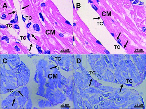
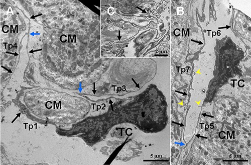
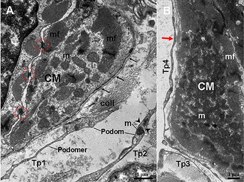
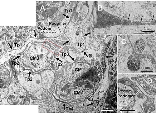
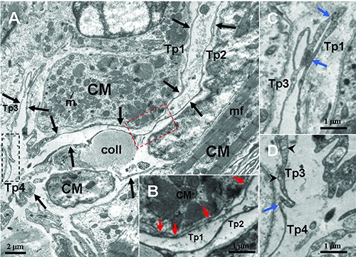
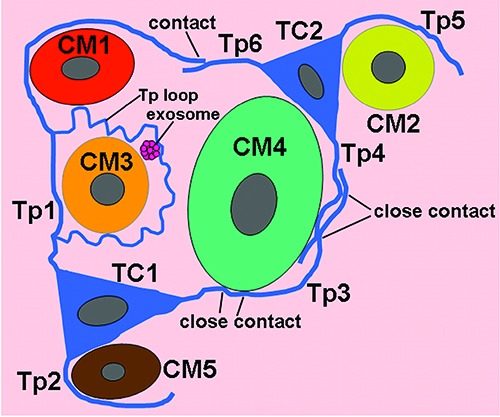
Telocytes (TCs) are new interstitial cells, and they are involved in tissue regeneration, particularly in heart. Therefore, TCs are suggested to be a promising cell in regenerative medicine. However, the information of location structural characteristics and functions of TCs is still limited. In this study, cardiac TCs of the Chinese giant salamanders (Andrias davidianus) were identified by transmission electron microscopy. TCs were located in the interstitium between cardiomyocytes (CM). TCs possessed distinctive ultrastructural characteristics, including one to two very long and thin moniliform telopodes (Tps), emerging points from the cell body, caveolae, dichotomous branchings, labyrinthic systems, neighbouring exosomes and homo-cellular contacts between Tps. TCs/Tps were frequently observed in close proximity to cardiomyocytes. Moreover, Tps established hetero-cellular contacts with cardiomyocytes. Our results confirm the presence of TCs in the myocardium of the A. davidianus. This will help us to better understand roles of TCs in amphibian hearts.
Conflict of interest statement
Conflict of interest: The authors declare no conflict of interest.
Figures
Similar articles
-
Telocytes in pancreas of the Chinese giant salamander (Andrias davidianus).J Cell Mol Med. 2016 Nov;20(11):2215-2219. doi: 10.1111/jcmm.12948. Epub 2016 Sep 20. J Cell Mol Med. 2016. PMID: 27650046 Free PMC article.
-
Telocytes in gastric lamina propria of the Chinese giant salamander, Andrias davidianus.Sci Rep. 2016 Sep 15;6:33554. doi: 10.1038/srep33554. Sci Rep. 2016. PMID: 27629815 Free PMC article.
-
Telocytes in ileum of the Chinese giant salamander: ultrastructural evidence.J Cell Mol Med. 2016 Mar;20(3):568-74. doi: 10.1111/jcmm.12741. Epub 2016 Jan 25. J Cell Mol Med. 2016. PMID: 26805522 Free PMC article.
-
Telocytes in cardiac regeneration and repair.Semin Cell Dev Biol. 2016 Jul;55:14-21. doi: 10.1016/j.semcdb.2016.01.037. Epub 2016 Jan 28. Semin Cell Dev Biol. 2016. PMID: 26826525 Review.
-
Cardiac Telocyte-Derived Exosomes and Their Possible Implications in Cardiovascular Pathophysiology.Adv Exp Med Biol. 2017;998:237-254. doi: 10.1007/978-981-10-4397-0_16. Adv Exp Med Biol. 2017. PMID: 28936744 Review.
Cited by
-
Twenty years of histochemistry in the third millennium, browsing the scientific literature.Eur J Histochem. 2020 Dec 29;64(4):3213. doi: 10.4081/ejh.2020.3213. Eur J Histochem. 2020. PMID: 33478199 Free PMC article.
-
Telocytes and their structural relationships with surrounding cell types in the skin of silky fowl by immunohistochemistrical, transmission electron microscopical and morphometric analysis.Poult Sci. 2021 Sep;100(9):101367. doi: 10.1016/j.psj.2021.101367. Epub 2021 Jun 29. Poult Sci. 2021. PMID: 34325111 Free PMC article.
References
-
- Cretoiu D, Cretoiu SM, Simionescu AA, Popescu LM. Telocytes, a distinct type of cell among the stromal cells present in the lamina propria of jejunum. Histol Histopathol 2012;27: 1067-78. - PubMed
-
- Díaz-Flores L, Gutiérrez R, Díaz-Flores L, Goméz MG, Sáez FJ, Madrid JF. Behaviour of telocytes during physiopathological activation. Semin Cell Dev Biol 2016;55:50-61. - PubMed
MeSH terms
LinkOut - more resources
Full Text Sources
