

Production, characterization, and in vivo half-life extension of polymeric IgA molecules in mice
- PMID: 31122132
- PMCID: PMC6748581
- DOI: 10.1080/19420862.2019.1622940
Production, characterization, and in vivo half-life extension of polymeric IgA molecules in mice
Abstract
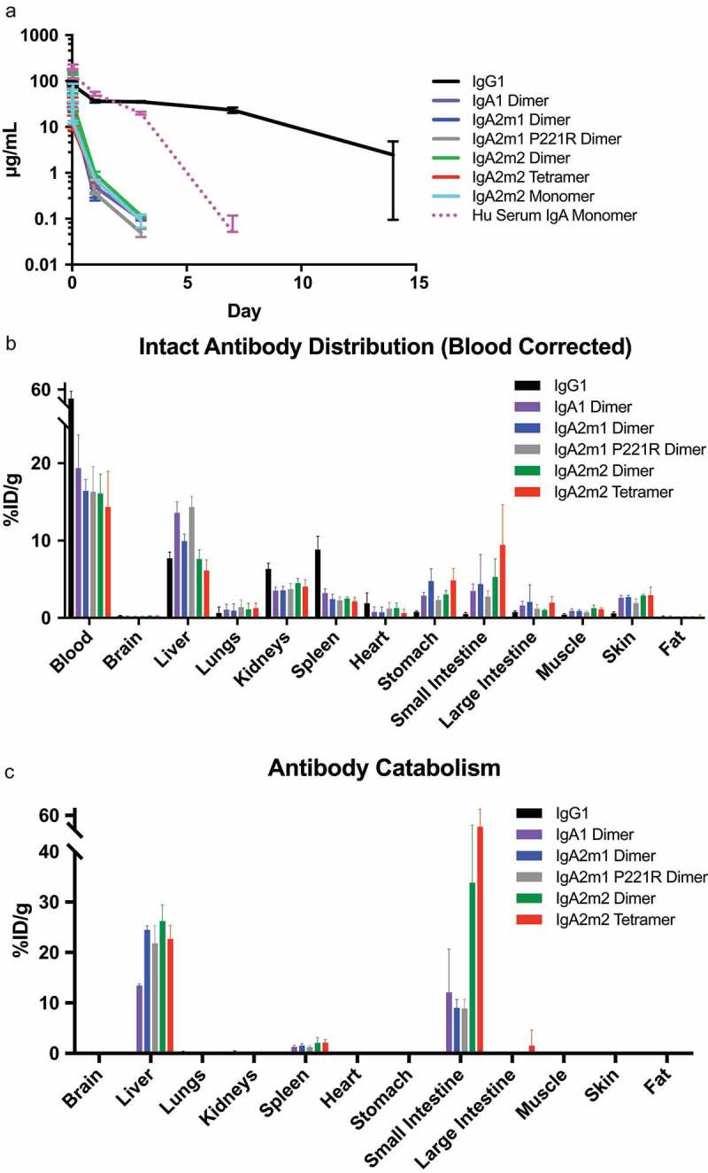
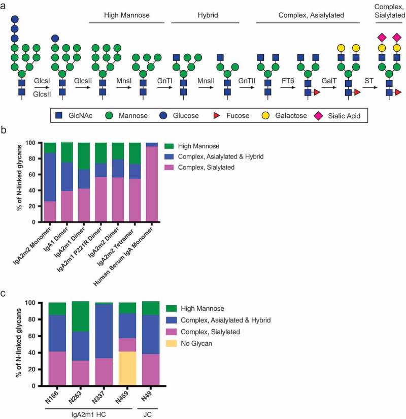
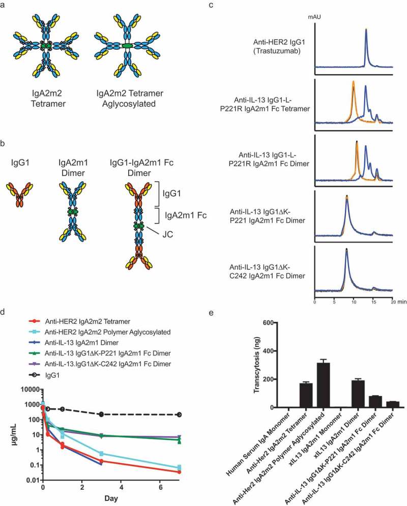
IgA antibodies have broad potential as a novel therapeutic platform based on their superior receptor-mediated cytotoxic activity, potent neutralization of pathogens, and ability to transcytose across mucosal barriers via polymeric immunoglobulin receptor (pIgR)-mediated transport, compared to traditional IgG-based drugs. However, the transition of IgA into clinical development has been challenged by complex expression and characterization, as well as rapid serum clearance that is thought to be mediated by glycan receptor scavenging of recombinantly produced IgA monomer bearing incompletely sialylated N-linked glycans. Here, we present a comprehensive biochemical, biophysical, and structural characterization of recombinantly produced monomeric, dimeric and polymeric human IgA. We further explore two strategies to overcome the rapid serum clearance of polymeric IgA: removal of all N-linked glycosylation sites creating an aglycosylated polymeric IgA and engineering in FcRn binding with the generation of a polymeric IgG-IgA Fc fusion. While previous reports and the results presented in this study indicate that glycan-mediated clearance plays a major role for monomeric IgA, systemic clearance of polymeric IgA in mice is predominantly controlled by mechanisms other than glycan receptor clearance, such as pIgR-mediated transcytosis. The developed IgA platform now provides the potential to specifically target pIgR expressing tissues, while maintaining low systemic exposure.
Keywords: Asialoglycoprotein receptor (ASGPR); N-linked glycan; polymeric IgG receptor (pIgR); serum half-life; transcytosis.
Figures
Similar articles
-
Enhancing IgG distribution to lung mucosal tissue improves protective effect of anti-Pseudomonas aeruginosa antibodies.JCI Insight. 2018 Jun 21;3(12):e97844. doi: 10.1172/jci.insight.97844. eCollection 2018 Jun 21. JCI Insight. 2018. PMID: 29925682 Free PMC article.
-
Epithelial transcytosis of monomeric IgA and IgG cross-linked through antigen to polymeric IgA. A role for monomeric antibodies in the mucosal immune system.J Immunol. 1994 Jan 1;152(1):72-6. J Immunol. 1994. PMID: 8254208
-
Targeting mucosal sites by polymeric immunoglobulin receptor-directed peptides.J Exp Med. 2002 Aug 19;196(4):551-5. doi: 10.1084/jem.20020581. J Exp Med. 2002. PMID: 12186846 Free PMC article.
-
Regulation of the polymeric immunoglobulin receptor and IgA transport: new advances in environmental factors that stimulate pIgR expression and its role in mucosal immunity.Mucosal Immunol. 2011 Nov;4(6):598-602. doi: 10.1038/mi.2011.37. Epub 2011 Sep 28. Mucosal Immunol. 2011. PMID: 21956244 Free PMC article. Review.
-
Polymeric immunoglobulin receptor.J Oral Sci. 2011 Jun;53(2):147-56. doi: 10.2334/josnusd.53.147. J Oral Sci. 2011. PMID: 21712618 Review.
Cited by
-
Targeting Myeloid Checkpoint Molecules in Combination With Antibody Therapy: A Novel Anti-Cancer Strategy With IgA Antibodies?Front Immunol. 2022 Jul 5;13:932155. doi: 10.3389/fimmu.2022.932155. eCollection 2022. Front Immunol. 2022. PMID: 35865547 Free PMC article. Review.
-
Structural and Biochemical Requirements for Secretory Component Interactions with Dimeric IgA.J Immunol. 2024 Jul 15;213(2):226-234. doi: 10.4049/jimmunol.2300717. J Immunol. 2024. PMID: 38809110 Free PMC article.
-
Polymeric immunoglobulin receptor (pIgR) in cancer progression: a critical role and potential therapeutic target.Apoptosis. 2025 Aug;30(7-8):1751-1775. doi: 10.1007/s10495-025-02116-x. Epub 2025 May 26. Apoptosis. 2025. PMID: 40415061 Review.
-
Development and Evaluation of a Robust Sandwich Immunoassay System Detecting Serum WFA-Reactive IgA1 for Diagnosis of IgA Nephropathy.Int J Mol Sci. 2022 May 5;23(9):5165. doi: 10.3390/ijms23095165. Int J Mol Sci. 2022. PMID: 35563555 Free PMC article.
-
Single B cell transcriptomics identifies multiple isotypes of broadly neutralizing antibodies against flaviviruses.PLoS Pathog. 2023 Oct 9;19(10):e1011722. doi: 10.1371/journal.ppat.1011722. eCollection 2023 Oct. PLoS Pathog. 2023. PMID: 37812640 Free PMC article.
References
-
- Pascal V, Laffleur B, Debin A, Cuvillier A, van Egmond M, Drocourt D, Imbertie L, Pangault C, Tarte K, Tiraby G, et al. Anti-CD20 IgA can protect mice against lymphoma development: evaluation of the direct impact of IgA and cytotoxic effector recruitment on CD20 target cells. Haematologica. 2012;97:1686–94. doi:10.3324/haematol.2011.061408. - DOI - PMC - PubMed
MeSH terms
Substances
LinkOut - more resources
Full Text Sources
Other Literature Sources
Miscellaneous