

Evaluation of the Efficacy of Rotational Corrections for Standard-Fractionation Head and Neck Image-Guided Radiotherapy
- PMID: 31122178
- PMCID: PMC6535727
- DOI: 10.1177/1533033819853824
Evaluation of the Efficacy of Rotational Corrections for Standard-Fractionation Head and Neck Image-Guided Radiotherapy
Abstract
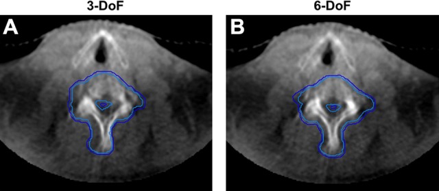
Purpose: Modern linear accelerators are equipped with cone beam computed tomography and robotic couches that can correct for errors in the translational (X, Y, Z) and rotational (α, β, γ) axes prior to treatment delivery. Here, we compared the positional accuracy of 2 cone beam registration approaches: (1) employing translational shifts only in 3 degrees of freedom (X, Y, Z), versus; (2) using translational-rotational shifts in 6 degrees of freedom (X, Y, Z, α, β, γ).
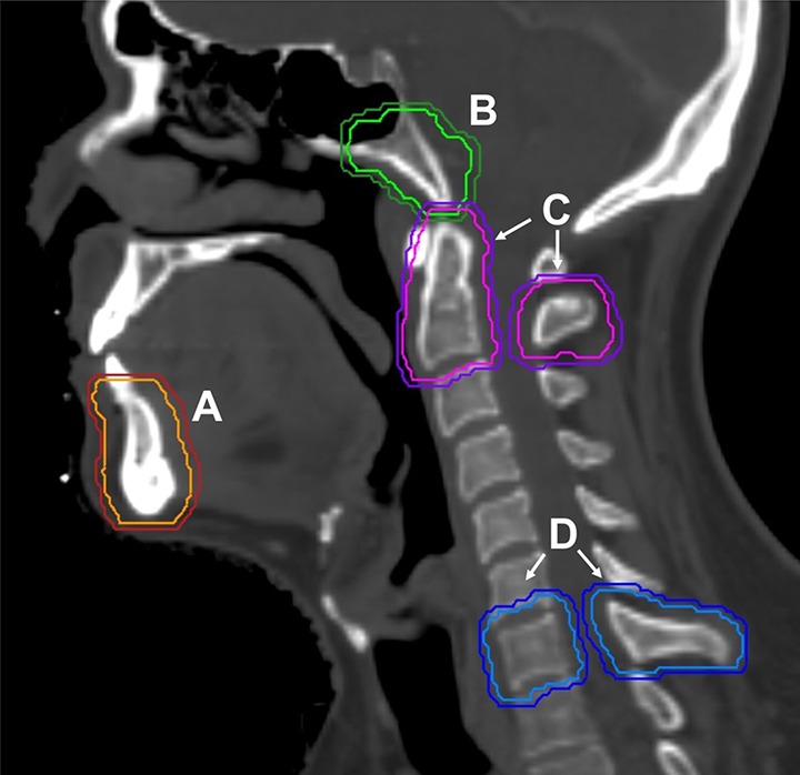
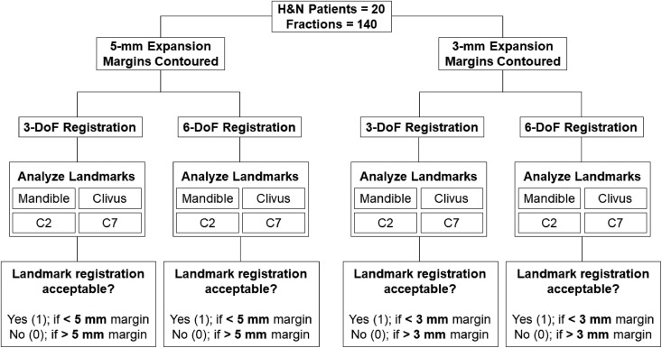
Methods: This retrospective study examined 140 interfraction cone beam images from 20 patients with head and neck cancer treated with standard intensity-modulated radiation therapy. The cone beam images were matched to planning simulation scans in 3, then in 6 degrees of freedom, using the mandible, clivus, and C2 and C7 vertebrae as surrogate volumes. Statistical analyses included a generalized mixed model and was used to assess whether there were significant differences in acceptable registrations between the 2 correction methods.
Results: The rates of improvement with corrections in 6 degrees of freedom for the mandible with a 5-mm expansion margin were 54.55% ( P = .793), for the clivus 85.71% ( P = .222), and for C7 87.50% ( P = .015). There was a 100% increase in acceptability for the C2 vertebra within the 5-mm margin ( P < .001). For the 3-mm expansion margin, the rates of improvement for the mandible, clivus, C2, and C7 were 63.16% ( P = .070), 91.30% ( P = .011), 84.21% ( P = .027), and 76.92% ( P < .001), respectively.
Conclusions: Significant registration improvements with the use of rotational corrections with a 5-mm expansion margin are only seen in the C7 vertebra. At the 3-mm margin, significant improvements are found for the C2, C7, and clivus registrations, suggesting that intensity-modulated radiotherapy treatments for head and neck cancers with 3-mm planning target volume margins may benefit from corrections in 6 degrees of freedom.
Keywords: 6 degrees of freedom; cone beam computed tomography; head and neck cancer; image guidance; intensity-modulated radiation therapy; rotational corrections.
Conflict of interest statement
Figures
References
-
- Pouliot J, Bani-Hashemi A, Josephine Chen, et al. Low-dose megavoltage cone-beam CT for radiation therapy. Int J Radiat Oncol Biol Phys. 2005;61(2):552–560. doi:10.1016/j.ijrobp.2004.10.011. - PubMed
-
- Chen AM, Farwell DG, Luu Q, Donald PJ, Perks J, Purdy JA. Evaluation of the planning target volume in the treatment of head and neck cancer with intensity-modulated radiotherapy: what is the appropriate expansion margin in the setting of daily image guidance? Int J Radiat Oncol Biol Phys. 2011;81(4):943–949. doi:10.1016/j.ijrobp.2010.07.017. - PubMed
-
- Guckenberger M, Meyer J, Wilbert J, Baier K, Sauer O, Flentje M. Precision of image-guided radiotherapy (IGRT) in six degrees of freedom and limitations in clinical practice. Strahlentherapie und Onkol. 2007;183(6):307–313. doi: 10.1007/s00066-007-1695-0. - PubMed
-
- Bertholet J, Worm E, Høyer M, Poulsen P. Cone beam CT-based set-up strategies with and without rotational correction for stereotactic body radiation therapy in the liver. Acta Oncol (Madr). 2017;56(6):860–866. doi: 10.1080/0284186X.2017.1288925. - PubMed
-
- Spadea MF, Baroni G, Riboldi M, et al. Benefits of six degrees of freedom for optically driven patient set-up correction in SBRT. Technol Cancer Res Treat. 2008;7(3):187–195. doi:10.1177/153303460800700304. - PubMed
Publication types
MeSH terms
LinkOut - more resources
Full Text Sources
Medical
Miscellaneous
