Review
. 2020 Apr:77:15-28.
doi: 10.1016/j.seizure.2019.05.008. Epub 2019 May 11.

Utility of MRI, PET, and ictal SPECT in presurgical evaluation of non-lesional pediatric epilepsy

Affiliations
Review

Utility of MRI, PET, and ictal SPECT in presurgical evaluation of non-lesional pediatric epilepsy

Csaba Juhász et al. Seizure. 2020 Apr.

Abstract

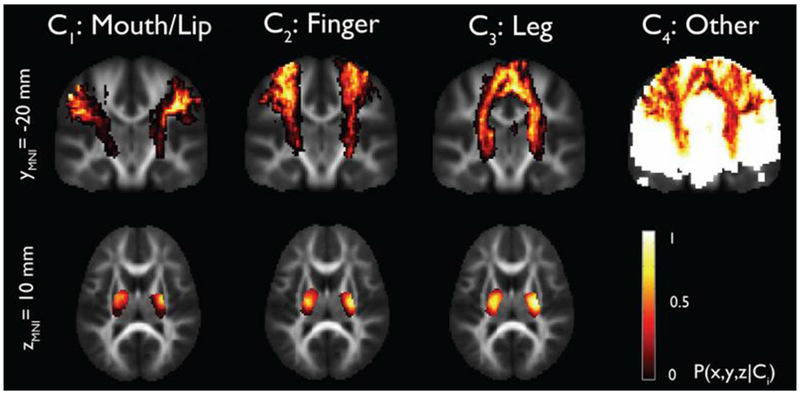
Children with epilepsy and normal structural MRI pose a particular challenge in localization of epileptic foci for surgical resection. Many of these patients have subtle structural lesions such as mild cortical dysplasia that can be missed by conventional MRI but may become detectable by optimized and advanced MRI acquisitions and post-processing. Specificity of objective analytic techniques such as voxel-based morphometry remains an issue. Combination of MRI with functional imaging approaches can improve the accuracy of detecting epileptogenic brain regions. Analysis of glucose positron emission tomography (PET) combined with high-resolution MRI can optimize detection of hypometabolic cortex associated with subtle cortical malformations and can also enhance presurgical evaluation in children with epileptic spasms. Additional PET tracers may detect subtle epileptogenic lesions and cortex with enhanced specificity in carefully selected subgroups with various etiologies; e.g., increased tryptophan uptake can identify epileptogenic cortical dysplasia in the interictal state. Subtraction ictal SPECT can be also useful to delineate ictal foci in those with non-localizing PET or after failed surgical resection. Presurgical delineation of language and motor cortex and the corresponding white matter tracts is increasingly reliable by functional MRI and DTI techniques; with careful preparation, these can be useful even in young and sedated children. While evidence-based pediatric guidelines are still lacking, the data accumulated in the last decade strongly indicate that multimodal imaging with combined analysis of MRI, PET, and/or ictal SPECT data can optimize the detection of subtle epileptogenic lesions and facilitate seizure-free outcome while minimizing the postsurgical functional deficit in children with normal conventional MRI.

Keywords: Cortical dysplasia; Epilepsy surgery; Magnetic resonance imaging; Pediatric epilepsy; Positron emission tomography; Single photon emission computed tomography.


Conflict of interest statement

Declarations of interest: none

Figures

Figure 1.
Figure 1.
Detection of a left inferior frontal epileptic focus in a 12-year old child whose MRI was read as normal originally. FDG PET showed a clearly hypometabolic area (arrow), best appreciated on coronal planes, in the inferior frontal cortex. A secondary review of T1-weighted and FLAIR images, co-registered with the PET images, showed a small suspicious area of cortical malformation in the same region (asterisks on images in the bottom row). Subsequent intracranial EEG showed seizure onset from this region, which was resected.
Figure 2.
Figure 2.
Detection of an epileptogenic tuber (A-C) and focal cortical dysplasia (D-F) by high α-[11C]methyl-L-tryptophan (AMT) uptake on PET imaging. (A) Axial FLAIR MRI showed multiple lesions in both hemispheres (arrows indicate six of the lesions). (B) All of these lesions were severely hypometabolic on FDG-PET. (C) AMT-PET showed increased tracer uptake in the left frontal hypometabolic tuber (orange arrows), suggesting epileptogenicity in this lesion. In a young child with suspected frontal lobe seizures but no clear lesion detected by MRI, although mild blurring of the gray/white matter junction was noticed in the left frontal lobe (D), FDG-PET (E) showed mild hypometabolism in the left frontal cortex (arrow); in the same area, AMT-PET showed increased interictal uptake (arrow) suggesting epileptogenic cortex.
Figure 3.
Figure 3.
Localization of a right frontal epileptic focus by SISCOM (subtraction ictal SPECT co-registered to MRI) in a patient with a large fronto-parietal infarct due to a childhood head injury. FDG-PET showed severe hypometabolism in the infarct region and mild hypometabolism inferior to that region but did not localize the epileptic cortex located in front of the infarcted area.
Figure 4.
Figure 4.
Accurate co-localization of an fMRI activation cluster with the location of depth electrodes detecting language disruption (yellow circles) in the pars opercularis of the left inferior frontal gyrus (A-C). Postoperative MRI showing the resection with the pars opercularis intact. The child was seizure-free and had no language deficit 1 year after surgery (From Ribaupierre et al. [98] with permission).
Figure 5.
Figure 5.
BOLD signal increases in the left hemisphere in response to a speech-based auditory task in a child under anesthesia with chloral hydrate. (From Ives-Deliperi et al. [106] with permission).
Figure 6.
Figure 6.
(A) Axial MR images showing the overlay of cortical fMRI activation maps for passive movement of the left lower extremity (pink) in a 16-year-old girl with a history of left functional hemispherectomy showing orthotopic cortical activation in the posterior/superior aspect of the right paracentral lobule. (B) Activation maps for passive movement of the right lower extremity (red) are in a more posterior and inferior location in the right (ipsilateral) hemisphere. Parasagittal (C) and coronal-oblique (D) images show the relationship between the area of activation for both lower extremities. (From Choudri et al. [108] with permission).
Figure 7.
Figure 7.
Stereotaxic probability maps of “mouth/lip”, “fingers”, and “leg” pathways from ICA+BSM tractography. Each map shows the probability of a voxel that belongs to mouth/lip, finger, and leg pathways in a group of healthy children (n=17, age 4.3-17.8 years). (Reproduced with permission from Jeong et al. [138]).
Figure 8.
Figure 8.
Example of a residual transcallosal connection documented by DTI fiber tracking (and superimposed on an axial T1-weighted image) in a right-handed boy with Sturge-Weber syndrome who underwent right functional hemispherectomy at age 1 year due to intractable seizures. He became seizure-free for 6 years but then started having seizures emerging from the right centro-temporal region. DTI 9 years after the initial surgery showed residual interhemispheric connections through the genu of the corpus callosum (arrow). He underwent a subsequent right anatomical hemispherectomy that resulted in seizure freedom.

References

    1. Jayakar P, Gaillard WD, Tripathi M, Libenson MH, Mathern GW, Cross JH; Task Force for Paediatric Epilepsy Surgery, Commission for Paediatrics, and the Diagnostic Commission of the International League Against Epilepsy. Diagnostic test utilization in evaluation for resective epilepsy surgery in children. Epilepsia. 2014;55:507–18. - PubMed
    1. Hyslop A, Miller I, Bhatia S, Resnick T, Duchowny M, Jayakar P. Minimally resective epilepsy surgery in MRI-negative children. Epileptic Disord. 2015;17:263–74. - PubMed
    1. Craven IJ, Griffiths PD, Bhattacharyya D, Grunewald RA, Hodgson T, Connolly DJ, et al. 3.0 T MRI of 2000 consecutive patients with localisation-related epilepsy. Br J Radiol. 2012;85:1236–42. - PMC - PubMed
    1. Gaillard WD, Chiron C, Cross JH, Harvey AS, Kuzniecky R, Hertz-Pannier L, et al. Guidelines for imaging infants and children with recent-onset epilepsy. Epilepsia. 2009;50:2147–2153. - PubMed
    1. Huppertz HJ, Wellmer J, Staack AM, Altenmuller DM, Urbach H, Kroll J. Voxel-based 3D MRI analysis helps to detect subtle forms of subcortical band heterotopia. Epilepsia. 2008;49:772–85. - PubMed

MeSH terms

Supplementary concepts