

Susceptibility-Weighted MR Imaging Hypointense Rim in Progressive Multifocal Leukoencephalopathy: The End Point of Neuroinflammation and a Potential Outcome Predictor
- PMID: 31122919
- PMCID: PMC7028608
- DOI: 10.3174/ajnr.A6072
Susceptibility-Weighted MR Imaging Hypointense Rim in Progressive Multifocal Leukoencephalopathy: The End Point of Neuroinflammation and a Potential Outcome Predictor
Abstract
Background and purpose: Progressive multifocal leukoencephalopathy (PML) represents a life-threatening demyelinating disorder of the brain caused by reactivation of a rare opportunistic infection with JC Polyomavirus. The aims of this study were to describe the incidence of a susceptibility-weighted imaging hypointense rim in patients with multifocal leukoencephalopathy and to explore the histologic correlates and prognostic value of the rim with regard to the clinical outcome.
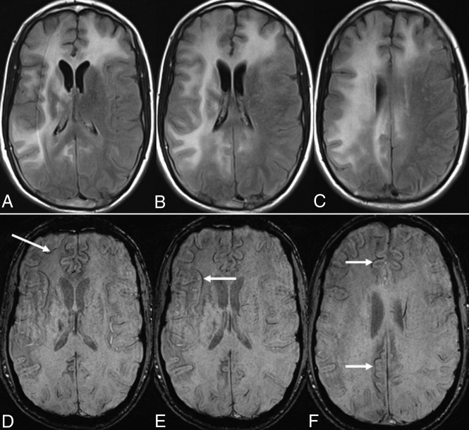
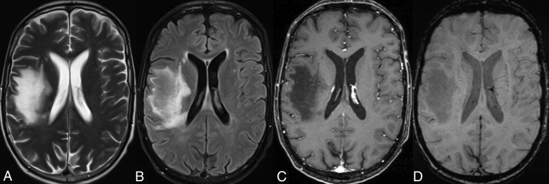
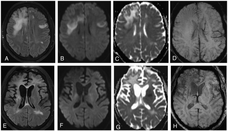
Materials and methods: This retrospective study included 18 patients with a definite diagnosis of progressive multifocal leukoencephalopathy. Ten patients were HIV-positive, 3 patients had natalizumab-associated progressive multifocal leukoencephalopathy, 1 patient had multiple myeloma, 3 patients had a history of lymphoma, and 1 was diagnosed with acute myeloid leukemia. Patients were divided into short- (up to 12 months) and long-term (>12 months) survivors. A total of 93 initial and follow-up MR imaging examinations were reviewed. On SWI, the presence and development of a hypointense rim at the periphery of the progressive multifocal leukoencephalopathy lesions were noted. A postmortem histologic examination was performed in 2 patients: A rim formed in one, and in one, there was no rim.
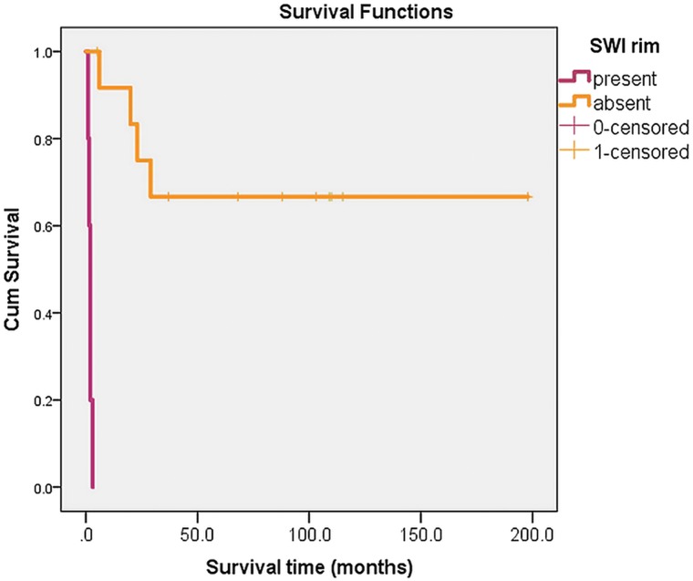
Results: A total of 73 progressive multifocal leukoencephalopathy lesions were observed. In 13 (72.2%) patients, a well-defined thin, linear, hypointense rim at the periphery of the lesion toward the cortical side was present, while in 5 (27.8%) patients, it was completely absent. All 11 long-term survivors and 2 short-term survivors presented with a prominent SWI-hypointense rim, while 5/7 short-term survivors did not have this rim.
Conclusions: The thin, uniformly linear, gyriform SWI-hypointense rim in the paralesional U-fibers in patients with definite progressive multifocal leukoencephalopathy might represent an end-point stage of the neuroinflammatory process in long-term survivors.
© 2019 by American Journal of Neuroradiology.
Figures
Similar articles
-
Cortical hypointensity in T2-weighted gradient-echo sequences in patients with progressive multifocal leukoencephalopathy.Radiologia (Engl Ed). 2020 Jan-Feb;62(1):59-66. doi: 10.1016/j.rx.2019.06.003. Epub 2019 Jul 30. Radiologia (Engl Ed). 2020. PMID: 31375267 Review. English, Spanish.
-
Pomalidomide-associated progressive multifocal leukoencephalopathy in multiple myeloma: cortical susceptibility-weighted imaging hypointense findings prior to clinical deterioration.J Neurovirol. 2020 Jun;26(3):452-455. doi: 10.1007/s13365-020-00845-0. Epub 2020 May 11. J Neurovirol. 2020. PMID: 32394398
-
High b-value diffusion-weighted imaging in progressive multifocal leukoencephalopathy in HIV patients.Eur Radiol. 2017 Sep;27(9):3593-3599. doi: 10.1007/s00330-017-4761-8. Epub 2017 Feb 6. Eur Radiol. 2017. PMID: 28168372 Free PMC article.
-
Use of quantitative susceptibility mapping (QSM) in progressive multifocal leukoencephalopathy.J Neuroradiol. 2016 Feb;43(1):6-10. doi: 10.1016/j.neurad.2015.08.001. Epub 2015 Oct 23. J Neuroradiol. 2016. PMID: 26475668
-
Clinical and Radiological Characterization of Progressive Multifocal Leukoencephalopathy in HIV-Infected Patients: A Retrospective Analysis and Review of the Literature.Acta Med Port. 2015 May-Jun;28(3):286-96. doi: 10.20344/amp.5950. Epub 2015 Jun 30. Acta Med Port. 2015. PMID: 26421780 Review.
Cited by
-
Adoptive Transfer of JC Virus-Specific T Lymphocytes for the Treatment of Progressive Multifocal Leukoencephalopathy.Ann Neurol. 2021 Apr;89(4):769-779. doi: 10.1002/ana.26020. Epub 2021 Feb 10. Ann Neurol. 2021. PMID: 33459417 Free PMC article.
-
MRI imaging features of HIV-related central nervous system diseases: diagnosis by pattern recognition in daily practice.Jpn J Radiol. 2021 Nov;39(11):1023-1038. doi: 10.1007/s11604-021-01150-4. Epub 2021 Jun 14. Jpn J Radiol. 2021. PMID: 34125369 Free PMC article. Review.
-
Primary central nervous system lymphoma: Imaging features and differential diagnosis.Neuroradiol J. 2024 Dec;37(6):705-722. doi: 10.1177/19714009241252625. Epub 2024 May 4. Neuroradiol J. 2024. PMID: 38703015 Review.
-
The neuroradiology of progressive multifocal leukoencephalopathy: a clinical trial perspective.Brain. 2022 Apr 18;145(2):426-440. doi: 10.1093/brain/awab419. Brain. 2022. PMID: 34791056 Free PMC article. Review.
-
Secondary Central Nervous System Demyelinating Disorders in the Elderly: A Narrative Review.Healthcare (Basel). 2023 Jul 25;11(15):2126. doi: 10.3390/healthcare11152126. Healthcare (Basel). 2023. PMID: 37570367 Free PMC article. Review.
References
-
- Carson KR, Focosi D, Major EQ, et al. . Monoclonal antibody-associated progressive multifocal leucoencephalopathy in patients with rituximab, natalizumab, and efalizumab: a review from the Research on Adverse Drug Events and Reports (RADAR) project. Lancet Oncol 2009;10:816–24 10.1016/S1470-2045(09)70161-5 - DOI - PubMed
MeSH terms
LinkOut - more resources
Full Text Sources