Review
. 2019 Aug 1;33(15-16):903-935.
doi: 10.1101/gad.325050.119. Epub 2019 May 23.

PRC2 is high maintenance

Affiliations
Review

PRC2 is high maintenance

Jia-Ray Yu et al. Genes Dev. .

Abstract

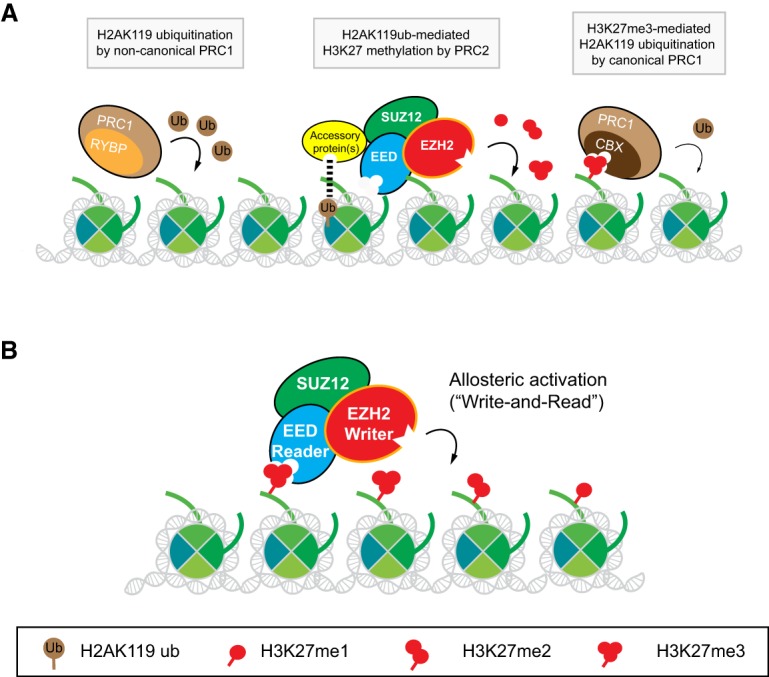
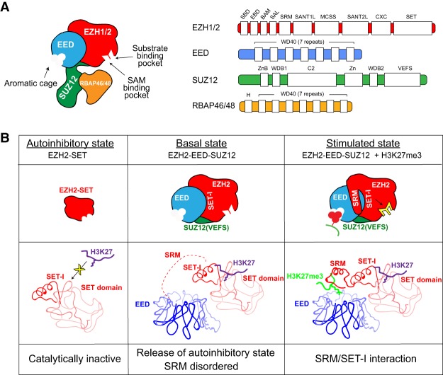
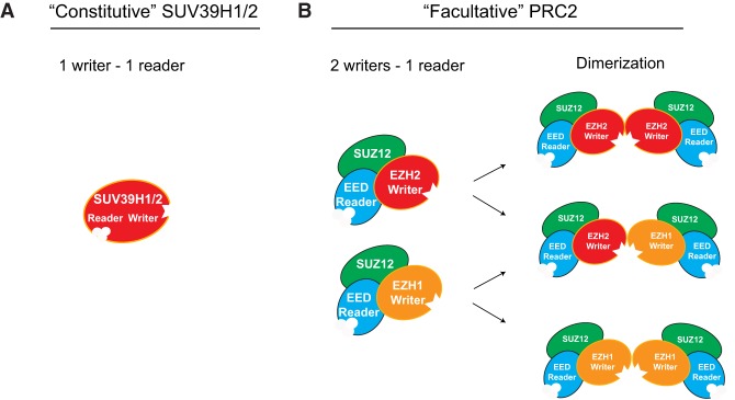
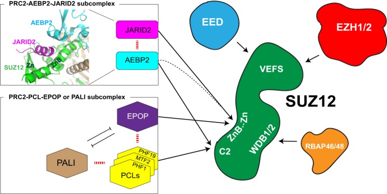
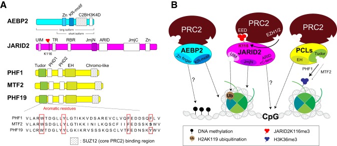
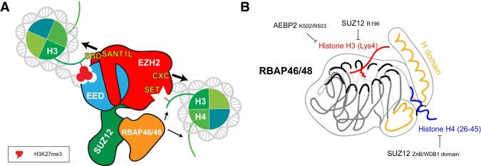
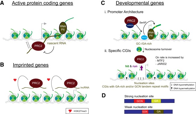
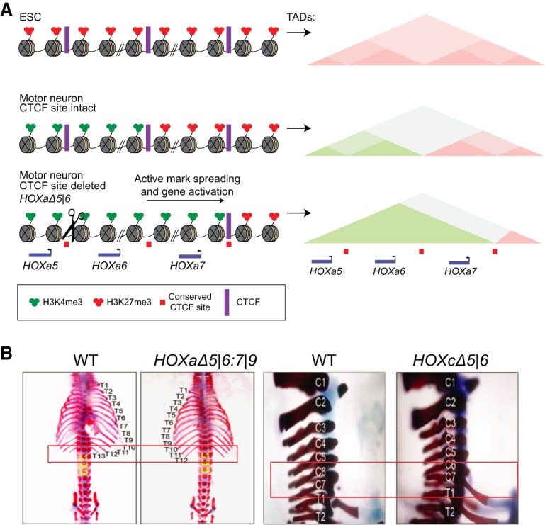
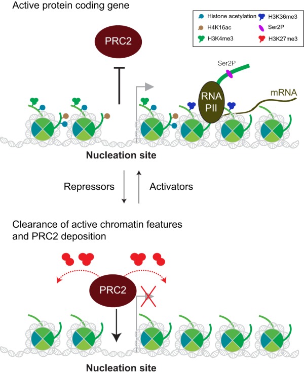
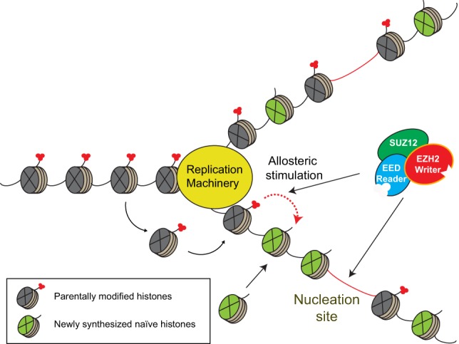
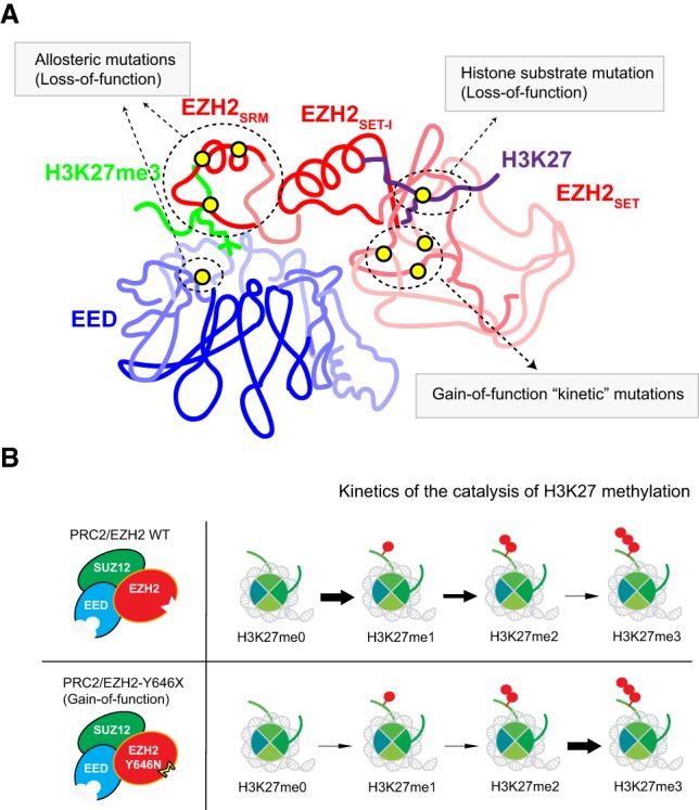
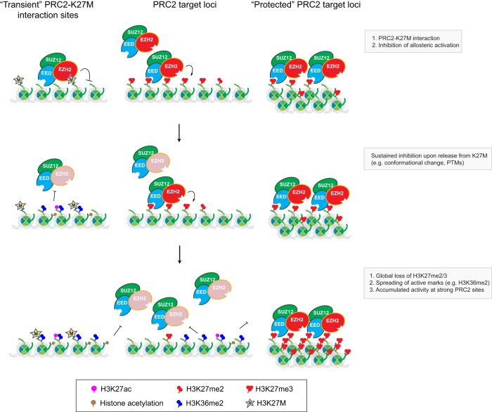
As the process that silences gene expression ensues during development, the stage is set for the activity of Polycomb-repressive complex 2 (PRC2) to maintain these repressed gene profiles. PRC2 catalyzes a specific histone posttranslational modification (hPTM) that fosters chromatin compaction. PRC2 also facilitates the inheritance of this hPTM through its self-contained "write and read" activities, key to preserving cellular identity during cell division. As these changes in gene expression occur without changes in DNA sequence and are inherited, the process is epigenetic in scope. Mutants of mammalian PRC2 or of its histone substrate contribute to the cancer process and other diseases, and research into these aberrant pathways is yielding viable candidates for therapeutic targeting. The effectiveness of PRC2 hinges on its being recruited to the proper chromatin sites; however, resolving the determinants to this process in the mammalian case was not straightforward and thus piqued the interest of many in the field. Here, we chronicle the latest advances toward exposing mammalian PRC2 and its high maintenance.

Keywords: PRC2; Polycomb; chromatin; epigenetics.


Figures

Figure 1.
Figure 1.
Interplay between Polycomb proteins: PRC1 and PRC2. (A) A model for reciprocal recruitment between PRC1 and PRC2. RYBP-containing PRC1 (noncanonical PRC1) catalyzes H2AK119ub, which in turn putatively recruits PRC2 through its accessory protein (i.e., JARID2) interaction with H2AK119ub. The catalysis of H3K27me3 by PRC2 recruits CBX-containing PRC1 (canonical PRC1). CBX-containing PRC1 harbors an intrinstic activity for chromatin compaction and a relatively low activity for the catalysis of H2AK119ub. (B) Schematic illustration of allosteric activation of PRC2 involving its “write and read” mechanism.
Figure 2.
Figure 2.
PRC2 architecture. (A) Schematic representation of the PRC2 complex. (Left) Displayed are the composition of core PRC2 and association of its subunits derived from structural and biochemical studies. The aromatic cage of EED and the catalytic sites of EZH1/2 (substrate-binding pocket and SAM-binding pocket) are indicated. (Right) Domains within PRC2 core subunits are indicated. (B) Schematic representation of the PRC2 states: autoinhibitory (left), basal (middle), and “allosterically” stimulated (right). For simplicity, only key features of the PRC2 structure are illustrated. (Middle) The EZH2-SET domain is catalytically inactive and this autoinhibitory state is released by forming the EZH2-EED-SUZ12 complex. In this basal state, the stimulatory-responsive motif (SRM) is disordered (middle; dashed red line) but can align with SET-I when H3K27me3 is recognized by the aromatic cage of EED (right).
Figure 3.
Figure 3.
Reader and writer modules within the PRC2 complex and SUV39H1/2 (yeast CLR4). The “writer” and “reader” modules comprise one protein in the case of SUV39H1/2 (yeast CLR4) (A) but are segregated into two distinct subunits (EZH1/EZH2 and EED, respectively) in the case of PRC2 (B, left). (B, right) PRC2 comprises one of two distinct “writers.” (Middle) EZH1 or EZH2 and can form homodimers or heterodimers, likely providing unknown regulatory roles that are cell type-specific.
Figure 4.
Figure 4.
Assembly of core PRC2 and accessory proteins. Schematic representation illustrating the means by which SUZ12 (green) functions as a structural platform. PRC2 subunits and accessory proteins interact with different domains of SUZ12, as indicated (see arrows). (Left) Two major PRC2 subcomplexes are illustrated (square boxes). Interactions and antagonisms among accessory factors are illustrated by dashed red lines and blocked arrows, respectively. (Top left) Structure showing AEBP2 and JARID2 cooperative interaction with SUZ12. AEBP2 and SUZ12 (ZnB and Zn domains) create a groove that fits the TR (transrepression) domain of JARID2 (modified from Protein Data Bank: 5WAI). This three-way junction formed by JARID2, AEBP2, and SUZ12 creates a stable and unique PRC2 subcomplex PRC2–AEBP2–JARID2. Note that AEBP2 interacts with both the C2 and ZnB domains. Although AEBP2 binds to the ZnB domain, it cooperatively interacts with JARID2 (dashed black arrow), while competing with PCLs.
Figure 5.
Figure 5.
Biochemical characterization of PRC2 accessory factors. (A) Domains within PRC2 accessory proteins are indicated. SUZ12 binding domains are highlighted by gray dots. (Bottom) Sequence alignment of the Tudor domains within three mammalian PCL proteins. The critical aromatic residues that can form a cage are highlighted by red squares. The cage within the PCLtudor are bound to H3K36me3 (and, to a lesser extent, H3K36me2) in vitro. (B) PRC2 accessory proteins regulate its activity through several means: increasing its affinity for nucleosome binding (e.g., AEBP2KR motif and JARID2JmjN) and DNA (e.g., AEBP2Zn, JARID2C-term, and PCLEH), interacting with histone posttranslational modifications (hPTMs) (e.g., JARID2UIM [H2AK119ub] and PCLTudor [H3K36me2/me3]), and/or regulating its allosteric activation (e.g., JARID2-K116me3). (C2B) C2-binding domain; (TR) transrepression; (RBR) RNA-binding region; (JmjN) Jumonji N; (JmjC) Jumonji C; (PHD1/2): plant homeodomain 1/2.
Figure 6.
Figure 6.
Interaction between the PRC2 complex and chromatin. (A) Schmatic representation of the PRC2 complex bound to a dinucleosome of which one comprises trimethylated H3K27 and the other unmodified H3K27. (Left) The SBD and SANT1L domains of EZH2 are in contact with the H3K27me3-containing nucleosome and the CXC and SET domains of EZH2 interact with the unmethylated nucleosome, thereby bridging the two (big arrows). H3K27me3 resides in the EED aromatic cage (left), while unmethylated H3K27 is at the EZH2 active site (right). RBAP46/48 interacts with the histone H3 and H4 tails (small arrows). (B) Schematic representation of RBAP46/48 interacting with the histone H3 and H4 tails. These interactions are competitive with RBAP46/48 interaction with SUZ12 and AEBP2.
Figure 7.
Figure 7.
Three modes of PRC2 recruitment to chromatin in mammals. (A) At active protein coding genes, the 5′ region of the nascent RNA recruits PRC2 through interaction with its core EZH2 subunit. This interaction precludes PRC2 activity. (B) At imprinted genes, long ncRNAs (lncRNAs) produced from the same loci recruit PRC2 in cis. PRC2 deposits H3K27me3 on these transcriptionally silent genes. (C) At developmental genes, promoter architecture and specific CGIs recruit PRC2. (Panel i) Erk1/2 localizes to GC-/GA-rich regions on the genome, mediating nucleosome turnover as well as phosphorylation of RNAPII at its C-terminal domain (CTD)-Ser5 residue. These events provide a promoter architecture conducive to PRC2/JARID2 recruitment. (Panel ii) Through its low-affinity interactions with chromatin, PRC2 can recognize nucleation sites that contain hypomethylated CGIs with GA-rich and/or GCN tandem repeat motifs via a “hit and run” mechanism, but its on rate is lower than its off rate. The on rate is increased by PRC2 interaction with MTF2/PCL2 and/or JARID2. (D) Nucleation sites (both strong and weak) are enriched for dense CGIs, which have a high number of CG dinucleotides within the island (blue boxes). Both types of nucleation sites are enriched for “GA” and/or “GCN” tandem repeat motifs; however, strong nucleation sites have longer GCN tandem repeats (red boxes) and a different distribution of GA content (light-green and dark-green boxes) compared with weak ones.
Figure 8.
Figure 8.
Spreading of PRC2 activity after initial recruitment. (A) PRC2 targets are engaged in a network of interactions wherein nucleation sites are concentrated, forming Polycomb foci. Following the nucleation event, PRC2 spreads H3K27me2/3 domains across the genome proximally as well as distally via long-range 3D contacts, all within Polycomb foci. (B) Detailed mechanism by which PRC2 spreads the products of its catalysis. PRC2 first catalyzes H3K27me2 at the nucleation sites (strong or weak), which are then converted to H3K27me3 once PRC2 reaches sufficient concentrations. Through binding to H3K27me3, PRC2 is allosterically stimulated and rapidly spreads H3K27me2 to adjacent chromatin. H3K27me2 is then converted to H3K27me3, and, as PRC2 moves further from the nucleation sites, its stability on chromatin decreases such that H3K27me3 domains remain proximal and H3K27me2 domains remain distal to its nucleation sites. The strong and weak nucleation sites engage in long-range interactions.
Figure 9.
Figure 9.
How does PRC2 spreading stop? (A) Upon differentiation of ESCs to motor neurons, a tight boundary between transcriptionally active domains and PRC2-mediated repressive domains is maintained by CTCF binding to its cognate sites in the HoxA cluster. In this case, active loci are sequestered in a TAD independent from that sequestering repressed domains. (B) Deletion of CTCF-binding sites result in homeotic transformation in mice. (Left) Proper ribs do not protrude from the T13 position. (Right) An extra rib aberrantly protrudes from the C7 position (adapted from Narendra et al. 2016).
Figure 10.
Figure 10.
Establishment of PRC2 on chromatin during cellular state transitions. When a given nucleation site is occupied by active chromatin modifications within an actively transcribing gene, PRC2 binding is precluded. Should this gene be bound by transcriptional repressors in response to a change in the cellular state, histone deacetylases and demethylases would then clear all of the active chromatin features. PRC2 can now bind to the cleared nucleation site and maintain repression of this gene in this specific cellular lineage. This process is conceivably reversible, as binding of transcriptional activatiors and histone-modifiying enzymes such as histone acetyltransferases and methyltransferases could evict PRC2 and reactivate this gene in response to a reversal of the cellular state.
Figure 11.
Figure 11.
Two distinct catalytic subunits, EZH1 and EZH2. (A) Schematic representation depicting the residues/regions that distinguish EZH2 and EZH1. While the SAL, SRM, CXC, and SET domains are well conserved, the SBD, EBD, BAM, SANT1L, and SANT2L domains are less conserved (highlighted in red and pink). Distinct residues within the SRM and SET domains are indicated. The EZH1-specific nucleosome-binding regions (SANT1L, MCSS, and SANT2L) (Son et al. 2013) are poorly conserved with EZH2. (B) The distinct functions of PRC2/EZH2 and PRC2/EZH1 during development are indicated. (C) A summary of the comparsion between PRC2/EZH2 and PRC2/EZH1 activities.
Figure 12.
Figure 12.
Maintenance of Polycomb repression after DNA replication. Following replication of an H3K27me3-modified region, parentally modified (dark gray) and newly synthesized naïve octamers (green) are randomly deposited to daughter DNA strands. The EED subunit of PRC2 can recognize H3K27me3-modified nucleosomes, with resultant allosteric activation of PRC2. This “write and read” mechanism stimulates catalysis of H3K27me3 to adjacent nucleosomes. In parallel, PRC2 could recognize a nucleation site and spread the modification accordingly.
Figure 13.
Figure 13.
Mutations of PRC2 and its substrate in cancer. (A) Major groups of mutations that influence PRC2 function in cancer. Mutations that are found in the EED cage (EED I363M) (Ueda et al. 2016) and SRM domain of EZH2 (P132S, D142V, and F145L) inhibit allosteric activation of PRC2 (Lee et al. 2018c). Mutations that are found in the catalytic SET domain (Y646X [X = S, N, F, C, or H], A682G, A682V, and A692V) are gain-of-function “kinetic” mutations. Histone H3K27M is a dominant-negative substrate mutation that globally inhibits PRC2 activity. (B) Illustration depicting the kinetics of catalysis of each H3K27 methylation state. EZH2 mutants in Y646 specifically promote the catalysis of H3K27me3 from H3K27me2 but dampen catalysis of the lower methylation states.
Figure 14.
Figure 14.
Diverse modes of PRC2 inhibition by H3K27M. (Top) When H3K27M is first expressed in a cell, PRC2 becomes trapped due to its higher affinity for nucleosomes comprising H3K27M relative to the wild-type case. (Middle) Interaction between PRC2 and H3K27M is transient, but freed PRC2 is compromised, being less active. The less active PRC2 is then recruited to loci independent of H3K27M, where it exhibits altered activity. (Bottom) The decrease in H3K27me2/3 deposition in H3K27M cells results in a progressive gain in the chromatin deposition of histone posttranslational modifications (hPTMs), such as bulk acetylated histones, including H3K27 acetylation and H3K36me2. Such hPTMs have the potential to directly repel PRC2. Thus, the gain in these hPTMs in H3K27M cells lead to wide-scale effects on PRC2 activity and the epigenetic landscape of chromatin. On the other hand, “protected” PRC2 target loci that are largely devoid of H3K27M exhibit focal gains of H3K27me2/3.

References

    1. Ai S, Yu X, Li Y, Peng Y, Li C, Yue Y, Tao G, Li C, Pu WT, He A. 2017. Divergent requirements for EZH1 in heart development versus regeneration. Circ Res 121: 106–112. 10.1161/CIRCRESAHA.117.311212 - DOI - PMC - PubMed
    1. Alabert C, Barth TK, Reveron-Gomez N, Sidoli S, Schmidt A, Jensen ON, Imhof A, Groth A. 2015. Two distinct modes for propagation of histone PTMs across the cell cycle. Genes Dev 29: 585–590. 10.1101/gad.256354.114 - DOI - PMC - PubMed
    1. Alekseyenko AA, Gorchakov AA, Kharchenko P V, Kuroda MI. 2014. Reciprocal interactions of human C10orf12 and C17orf96 with PRC2 revealed by BioTAP-XL cross-linking and affinity purification. Proc Natl Acad Sci 111: 2488–2493. 10.1073/pnas.1400648111 - DOI - PMC - PubMed
    1. Almeida M, Pintacuda G, Masui O, Koseki Y, Gdula M, Cerase A, Brown D, Mould A, Innocent C, Nakayama M, et al. 2017. PCGF3/5-PRC1 initiates Polycomb recruitment in X chromosome inactivation. Science 356: 1081–1084. 10.1126/science.aal2512 - DOI - PMC - PubMed
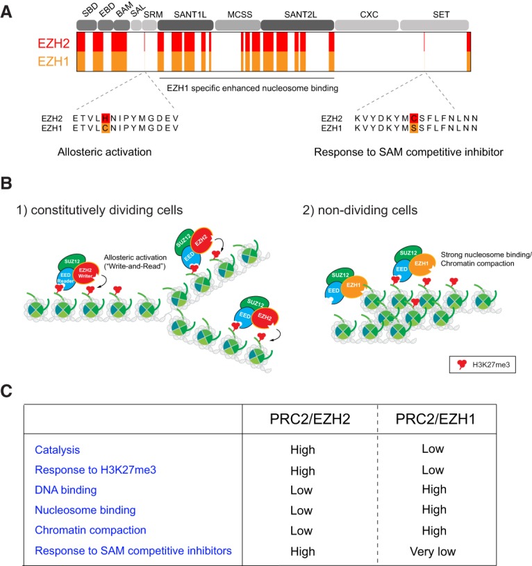
    1. Antonysamy S, Condon B, Druzina Z, Bonanno JB, Gheyi T, Zhang F, MacEwan I, Zhang A, Ashok S, Rodgers L, et al. 2013. Structural context of disease-associated mutations and putative mechanism of autoinhibition revealed by X-ray crystallographic analysis of the EZH2-SET domain. PLoS One 8: e84147 10.1371/journal.pone.0084147 - DOI - PMC - PubMed

Publication types