. 2019 May 24;364(6442):787-792.
doi: 10.1126/science.aaw7446.

Bacterial pseudokinase catalyzes protein polyglutamylation to inhibit the SidE-family ubiquitin ligases

Affiliations

Bacterial pseudokinase catalyzes protein polyglutamylation to inhibit the SidE-family ubiquitin ligases

Miles H Black et al. Science. .

Abstract

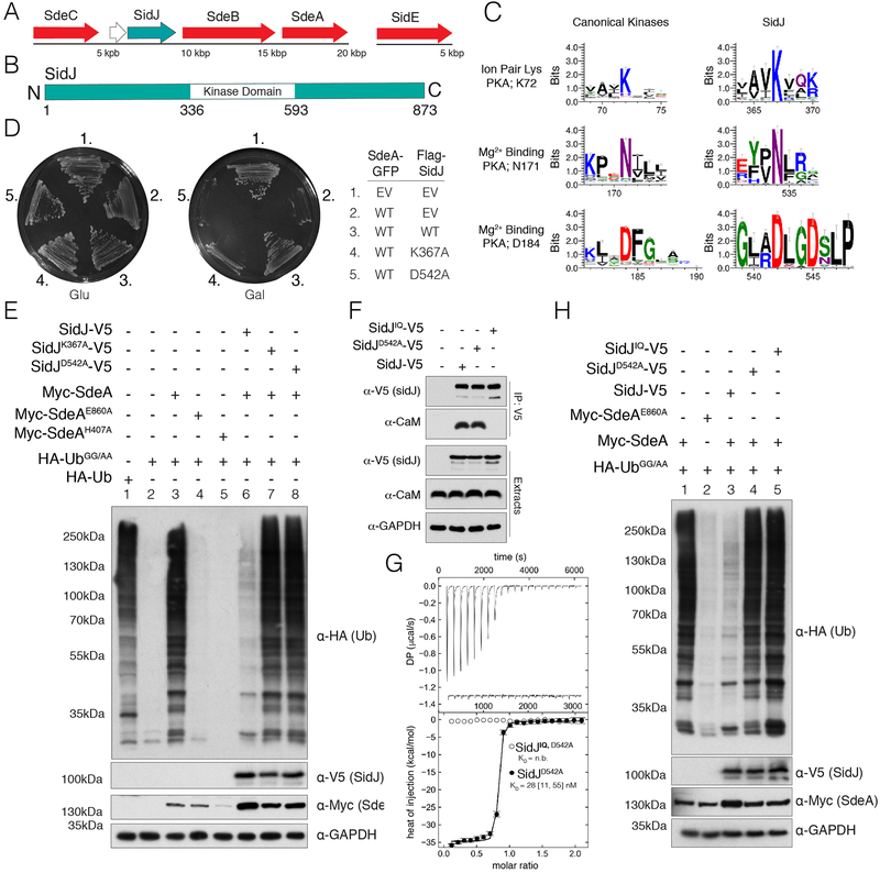
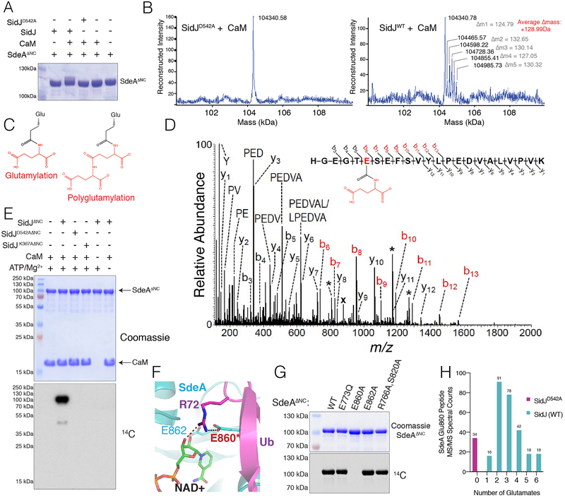
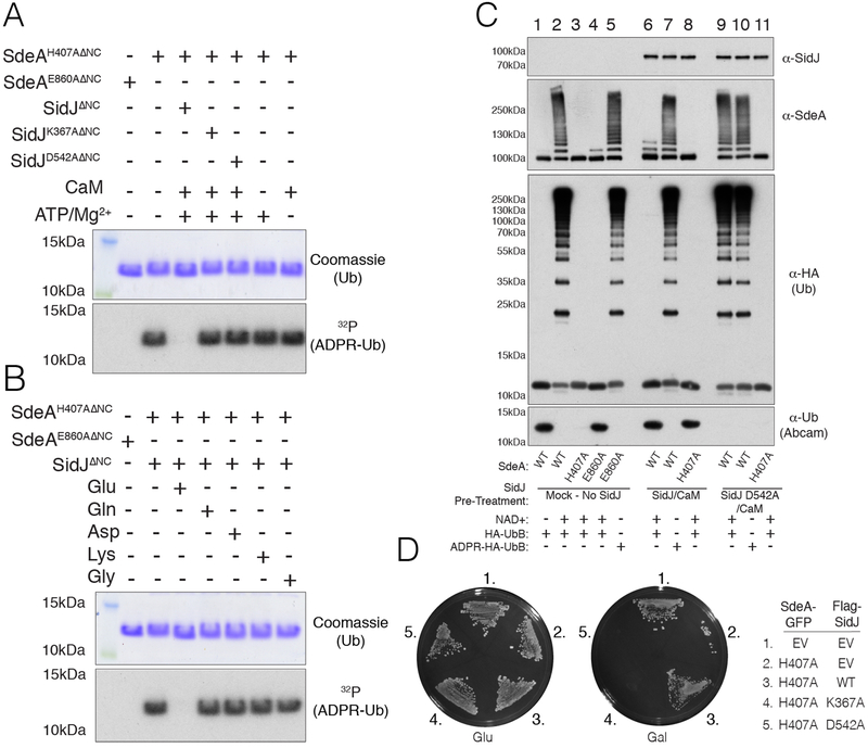
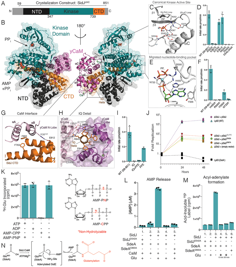
Enzymes with a protein kinase fold transfer phosphate from adenosine 5'-triphosphate (ATP) to substrates in a process known as phosphorylation. Here, we show that the Legionella meta-effector SidJ adopts a protein kinase fold, yet unexpectedly catalyzes protein polyglutamylation. SidJ is activated by host-cell calmodulin to polyglutamylate the SidE family of ubiquitin (Ub) ligases. Crystal structures of the SidJ-calmodulin complex reveal a protein kinase fold that catalyzes ATP-dependent isopeptide bond formation between the amino group of free glutamate and the γ-carboxyl group of an active-site glutamate in SidE. We show that SidJ polyglutamylation of SidE, and the consequent inactivation of Ub ligase activity, is required for successful Legionella replication in a viable eukaryotic host cell.


Conflict of interest statement

Competing Interests:N/A.

Figures

Fig. 1.
Fig. 1.. A putative kinase domain and CaM binding are required for SidJ suppression of SdeA toxicity and non-canonical ubiquitination.
(A) Organization of the SidE family (red) and SidJ (teal) effectors in the genome of L. pneumophila. (B) Domain architecture of L. pneumophila SidJ depicting the location of the predicted kinase domain. (C) Sequence logos (weblogos) highlighting conserved kinase active site residues in 106 SidJ homologs and 3,998 homologs of typical protein kinases (Pfam domain PF00069). The height of the amino acid stack is proportional to the sequence conservation at that position. (D) Growth inhibition assay depicting the growth of S. cerevisiae expressing SdeA-GFP and Flag-SidJ, or the predicted inactive K367A and D542A mutants. EV; empty vector. (E) Protein immunoblotting of total extracts from HEK293A cells expressing HA-Ub, HA-UbGG/AA, Myc-SdeA and SidJ-V5, or the indicated mutants. GAPDH is shown as a loading control. (F) Protein immunoblotting of V5-immunoprecipitates and cell extracts from HEK293A cells expressing V5 tagged SidJ, SidJD542A, or the SidJIQ mutant. Immunoprecipitates and cell extracts were analyzed for CaM and SidJ. GAPDH is shown as a loading control. (G) Isotherms depicting the binding of human CaM to SidJD542AΔNC. The top panel shows the SVD-reconstructed thermograms (DP=differential power), the lower panel shows the isotherms. Results are reported as best fit with boundaries of 68.3% confidence interval. (H) Protein immunoblotting of HEK293A total cell extracts expressing HA-tagged UbGG/AA, Myc-SdeA, V5 tagged SidJ, SidJD542A, or the SidJIQ mutant. GAPDH is shown as a loading control.
Fig. 2.
Fig. 2.. SidJ polyglutamylates the SidE effectors.
(A) SDS-PAGE and Coomassie staining of SdeAΔNC isolated from E. coli following co-expression with CaM, SidJ, or the indicated mutants. (B) Intact mass LC/MS spectra of SdeAΔNC from (A). (C) Structures depicting glutamylation and polyglutamylation. (D) MS/MS spectrum of monoglutamylated (Glu) SdeAΔNC peptide ion HGEGTE(Glu)SEFSVYLPEDVALVPVK. The precursor ion, m/z 878.10 (3+) and labeled with (x), was subjected to HCD fragmentation to generate the MS/MS spectrum shown. Fragment b-ions containing the modified glutamate residue show a mass shift consistent with the addition of one Glu group (+129.043 Da) (red labels). Peaks labeled with a single asterisk (*) correspond to loss of water (−18 Da) from fragment ions. (E) Incorporation of 14C-Glu into SdeAΔNC by SidJΔNC. Reaction products were separated by SDS PAGE and visualized by Coomassie staining (upper) and autoradiography (lower). (F) Close-up of the NAD+ binding pocket of SdeA (PDB 5YIJ (16)). (G) Incorporation of 14C-Glu into WT SdeAΔNC, but not the E860A mutant. Reaction products analyzed as in (E). (H) Histogram depicting the MS/MS spectral matches to unmodified, glutamylated, and polyglutamylated E860-containing tryptic peptides of Myc-tagged SdeA immunopurified from HEK293 cells expressing SidJ-V5 or the D542A mutant.
Fig. 3.
Fig. 3.. SidJ inactivates SdeA Ub ligase activity by polyglutamylating an active site residue in the ART domain.
(A) SDS PAGE and autoradiography depicting the incorporation of 32P-ADPR into HA-Ub from [α−32P]NAD+ by SdeAH407AΔNC. SdeAH407AΔNC or the E860A mutant were pretreated in glutamylation assays with SidJΔNC or the indicated mutants (−/+ ATP/Mg2+ or CaM) and SdeAH407AΔNC activity was subsequently analyzed. Reaction products were analyzed as in Fig. 2E. (B) SDS PAGE and autoradiography depicting the incorporation of 32P-ADPR into HA-Ub from [α−32P]NAD+ by SdeAH407AΔNC. SdeAH407AΔNC or the E860A mutant were pretreated with SidJΔNC, CaM, ATP/Mg2+ or Glu, Gln, Asp, Lys or Gly and SdeAH407AΔNC activity was analyzed as in A. (C) Protein immunoblotting following in vitro ubiquitination assays with the indicated mutants of SdeAΔNC. SdeAΔNC and mutants were pretreated in glutamylation reactions in the absence (lanes 1–5) or presence (lanes 6–8) of SidJΔNC/CaM, or SidJD542AΔNC/CaM (lanes 9–11). Ubiquitination reactions were started by the addition of NAD+ and HA-Ub or HA-ADPR Ub. The reaction components were resolved by SDS-PAGE and SidJΔNC, SdeAΔNC and Ub (using anti-HA or Abcam antibodies. which do not recognize Arg42 modified Ub) were detected by immunoblotting. (D) Growth inhibition assay depicting the growth of S. cerevisiae expressing SdeAH407A-GFP and Flag-SidJ, or the predicted catalytically inactive K367A and D542A mutants. EV; empty vector.
Fig. 4.
Fig. 4.. Crystal structure of the SidJ-CaM complex uncovers insight into the mechanism of SidJ-catalyzed polyglutamylation.
(A) Domain architecture of SidJ depicting the N-terminal domain (NTD; black), the C-terminal domain (CTD; orange) and the kinase domain (KD; teal). Residues corresponding to SidJΔNC are indicated above the schematic. (B) Overall structure of the SidJΔNC-yCaM complex. yCaM is in magenta, and SidJ is colored as in A. (C) Close-up view of the canonical kinase active site of SidJ showing the interactions (dashed lines) involved in PPi binding. The PPi is shown in stick and the Mg2+ ions are in light blue spheres. (D) Glutamylation activity of SidJ and mutants using SdeAΔNC and [3H]Glu as substrates. Reaction products were resolved by SDS PAGE and radioactive gel bands were excised and 3H incorporation into SdeAΔNC was quantified by scintillation counting. (E) Close-up view of the migrated SidJ nucleotide-binding site depicting the interactions involved in AMP and PPi binding. The AMP and PPi are shown in stick and the Mg2+ ion as a light blue sphere. (F) Glutamylation activity of SidJ and kinase active site mutants using SdeAΔNC and [3H]Glu as substrates. Reaction products were analyzed as in D. (G) Close-up view of the interaction between the N-lobe of CaM and residues in the CTD of SidJ. SidJ is colored as in A and the yCaM N-lobe and C lobes are in pink and magenta, respectively. (H) Close-up view of the IQ helix of SidJ and its interactions with the N- and C-lobes of yCaM. Color coding as in G. (I) Glutamylation activity of SidJ and CaM binding mutants. Reaction products were analyzed as in D. (J) Replication of L. pneumophila strains in A. castellanii. Infected amoeba cells were lysed at the indicated timepoints and bacterial replication was quantified by plating serial dilutions of lysates. Results are representative of three independent experiments. (K) Glutamylation activity of SidJ using SdeAΔNC and [3H]Glu as substrates with different nucleotide analogs. Reactions went to completion and products were analyzed as in (D). The chemical structures of AMP-PNP and AMP-CPP are also shown with the non-hydrolysable bonds in red. (L) HPLC-MS/MS quantification of AMP released during SidJ-catalyzed glutamylation of SdeAΔNC. (M) Quantification of acyl-adenylate formation following reactions with SidJΔNC, SdeAΔNC and [α−32P]ATP. The reactions were terminated by the addition of TCA and the SdeA acyl-adenylate was detected by scintillation counting of the acid-insoluble material. In lanes 4 and 5, Glu was added at time 0 or after 30 minutes. (N) Schematic representation of the proposed SidJ-catalyzed glutamylation reaction with the acyl-AMP SidE intermediate in brackets.

Comment in

References

    1. Cornejo E, Schlaermann P, Mukherjee S, How to rewire the host cell: A home improvement guide for intracellular bacteria. J Cell Biol 216, 3931–3948 (2017). - PMC - PubMed
    1. Sherwood RK, Roy CR, Autophagy Evasion and Endoplasmic Reticulum Subversion: The Yin and Yang of Legionella Intracellular Infection. Annu Rev Microbiol 70, 413–433 (2016). - PubMed
    1. Isberg RR, O’Connor TJ, Heidtman M, The Legionella pneumophila replication vacuole: making a cosy niche inside host cells. Nat Rev Microbiol 7, 13–24 (2009). - PMC - PubMed
    1. Xu D, Jaroszewski L, Li Z, Godzik A, FFAS-3D: improving fold recognition by including optimized structural features and template re-ranking. Bioinformatics 30, 660–667 (2014). - PMC - PubMed
    1. Havey JC, Roy CR, Toxicity and SidJ-Mediated Suppression of Toxicity Require Distinct Regions in the SidE Family of Legionella pneumophila Effectors. Infect Immun 83, 3506–3514 (2015). - PMC - PubMed

Publication types

MeSH terms