Thalamic arousal network disturbances in temporal lobe epilepsy and improvement after surgery
- PMID: 31123139
- PMCID: PMC6744309
- DOI: 10.1136/jnnp-2019-320748
Thalamic arousal network disturbances in temporal lobe epilepsy and improvement after surgery
Erratum in
-
Corrigendum: Oral Presentations in Clinical Neurosurgery 2019.Neurosurgery. 2020 Feb 1;86(2):315. doi: 10.1093/neuros/nyz486. Neurosurgery. 2020. PMID: 31612962 No abstract available.
Abstract
Objective: The effects of temporal lobe epilepsy (TLE) on subcortical arousal structures remain incompletely understood. Here, we evaluate thalamic arousal network functional connectivity in TLE and examine changes after epilepsy surgery.
Methods: We examined 26 adult patients with TLE and 26 matched control participants and used resting-state functional MRI (fMRI) to measure functional connectivity between the thalamus (entire thalamus and 19 bilateral thalamic nuclei) and both neocortex and brainstem ascending reticular activating system (ARAS) nuclei. Postoperative imaging was completed for 19 patients >1 year after surgery and compared with preoperative baseline.
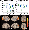
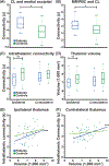
Results: Before surgery, patients with TLE demonstrated abnormal thalamo-occipital functional connectivity, losing the normal negative fMRI correlation between the intralaminar central lateral (CL) nucleus and medial occipital lobe seen in controls (p < 0.001, paired t-test). Patients also had abnormal connectivity between ARAS and CL, lower ipsilateral intrathalamic connectivity, and smaller ipsilateral thalamic volume compared with controls (p < 0.05 for each, paired t-tests). Abnormal brainstem-thalamic connectivity was associated with impaired visuospatial attention (ρ = -0.50, p = 0.02, Spearman's rho) while lower intrathalamic connectivity and volume were related to higher frequency of consciousness-sparing seizures (p < 0.02, Spearman's rho). After epilepsy surgery, patients with improved seizures showed partial recovery of thalamo-occipital and brainstem-thalamic connectivity, with values more closely resembling controls (p < 0.01 for each, analysis of variance).
Conclusions: Overall, patients with TLE demonstrate impaired connectivity in thalamic arousal networks that may be involved in visuospatial attention, but these disturbances may partially recover after successful epilepsy surgery. Thalamic arousal network dysfunction may contribute to morbidity in TLE.
Keywords: epilepsy surgery; functional connectivity; functional neuroimaging; partial seizures; temporal lobe epilepsy.
© Author(s) (or their employer(s)) 2019. No commercial re-use. See rights and permissions. Published by BMJ.
Conflict of interest statement
Competing interests: None declared.
Figures


Similar articles
-
People with mesial temporal lobe epilepsy have altered thalamo-occipital brain networks.Epilepsy Behav. 2021 Feb;115:107645. doi: 10.1016/j.yebeh.2020.107645. Epub 2020 Dec 15. Epilepsy Behav. 2021. PMID: 33334720 Free PMC article.
-
Functional connectivity disturbances of the ascending reticular activating system in temporal lobe epilepsy.J Neurol Neurosurg Psychiatry. 2017 Nov;88(11):925-932. doi: 10.1136/jnnp-2017-315732. Epub 2017 Jun 19. J Neurol Neurosurg Psychiatry. 2017. PMID: 28630376 Free PMC article.
-
Arousal and salience network connectivity alterations in surgical temporal lobe epilepsy.J Neurosurg. 2022 Jul 8;138(3):810-820. doi: 10.3171/2022.5.JNS22837. Print 2023 Mar 1. J Neurosurg. 2022. PMID: 35901709 Free PMC article.
-
Impaired vigilance networks in temporal lobe epilepsy: Mechanisms and clinical implications.Epilepsia. 2020 Feb;61(2):189-202. doi: 10.1111/epi.16423. Epub 2020 Jan 4. Epilepsia. 2020. PMID: 31901182 Free PMC article. Review.
-
Neuroscience of coma.Handb Clin Neurol. 2025;207:29-47. doi: 10.1016/B978-0-443-13408-1.00010-5. Handb Clin Neurol. 2025. PMID: 39986726 Review.
Cited by
-
Altered Resting State Networks Before and After Temporal Lobe Epilepsy Surgery.Brain Topogr. 2022 Nov;35(5-6):692-701. doi: 10.1007/s10548-022-00912-1. Epub 2022 Sep 8. Brain Topogr. 2022. PMID: 36074203
-
Thalamic neural activity and epileptic network analysis using stereoelectroencephalography: a prospective study protocol.BMJ Open. 2025 Jun 10;15(6):e097957. doi: 10.1136/bmjopen-2024-097957. BMJ Open. 2025. PMID: 40499969 Free PMC article.
-
Thalamus and consciousness: a systematic review on thalamic nuclei associated with consciousness.Front Neurol. 2025 Jun 18;16:1509668. doi: 10.3389/fneur.2025.1509668. eCollection 2025. Front Neurol. 2025. PMID: 40642212 Free PMC article.
-
Profiles of resting state functional connectivity in temporal lobe epilepsy associated with post-laser interstitial thermal therapy seizure outcomes and semiologies.Front Neuroimaging. 2023 Nov 9;2:1201682. doi: 10.3389/fnimg.2023.1201682. eCollection 2023. Front Neuroimaging. 2023. PMID: 38025313 Free PMC article.
-
SUDEP: The Worst in Epilepsy and the Hardest to Image.Epilepsy Curr. 2020 Mar;20(2):73-74. doi: 10.1177/1535759719896791. Epub 2019 Dec 30. Epilepsy Curr. 2020. PMID: 31884816 Free PMC article.
References
-
- Witt JA, Helmstaedter C. Cognition in epilepsy: current clinical issues of interest. Curr Opin Neurol 2017;30:174–9. - PubMed
Publication types
MeSH terms
Grants and funding
- R01 NS075270/NS/NINDS NIH HHS/United States
- R01 NS112252/NS/NINDS NIH HHS/United States
- F31 NS106735/NS/NINDS NIH HHS/United States
- T32 GM007347/GM/NIGMS NIH HHS/United States
- R00 NS097618/NS/NINDS NIH HHS/United States
- R01 NS108445/NS/NINDS NIH HHS/United States
- R01 NS095291/NS/NINDS NIH HHS/United States
- K99 NS097618/NS/NINDS NIH HHS/United States
- U54 HD083211/HD/NICHD NIH HHS/United States
- S10 OD021771/OD/NIH HHS/United States
- T32 EB021937/EB/NIBIB NIH HHS/United States
- R01 NS110130/NS/NINDS NIH HHS/United States
LinkOut - more resources
Full Text Sources
