. 2019 May 23;9(1):7772.
doi: 10.1038/s41598-019-44253-9.

Engineering of anti-human interleukin-4 receptor alpha antibodies with potent antagonistic activity

Affiliations

Engineering of anti-human interleukin-4 receptor alpha antibodies with potent antagonistic activity

Jung-Eun Kim et al. Sci Rep. .

Abstract

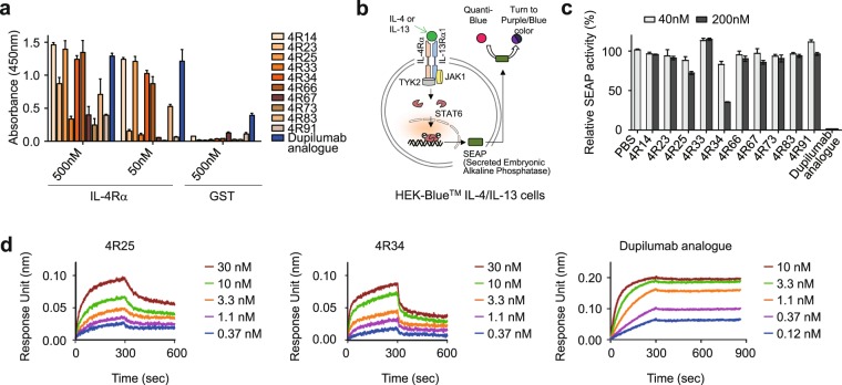
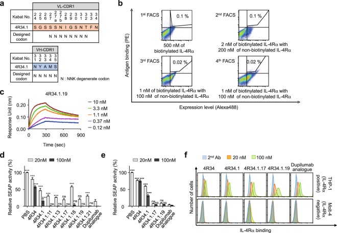
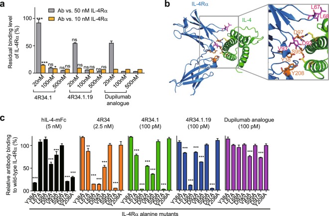
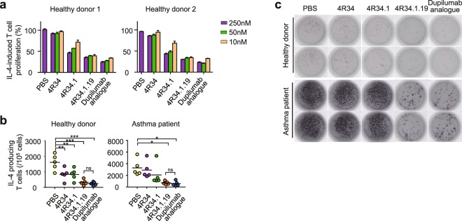
Development of antagonistic antibody (Ab) against interleukin-4 receptor alpha (IL-4Rα) subunit of IL-4/IL-13 receptors is a promising therapeutic strategy for T helper 2 (TH2)-mediated allergic diseases such as asthma and atopic dermatitis. Here we isolated anti-human IL-4Rα antagonistic Abs from a large yeast surface-displayed human Ab library and further engineered their complementarity-determining regions to improve the affinity using yeast display technology, finally generating a candidate Ab, 4R34.1.19. When reformatted as human IgG1 form, 4R34.1.19 specifically bound to IL-4Rα with a high affinity (KD ≈ 178 pM) and effectively blocked IL-4- and IL-13-dependent signaling in a reporter cell system at a comparable level to that of the clinically approved anti-IL-4Rα dupilumab Ab analogue. Epitope mapping by alanine scanning mutagenesis revealed that 4R34.1.19 mainly bound to IL-4 binding sites on IL-4Rα with different epitopes from those of dupilumab analogue. Further, 4R34.1.19 efficiently inhibited IL-4-dependent proliferation of T cells among human peripheral blood mononuclear cells and suppressed the differentiation of naïve CD4+ T cells from healthy donors and asthmatic patients into TH2 cells, the activities of which were comparable to those of dupilumab analogue. Our work demonstrates that both affinity and epitope are critical factors for the efficacy of anti-IL-4Rα antagonistic Abs.


Conflict of interest statement

Y.S.K., H.S.P., J.E.K. and J.K. are listed as inventors on pending patent applications (KR2018-0137199) for antibodies against IL-4Rα described in this work. J.A.K. and S.H.K. declare no financial competing interests. All authors declare no non-financial competing interests.

Figures

Figure 1
Figure 1
Isolation and characterization of human Abs directed against IL-4Rα. (a) Binding activity of the isolated anti-IL-4Rα Abs to plate-coated human IL-4Rα or GST, as determined by ELISA. Data represented as mean ± SD (n = 3). (b) Schematic diagram of the reporter HEK-BlueTM IL-4/IL-13 cell line to monitor the biological activity of anti-IL-4Rα Abs. The details are described in the text. (c) IL-4Rα-blocking activity of the indicated Abs, as determined by SEAP secretion levels from HEK-BlueTM IL-4/IL-13 cells after stimulation with rhIL-4 (100 pM) in the presence of the Abs (40 and 200 nM) for 24 h. Data are presented as percentage (mean ± SD (n = 3)) in SEAP levels relative to PBS-treated samples. (d) Binding isotherms of the immobilized anti-IL-4Rα Abs to soluble antigen IL-4Rα, measured by bio‐layer interferometry on OctetRED96 (Fortebio). The concentrations of IL-4Rα analysed are indicated (colored).
Figure 2
Figure 2
Affinity maturation of 4 R34 and characterization of the isolated clones. (a) Scheme of library construction and screening of 4R34 in the format of scFab using yeast surface display technology. The indicated residues in VL-CDR3, VH-CDR2, and VH-CDR3, highlighted by “X,” were randomly mutated, while maintaining the original amino acids at each residue of 4R34 at a frequency of approximately 50%, using designed spiked oligonucleotides. Numbering is according to the Kabat definition. (b) Flow cytometric analysis of antigen binding and expression levels of 4R34-based scFab yeast library in each round screening by FACS. The screening conditions of antigen and sorting gate used in each round are indicated. (c) Comparison of association and dissociation of soluble IL-4Rα antigen at 10 nM to immobilized anti-IL-4Rα antibodies (Abs), as measured by bio‐layer interferometry. (d) IL-4Rα-blocking activity of the indicated Abs, as determined by SEAP secretion levels from HEK-BlueTM IL-4/IL-13 cells after stimulation with rhIL-4 (100 pM) in the presence of the Abs (20 and 100 nM) for 24 h. Data are presented as percentage (mean ± SD (n = 3)) in SEAP levels relative to PBS-treated samples.
Figure 3
Figure 3
Engineering and characterization of 4 R34.1.19. (a) Library construction scheme of 4 R34.1, where the indicated residues in the VL-CDR1 and VH-CDR1 were randomized with NNK degenerate codon that encodes all 20 amino acids. (b) Flow cytometric analysis of antigen binding and expression levels of 4 R34.1-based scFab yeast library in each round screening by FACS. The screening conditions of antigen and sorting gate used in each round are indicated. (c) Binding isotherms of the immobilized anti-IL-4Rα Ab 4R34.1.19 to soluble antigen IL-4Rα, measured by bio‐layer interferometry. The concentrations of IL-4Rα analysed are indicated (colored). (d,e) IL-4Rα blocking activity of the indicated Abs, as determined by SEAP levels from HEK-BlueTM IL-4/IL-13 cells after stimulation with rhIL-4 (100 pM) (d) or rhIL-13 (1 nM) (e) in the presence of the Abs (20 and 100 nM) for 24 h. Data are presented as percentage (mean ± SD (n = 3)) in SEAP levels relative to phosphate buffer saline (PBS)-treated samples. Statistical analysis was performed using a two-way ANOVA followed by the Newman-Keuls post-test. *P < 0.05, **P < 0.01, ***P < 0.001; ns, not significant versus dupilumab analogue. (f) Binding specificity of the indicated Abs (20 and 100 nM) for cell surface expressed IL-4Rα, as analysed in IL-4Rα-expressing THP-1 cells and IL-4Rα-deficient Molt-4 cells by flow cytometry. Representative histograms from three independent experiments are shown.
Figure 4
Figure 4
Epitope mapping of anti-IL-4Rα Abs by alanine scanning mutagenesis. (a) Competitive ELISA showing the percentage of bound IL-4Rα (10 and 50 nM) to plate-coated hIL-4-mFc in the presence of the indicated Abs (20, 100, and 500 nM) compared to that without the Ab competitor. Data are represented as mean ± SD (n = 3). Statistical analyses were performed using a two-way ANOVA followed by the Newman-Keuls post-test. *P < 0.05, **P < 0.01, ***P < 0.001; ns, not significant versus dupilumab analogue. (b) Overall structure of the human IL-4Rα:IL-4 complex (PDB: 1IAR). Magnified section shows the residues of IL-4Rα putatively involved in IL-4 binding. (c) The percent relative binding of the indicated hIL-4-mFc (5 nM) and anti-IL-4Rα Abs (2.5 nM of 4 R34 and 100 pM of 4 R34.1, 4 R34.1.19 and dupilumab analogue) to IL-4Rα alanine mutants compared to that of wild-type IL-4Rα. Data are represented as mean ± SD (n = 3). Statistical analyses were performed using a one-way ANOVA followed by the Newman-Keuls post-test. *P < 0.05, **P < 0.01, ***P < 0.001 versus binding to wild-type IL-4Rα.
Figure 5
Figure 5
Inhibitory effects of anti-IL-4Rα Abs on IL-4-stimulated T cell proliferation and TH2 differentiation. (a) Dose-dependent blocking effects of anti-IL-4Rα Abs on the proliferation of T cells among PHA-activated PBMCs in response to rhIL-4 (500 pM), determined by CTG assay after 72 h culture. Data are represented as mean ± SD (n = 3). (b,c) Inhibitory effects of anti-IL-4Rα Abs (100 nM) on the TH2 differentiation of naïve CD4+CD45ROT cells from healthy donors or asthmatic patients after 7 days culture in TH2-skewing conditions in the presence of rhIL-4 (500 pM) and anti-IL-4Rα Abs (100 nM). The number of IL-4-producing TH2 cells were determined by ELISPOT. Quantification of spot forming T cells (b) and representative image both healthy donor and asthmatic patient (c) are shown. In (b), error bars, ± SD (n = 3). Statistical analyses were performed using a one-way ANOVA followed by the Newman-Keuls post-test. *P < 0.05, **P < 0.01, ***P < 0.001; ns, not significant versus PBS-treated group.

References

    1. Godar M, Blanchetot C, de Haard H, Lambrecht BN, Brusselle G. Personalized medicine with biologics for severe type 2 asthma: current status and future prospects. mAbs. 2018;10:34–45. doi: 10.1080/19420862.2017.1392425. - DOI - PMC - PubMed
    1. Kay AB. Allergy and allergic diseases. First of two parts. N Engl J Med. 2001;344:30–37. doi: 10.1056/NEJM200101043440106. - DOI - PubMed
    1. Gandhi NA, et al. Targeting key proximal drivers of type 2 inflammation in disease. Nature reviews. Drug discovery. 2016;15:35–50. doi: 10.1038/nrd4624. - DOI - PubMed
    1. Patel SS, Casale TB, Cardet JC. Biological therapies for eosinophilic asthma. Expert opinion on biological therapy. 2018;18:747–754. doi: 10.1080/14712598.2018.1492540. - DOI - PMC - PubMed
    1. Fajt ML, Wenzel SE. Development of New Therapies for Severe Asthma. Allergy Asthma Immunol Res. 2017;9:3–14. doi: 10.4168/aair.2017.9.1.3. - DOI - PMC - PubMed

Publication types

MeSH terms

Substances