

Histopathological and Molecular Evaluation of the Experimentally Infected Goats by the Larval Forms of Taenia multiceps
- PMID: 31123473
- PMCID: PMC6511590
Histopathological and Molecular Evaluation of the Experimentally Infected Goats by the Larval Forms of Taenia multiceps
Abstract
Background: Introduction of Taenia multiceps and T. gaigeri as two separate species have been recognized mainly on morphological grounds. This experimental study was conducted to test whether cerebral and non-cerebral forms of Coenurus cerebralis belong to one origin or they are originated from two different tape worms.
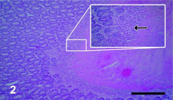
Methods: Two groups of dogs were infected with the cerebral and muscular sources of the coenuri cysts. About two months later the eggs were collected from the fecal samples to be used to experimentally infect other healthy goats. Histopathological and molecular evaluation was conducted in two groups of goats that were challenged with T. multiceps eggs obtained from the infected dogs by brain and muscular sources of coenuri cysts in School of Veterinary Medicine of Shiraz University, Shiraz, Iran in 2015. All aberrant sites of predilection of the metacestode in goats were muscles, heart, diaphragm and lungs. The brain and spinal cord were carefully dissected and examined but the cysts were not found in these locations. In addition, the molecular genetic markers of mitochondrial DNA (CO1 and ND1) were applied to resolve the questionable relationship between T. multiceps and T. gaigeri.
Results: The larval stages of T. multiceps in brain and in other aberrant sites, which showed similar morphological criteria, were monophyletic species.
Conclusion: Therefore, T. gaigeri must be considered taxonomically invalid.
Keywords: Coenurosis; Coenurus cerebralis; DNA (CO1 and ND1); Taenia gaigeri; Taenia multiceps.
Conflict of interest statement
Conflict of interest The authors declare no conflict of interest.
Figures


References
-
- Oryan A, Moghaddar N, Gaur SN. Metacestodes of sheep with special reference to their epidemiological status, pathogenesis and economic implications in Fars Province, Iran. Vet Parasitol. 1994;51(3–4):231–40. - PubMed
-
- Oryan A, Moazeni M, Amrabadi OR, Akbari M, Sharifiyazdi H. Comparison of distribution pattern, pathogenesis and molecularcharacteristics of larval stages of Taenia multiceps in sheep and goats. Small Ruminant Res. 2015;132:44–49.
-
- Akbari M, Moazeni M, Oryan A, et al. Experimental cerebral and non-cerebral coenurosis in goats: A comparative study on the morphological and molecular characteristics of the parasite. Vet Parasitol. 2015; 211(3–4):201–7. - PubMed
-
- Sharma DK, Chauhan PPS. Coenurosis status in Afro-Asian region: A review. Small Ruminant Res. 2006;64(3):197–202.
-
- Amrabadi O, Oryan A, Moazeni M, Sharifiyazdi H, Akbari M. Comparison of cerebral and non-cerebral coenurosis by genetic markers of glycolytic enzyme (enolase) and mitochondrial sequences in sheep and goats. Vet Parasitol. 2015; 214(3–4):333–6. - PubMed
LinkOut - more resources
Full Text Sources