Combined application of pharamcokinetic DCE-MRI and IVIM-DWI could improve detection efficiency in early diagnosis of ductal carcinoma in situ
- PMID: 31124276
- PMCID: PMC6612698
- DOI: 10.1002/acm2.12624
Combined application of pharamcokinetic DCE-MRI and IVIM-DWI could improve detection efficiency in early diagnosis of ductal carcinoma in situ
Abstract
Purpose: Ductal carcinoma in situ (DCIS) is a precursor of invasive ductal breast carcinoma (IDC). This study aimed to use pharamcokinetic dynamic contrast-enhanced magnetic resonance imaging (DCE-MRI) and intravoxel incoherent motion diffusion-weighted imaging (IVIM-DWI) for the early diagnosis of DCIS.
Methods: Forty-seven patients, including 25 with DCIS (age: 28-70 yr, mean age: 48.7 yr) and 22 with benign disease (age: 25-67 yr, mean age: 43.1 yr) confirmed by pathology, underwent pharamcokinetic DCE-MRI and IVIM-DWI in this study. The quantitative parameters Ktrans , Kep , Ve , Vp , and D, f, D* were obtained by processing of DCE-MRI and IVIM-DWI images with Omni-Kinetics and MITK-Diffusion softwares, respectively. Parameters were analyzed statistically using GraphPad Prism and MedCalc softwares.
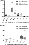
Results: All low-grade DCIS lesions demonstrated mass enhancement with clear boundaries, while most middle-grade and high-grade DCIS lesions showed non-mass-like enhancement (NMLE). DCIS lesions were significantly different from benign lesions in terms of Ktrans , Kep , and D (t = 5.959, P < 0.0001; t = 5.679, P < 0.0001; and t = 5.629, P < 0.0001, respectively). The AUC of Ktrans , Kep , D and the combined indicator of Ktrans , Kep, and D were 0.936, 0.902, 0.860, and 0.976, respectively. There was a significant difference in diagnostic efficacy only between D and the combined indicator (Z = 2.408, P = 0.016).
Conclusion: DCE-MRI and IVIM-DWI could make for the early diagnosis of DCIS, and reduce the misdiagnosis of DCIS and over-treatment of benign lesions.
Keywords: diffusion-weighted imaging; ductal carcinoma in situ; dynamic contrast-enhanced magnetic resonance imaging; intravoxel incoherent motion.
© 2019 The Authors. Journal of Applied Clinical Medical Physics published by Wiley Periodicals, Inc. on behalf of American Association of Physicists in Medicine.
Conflict of interest statement
The authors declare no competing financial interests.
Figures


Similar articles
-
Intravoxel incoherent motion MR imaging for breast lesions: comparison and correlation with pharmacokinetic evaluation from dynamic contrast-enhanced MR imaging.Eur Radiol. 2016 Nov;26(11):3888-3898. doi: 10.1007/s00330-016-4241-6. Epub 2016 Feb 10. Eur Radiol. 2016. PMID: 26863896
-
Intravoxel incoherent motion diffusion-weighted imaging as an adjunct to dynamic contrast-enhanced MRI to improve accuracy of the differential diagnosis of benign and malignant breast lesions.Magn Reson Imaging. 2017 Feb;36:175-179. doi: 10.1016/j.mri.2016.10.005. Epub 2016 Oct 11. Magn Reson Imaging. 2017. PMID: 27742437
-
Differentiating between benign and malignant sinonasal lesions using dynamic contrast-enhanced MRI and intravoxel incoherent motion.Eur J Radiol. 2018 Jan;98:7-13. doi: 10.1016/j.ejrad.2017.10.028. Epub 2017 Nov 1. Eur J Radiol. 2018. PMID: 29279173
-
Diagnostic accuracy of intravoxel incoherent motion (IVIM) and dynamic contrast-enhanced (DCE) MRI to differentiate benign from malignant breast lesions: A systematic review and meta-analysis.Eur J Radiol. 2023 Oct;167:111051. doi: 10.1016/j.ejrad.2023.111051. Epub 2023 Aug 16. Eur J Radiol. 2023. PMID: 37632999
-
Intravoxel Incoherent Motion Diffusion-Weighted Imaging for Quantitative Differentiation of Breast Tumors: A Meta-Analysis.Front Oncol. 2020 Oct 20;10:585486. doi: 10.3389/fonc.2020.585486. eCollection 2020. Front Oncol. 2020. PMID: 33194733 Free PMC article.
Cited by
-
Diagnosis of Cervical Cancer With Parametrial Invasion on Whole-Tumor Dynamic Contrast-Enhanced Magnetic Resonance Imaging Combined With Whole-Lesion Texture Analysis Based on T2- Weighted Images.Front Bioeng Biotechnol. 2020 Jun 11;8:590. doi: 10.3389/fbioe.2020.00590. eCollection 2020. Front Bioeng Biotechnol. 2020. PMID: 32596230 Free PMC article.
-
Predictive value of DCE-MRI and IVIM-DWI in osteosarcoma patients with neoadjuvant chemotherapy.Front Oncol. 2022 Oct 14;12:967450. doi: 10.3389/fonc.2022.967450. eCollection 2022. Front Oncol. 2022. PMID: 36313686 Free PMC article.
-
Intravoxel incoherent motion to differentiate spinal metastasis: A pilot study.Front Oncol. 2022 Oct 6;12:1012440. doi: 10.3389/fonc.2022.1012440. eCollection 2022. Front Oncol. 2022. PMID: 36276105 Free PMC article.
-
Looking through the imaging perspective: the importance of imaging necrosis in glioma diagnosis and prognostic prediction - single centre experience.Radiol Oncol. 2024 Feb 21;58(1):23-32. doi: 10.2478/raon-2024-0014. eCollection 2024 Mar 1. Radiol Oncol. 2024. PMID: 38378035 Free PMC article.
References
-
- Benson JR, Wishart GC. Predictors of recurrence for ductal carcinoma in situ after breast‐conserving surgery. Lancet Oncol. 2013;14:e348–e357. - PubMed
-
- Simmons A, Burrage PM, Nicolau DV Jr., Lakhani SR, Burrage K. Environmental factors in breast cancer invasion: a mathematical modelling review. Pathology. 2017;49:172–180. - PubMed
-
- Anderson WF, Chu KC. Ductal carcinoma in situ of the breast. N Engl J Med. 2004;351:399–402. - PubMed
-
- Frank GA, Danilova NV, Iu IuA, Nefedova NA. WHO classification of tumors of the breast, 2012. Arkh Patol. 2013;75:53. - PubMed
MeSH terms
Substances
Grants and funding
LinkOut - more resources
Full Text Sources
Medical

