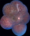
Multimodal retinal imaging of a 6-year-old male child with incontinentia pigmenti
- PMID: 31124522
- PMCID: PMC6552623
- DOI: 10.4103/ijo.IJO_192_19
Multimodal retinal imaging of a 6-year-old male child with incontinentia pigmenti
Keywords: Foveal hypoplasia; OCT angiography; incontinentia pigmenti.
Conflict of interest statement
None
Figures


Similar articles
-
Retinal imaging in incontinentia pigmenti.Indian J Ophthalmol. 2019 Jun;67(6):944-945. doi: 10.4103/ijo.IJO_417_19. Indian J Ophthalmol. 2019. PMID: 31124523 Free PMC article. No abstract available.
-
Optical Coherence Tomography Angiography and Ultra-Widefield Optical Coherence Tomography in a Child With Incontinentia Pigmenti.Ophthalmic Surg Lasers Imaging Retina. 2018 Apr 1;49(4):273-275. doi: 10.3928/23258160-20180329-11. Ophthalmic Surg Lasers Imaging Retina. 2018. PMID: 29664986 Free PMC article.
-
A 7-year-old female child of incontinentia pigmenti presenting with vitreous hemorrhage.Indian J Ophthalmol. 2017 Jun;65(6):533-535. doi: 10.4103/ijo.IJO_560_16. Indian J Ophthalmol. 2017. PMID: 28643725 Free PMC article.
-
Incontinentia pigmenti and the eye.Curr Opin Ophthalmol. 2022 Nov 1;33(6):525-531. doi: 10.1097/ICU.0000000000000863. Epub 2022 Jul 12. Curr Opin Ophthalmol. 2022. PMID: 35819905 Review.
-
Retinal imaging in infants.Surv Ophthalmol. 2021 Nov-Dec;66(6):933-950. doi: 10.1016/j.survophthal.2021.01.011. Epub 2021 Jan 29. Surv Ophthalmol. 2021. PMID: 33524458 Review.
Cited by
-
Avascular Peripheral Retina in Infants.Turk J Ophthalmol. 2023 Feb 24;53(1):44-57. doi: 10.4274/tjo.galenos.2022.76436. Turk J Ophthalmol. 2023. PMID: 36847634 Free PMC article. Review.
-
Optical Coherence Tomography Angiography in Pediatric Retinal Disorders.J Vitreoretin Dis. 2022 Jun 3;6(3):221-228. doi: 10.1177/24741264221083873. eCollection 2022 May-Jun. J Vitreoretin Dis. 2022. PMID: 37008546 Free PMC article. Review.
-
Optical Coherence Tomography and Optical Coherence Tomography Angiography in Pediatric Retinal Diseases.Diagnostics (Basel). 2023 Apr 18;13(8):1461. doi: 10.3390/diagnostics13081461. Diagnostics (Basel). 2023. PMID: 37189561 Free PMC article. Review.
-
Human Genetic Diseases Linked to the Absence of NEMO: An Obligatory Somatic Mosaic Disorder in Male.Int J Mol Sci. 2022 Jan 21;23(3):1179. doi: 10.3390/ijms23031179. Int J Mol Sci. 2022. PMID: 35163099 Free PMC article. Review.
References
-
- Minić S, Trpinac D, Obradović M. Incontinentiapigmenti diagnostic criteria update. Clin Genet. 2014;85:536–42. - PubMed
Publication types
MeSH terms
LinkOut - more resources
Full Text Sources

