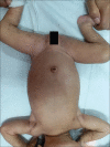
Retinal imaging in incontinentia pigmenti
- PMID: 31124523
- PMCID: PMC6552610
- DOI: 10.4103/ijo.IJO_417_19
Retinal imaging in incontinentia pigmenti
Keywords: Fundus fluorescein angiography; vasculature in incontinentia pigmenti.
Conflict of interest statement
None
Figures


Similar articles
-
Multimodal retinal imaging of a 6-year-old male child with incontinentia pigmenti.Indian J Ophthalmol. 2019 Jun;67(6):942-943. doi: 10.4103/ijo.IJO_192_19. Indian J Ophthalmol. 2019. PMID: 31124522 Free PMC article. No abstract available.
-
Fluorescein angiographic findings in a male infant with incontinentia pigmenti.J AAPOS. 2007 Oct;11(5):511-2. doi: 10.1016/j.jaapos.2007.03.006. Epub 2007 May 10. J AAPOS. 2007. PMID: 17498989
-
A 7-year-old female child of incontinentia pigmenti presenting with vitreous hemorrhage.Indian J Ophthalmol. 2017 Jun;65(6):533-535. doi: 10.4103/ijo.IJO_560_16. Indian J Ophthalmol. 2017. PMID: 28643725 Free PMC article.
-
Subthreshold Micropulse Laser Photocoagulation in the Management of Central Serous Chorioretinopathy.Int Ophthalmol Clin. 2016 Fall;56(4):165-74. doi: 10.1097/IIO.0000000000000140. Int Ophthalmol Clin. 2016. PMID: 27575766 Review. No abstract available.
-
Incontinentia pigmenti and the eye.Curr Opin Ophthalmol. 2022 Nov 1;33(6):525-531. doi: 10.1097/ICU.0000000000000863. Epub 2022 Jul 12. Curr Opin Ophthalmol. 2022. PMID: 35819905 Review.
Cited by
-
Avascular Peripheral Retina in Infants.Turk J Ophthalmol. 2023 Feb 24;53(1):44-57. doi: 10.4274/tjo.galenos.2022.76436. Turk J Ophthalmol. 2023. PMID: 36847634 Free PMC article. Review.
References
-
- Carney RG. Incontinentia pigmenti. A world statistical analysis. Arch Dermatol. 1976;112:535–42. - PubMed
-
- Minic S, Trpinac D, Obradovi’c M. Incontinentia pigmenti diagnostic’criteria update. Clin Genet. 2014;85:536–42. - PubMed
-
- Chen CJ, Han IC, Goldberg MF. Variable expression of retinopathy in a pedigree of patients with incontinentia pigmenti. Retina. 2015;35:2627–32. - PubMed
-
- Tzu JH, Murdock J, Parke DW, 3rd, Warman R, Hess DJ, Berrocal AM. Use of fluorescein angiography in incontinentia pigmenti: A case report. Ophthalmic Surg Lasers Imaging Retina. 2013;44:91–3. - PubMed
Publication types
MeSH terms
LinkOut - more resources
Full Text Sources

