. 2019 Jun;100(3):161-174.
doi: 10.1111/iep.12319. Epub 2019 May 24.

Murine experimental leprosy: Evaluation of immune response by analysis of peritoneal lavage cells and footpad histopathology

Affiliations

Murine experimental leprosy: Evaluation of immune response by analysis of peritoneal lavage cells and footpad histopathology

Fátima Regina Vilani-Moreno et al. Int J Exp Pathol. 2019 Jun.

Abstract

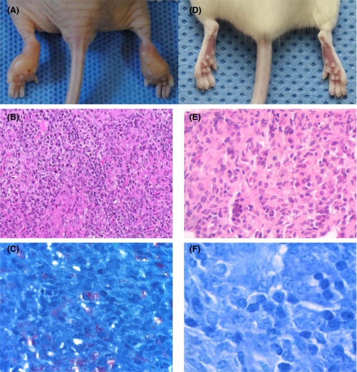
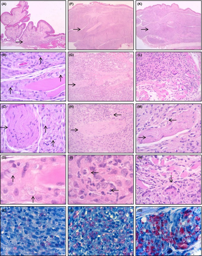
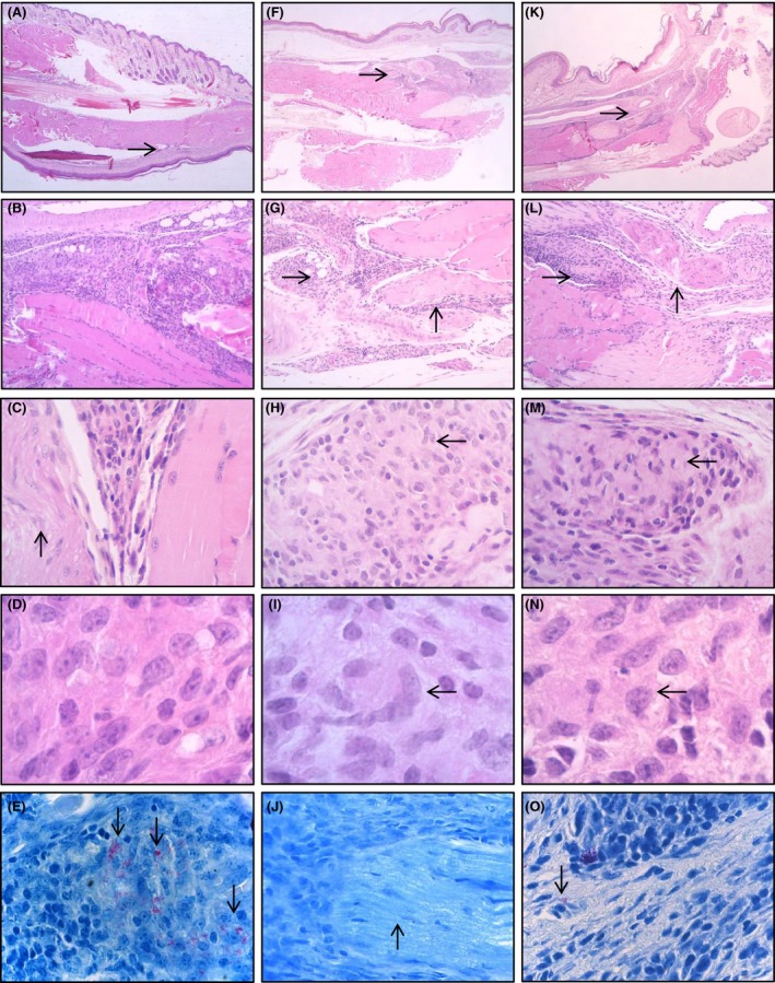
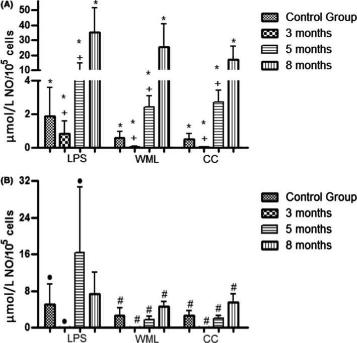
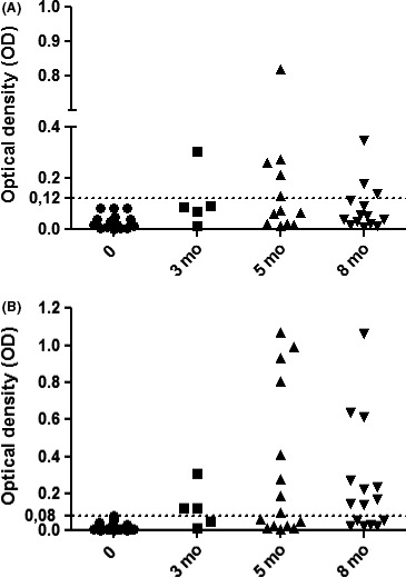
This study evaluated the immune response of nude and BALB/c mice inoculated in the footpads (FP) with Mycobacterium leprae after 3, 5 and 8 months. At each timepoint peritoneal cells, peripheral blood, FP and popliteal lymph nodes (PLN) were collected. Peritoneal cell cultures were performed to measure the H2 O2 , O2- , NO, IL-2, IL-4, IL-10, IL-12, IFN-γ and TNF levels. Serum levels of anti-PGL-I antibodies were also quantified. The results showed that the infection was progressive in nude mice with bacterial multiplication, development of macroscopic lesions in the FP and presence of bacilli in the PLN at 8 months. In BALB/c mice, the infection reached a plateau of bacillary multiplication at 5 months and regressed at 8 months. Histopathological analysis of FP revealed a mononuclear inflammatory infiltrate with a large number of neutrophils at 5 months, with a higher number in nude mice. At 8 months, the number of neutrophils decreased and the infiltrate was predominantly mononuclear in both mouse strains. There was no H2 O2, O2- , IL-2, IL-4, IL-10 and IFN-γ production in the course of infection in nude mice; however, in BALB/c, O2- and IL-12 production was higher at 5 months and NO, IFN-γ and TNF production was higher at 8 months when there was a decrease in the number of bacilli. The level of anti-PGL-I antibodies was higher in BALB/c mice. Thus, nude and BALB/c mice can be used as experimental models for the study of various aspects of leprosy.

Keywords: cytokines; histopathology; immune response; leprosy; mice; peritoneal lavage.


Conflict of interest statement

The authors report no conflicts of interest.

Figures

Figure 1
Figure 1
Nude mouse (A, B and C) and BALB/c (D, E and F) inoculated with Mycobacterium leprae. Macroscopic lesion in a nude mouse footpad 8 mo after inoculation (A). Lymph node with histiocytic infiltrate (B) with large number of bacilli inside the macrophages (C). Footpad without apparent macroscopic lesion of a BALB/c mouse inoculated with Mycobacterium leprae 8 mo after inoculation (D). Lymph node with lymphohistiocytic infiltrate (E) with few or no bacilli inside the macrophages (F). HE: B—200×, E—400×. Faraco‐Fite: C—400× and F—1000× [Colour figure can be viewed at wileyonlinelibrary.com]
Figure 2
Figure 2
Histological and bacilloscopic characteristics of the inflammatory infiltrate after inoculation of Mycobacterium leprae in the footpad of nude mice. Three months postinoculation samples (A, B, C, D and E). Five months postinoculation samples (F, G, H, I and J). Eight months postinoculation samples (K, L, M, N and O). Discrete inflammatory infiltrate (→) in 3‐mo samples (A). Moderate/intense inflammatory infiltrate in samples of 5 mo (F) and 8 mo (K). Inflammatory infiltrates mainly permeated the bundles of skeletal muscle, fibrotendinous tissue, vessels, nerves and subcutaneous tissue (B, G and L). The neural branches (→) were involved by the inflammatory infiltrate, but without the characteristics of neurotropism observed in leprosy lesions in human tissue (C, G, H and M). Granulomas were formed predominantly by multivacuolated macrophages (↑) without tuberculoid arrangement (C and D). Some samples contained focal areas with neutrophil clusters (←) (H and I) and rare multinucleated giant cells (↓) (N). Bacilloscopic index (Faraco‐Fite) ranged from 3+ to 6+ between the samples in the different groups (E, J and O), with the highest bacilloscopic index in the samples of 8 mo (↓) and lower in the 3 mo (↓). There was a predominance of solid bacilli with globi in all the groups. HE: A—20×, B—400×, C—400×, D—1000×, F—20×, G—40×, H—200×, I—1000×, K—20×, L—100×, M—400×, N—400×. Faraco‐Fite: E, J and O—400× [Colour figure can be viewed at wileyonlinelibrary.com]
Figure 3
Figure 3
Histological and bacilloscopic characteristics of the inflammatory infiltrate after inoculation of Mycobacterium leprae in the footpad of BALB/c mice. Three months postinoculation samples (A, B, C, D and E). Five months postinoculation samples (F, G, H, I and J). Eight months postinoculation samples (K, L, M, N and O). Discrete inflammatory infiltrate (→) in 3‐mo samples (A). Moderate inflammatory infiltrate in 5‐mo samples (F). Discrete/moderate infiltrate in 8‐mo samples (K). Inflammatory infiltrates mainly permeated the bundles of skeletal muscle, fibrotendinous tissues, vessels, nerves and subcutaneous tissue (B, C, G and L). The neural branches (↑) were involved by the inflammatory infiltrate, but without the characteristics of neurotropism as observed in leprosy lesions in human tissue (C, G, L and J). Granulomas were composed predominantly by epithelioid macrophages and lymphocytes (←) and show poorly organized tuberculoid arrangement (D, H, M, I and N). Bacilloscopy (Faraco‐Fite) ranged from 4+ to 0+, among samples in the different groups, with the highest bacilloscopic index in the samples of 3 mo (↓) and lower or negative at 8 mo (↓) (E, J and O). Prevalence of granulous bacilli in all groups. HE: A—20×, B—100×, C—400×, D—1000×, F—20×, G—100×, H—400×, I—1000×, K—20×, L—100×, M—400×, N—1000×. Faraco‐Fite: E, J and O—400× [Colour figure can be viewed at wileyonlinelibrary.com]
Figure 4
Figure 4
Nitric oxide (NO) production by peritoneal macrophages of nude (A) and BALB/c (B) mice at 3, 5 and 8 mo after inoculation with Mycobacterium leprae. CC: control culture (spontaneous production); LPS: lipopolysaccharide; WML: whole M. leprae antigen. * 8 mo > control group, 3 and 5 mo. + 5 mo > 3 mo (A) (< 0.05). • 5 mo > control group and 3 mo. # 8 mo > control group, 3 and 5 mo (B) (< 0.05)
Figure 5
Figure 5
Quantification of anti‐PGL‐I antibodies in the serum of nude (A) and BALB/c (B) mice at 3, 5 and 8 mo after inoculation with Mycobacterium leprae. The horizontal line is the cut‐off (mean + 3 SD) for sera from healthy animals

Similar articles

Cited by

  • An update on leprosy immunopathogenesis: systematic review.
    Silva MJA, Silva CS, Brasil TP, Alves AK, Dos Santos EC, Frota CC, Lima KVB, Lima LNGC. Silva MJA, et al. Front Immunol. 2024 Sep 6;15:1416177. doi: 10.3389/fimmu.2024.1416177. eCollection 2024. Front Immunol. 2024. PMID: 39308868 Free PMC article.

References

    1. Scollard DM, Adams LB, Gillis TP, et al. The continuing challenges of leprosy. Clin Microbiol Rev. 2006;19:338‐381. - PMC - PubMed
    1. Kaufmann SHE. Immunity to intracellular microbial pathogens. Immunol Today. 1995;16:338‐342. - PubMed
    1. Nathan CF, Murray HW, Wiebe ME, Rubin BY. Identification of interferon gamma as the lymphokine that activates human macrophage oxidative metabolism and anti‐microbial activity. J Exp Med. 1983;158:670‐674. - PMC - PubMed
    1. O'Neill LAJ, Pearce EJ. Immunometabolism governs dendritic cell and macrophage function. J Exp Med. 2016;213:15‐23. - PMC - PubMed
    1. Modlin RL. Th1‐Th2 paradigm: insights from leprosy. J Invest Dermatol. 1994;102:828‐832. - PubMed