Case Reports
. 2019 Aug;32(4):273-276.
doi: 10.1177/1971400919853789. Epub 2019 May 24.

Intraparenchymal extravasation of gadolinium mimicking an enhancing brain tumor

Affiliations
Case Reports

Intraparenchymal extravasation of gadolinium mimicking an enhancing brain tumor

David J Ritchie et al. Neuroradiol J. 2019 Aug.

Abstract

Gadolinium (Gd)-enhanced magnetic resonance imaging plays an essential role in the detection, characterization, and staging of intracranial neoplasms and vascular abnormalities. Although Gd is helpful in a majority of situations, it can lead to diagnostic misinterpretation in the setting of active vascular extravasation. Scarce reports of intracranial extravasation of Gd are present in the literature. Here, we report the first case of surgically proven spontaneous intraparenchymal extravasation of Gd mimicking an enhancing intra-axial neoplasm in a pediatric patient. Early and accurate recognition of Gd extravasation is critical in obtaining the accurate diagnosis and triaging patients expeditiously into proper avenues of care.

Keywords: Gadolinium; cavernous malformation; extravasation; hemorrhage; pediatrics.


Figures

Figure 1.
Figure 1.
Axial non-contrast head computed tomography (CT) through the level of the basal ganglia shows a large mass-occupying lesion with associated vasogenic edema centered on the left insula containing heterogeneously attenuating blood products. The mass results in local mass effect with effacement of the left lateral ventricle.
Figure 2.
Figure 2.
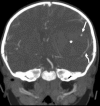
Coronal CT angiography maximum intensity projection of the head demonstrates a small focus of arterial-phase contrast enhancement, the so-called angiographic spot-sign (straight arrow), along the lateral aspect of the lesion (asterisk), which appears to be discontinuous from surrounding normal-appearing vessels (curved arrow).
Figure 3.
Figure 3.
Axial T2 FLAIR (a), T2 (b), T1 (c), and post-contrast fat-saturated T1 weighted (d) images show a predominantly T1-hypointense and T2-isointense lesion centered on the left insular lobe, with internal locules of blood at various stages of evolution (arrows). On the contrast-enhanced T1-weighted image, the lesion demonstrates heterogeneous and avid enhancement, which is most prominent in its lateral aspect (curved arrow), in the region where the focus of arterial-phase contrast enhancement was seen on the CTA (Figure 2).
Figure 4.
Figure 4.
Axial SWAN images (a–c) demonstrate extensive susceptibility artifact involving nearly the entire lesion. Additional smaller foci of susceptibility artifact are seen in the bilateral posterior parietal lobes (arrows) in (b) likely representing smaller cavernous hemangiomas. In (c), a small linear structure (curved arrow) with deep susceptibility artifact is seen adjacent to the dominant mass and likely represented a thrombosed developmental venous anomaly.
Figure 5.
Figure 5.
Intraoperative photograph shows a congealed hematoma (arrow) being suctioned out through a corticectomy in the inferior frontal lobe immediately superior to the Sylvian fissure. During surgery, the lesion was found entirely to be a hematoma, without evidence of an underlying mass.

References

    1. Cutsforth-Gregory JK, Black DF, Hoffman EM, et al. MRI spot sign. Neuro Clin Pract 2014; 4: 528–529. - PMC - PubMed
    1. Taydas O, Ogul H, Ozcan H, et al. Gadolinium-based contrast agent extravasation mimicking subarachnoid hemorrhage after electroconvulsive therapy. World Neurosurg 2018; 114: 130–133. - PubMed
    1. Mural Y, Ikeda Y, Teramoto A, et al. Magnetic resonance imaging-documented extravasation as an indicator of acute hypertensive intracerebral hemorrhage. J Neurosurg 1998; 88: 650–655. - PubMed
    1. Schindlbeck KA, Santaella A, Galinovic I, et al. Spot sign in acute intracerebral hemorrhage in dynamic T1-weighted magnetic resonance imaging. Stroke 2016; 2: 417–423. - PubMed
    1. Gross BA, Du R, Orbach DB, et al. The natural history of cerebral cavernous malformations in children. J Neurosurg Pediatr 2016; 17: 123–128. - PubMed

Publication types