

Supplementary abbreviated supine breast MRI following a standard prone breast MRI with single contrast administration: is it effective in detecting the initial contrast-enhancing lesions?
- PMID: 31124788
- PMCID: PMC6622433
- DOI: 10.5152/dir.2019.18167
Supplementary abbreviated supine breast MRI following a standard prone breast MRI with single contrast administration: is it effective in detecting the initial contrast-enhancing lesions?
Abstract
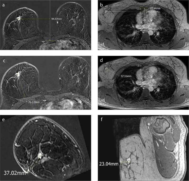
Purpose: We aimed to evaluate the detectability of contrast enhancing lesions, initially demonstrated in standard prone dynamic contrast-enhanced MRI (DCE-MRI), in a supplementary supine breast MRI examination performed following the standard prone DCE-MRI examination and to show the correlation of spatial displacement of the lesions with breast size and density.
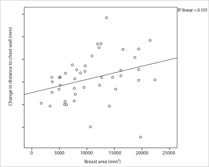
Methods: Forty-two patients with 45 lesions were prospectively evaluated. Supine breast MRI was acquired with a 6-channel body coil following a standard DCE-MRI in prone position after repositioning the patient. No additional contrast media was administered. Images were evaluated by two radiologists in consensus for the visibility of the lesions. Lesion localization relative to the sternal midline, chest wall and nipple was measured in both prone and supine positions. Correlations between lesion displacement and breast size or breast density were analyzed.
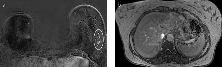
Results: Of 45 lesions, 23 (52.3%) were masses, 22 (47.7%) were nonmass enhancements (NME). Forty-four lesions (97.8%) could be detected on supine images. One linear NME of 33 mm in length could not be seen on supine images. Twenty (46.5%) of the detected lesions in supine position were equal to or smaller than 10 mm (11 NME [55%] and 9 masses [45%]). Lesion displacement relative to the chest wall increased with increasing breast size (P < 0.001).
Conclusion: An abbreviated supine sequence following a standard prone DCE-MRI with single contrast media administration is an effective method for defining the lesion location in supine position.
Conflict of interest statement
The authors declared no conflicts of interest.
Figures
Similar articles
-
Contrast enhanced breast MRI: spatial displacement from prone to supine patient's position. Preliminary results.Eur J Radiol. 2012 Jun;81(6):e771-4. doi: 10.1016/j.ejrad.2012.02.013. Epub 2012 Apr 12. Eur J Radiol. 2012. PMID: 22502792
-
Prediction of prone-to-supine tumor displacement in the breast using patient position change: investigation with prone MRI and supine CT.Breast Cancer. 2016 Jan;23(1):149-158. doi: 10.1007/s12282-014-0545-z. Epub 2014 Jun 10. Breast Cancer. 2016. PMID: 24913146
-
Feasibility of supine MRI (Magnetic Resonance Imaging)-navigated ultrasound in breast cancer patients.Asian J Surg. 2020 Aug;43(8):787-794. doi: 10.1016/j.asjsur.2019.09.007. Epub 2019 Dec 2. Asian J Surg. 2020. PMID: 31806212
-
Preliminary Results of a Simplified Breast MRI Protocol to Characterize Breast Lesions: Comparison with a Full Diagnostic Protocol and a Review of the Current Literature.Acad Radiol. 2017 Nov;24(11):1387-1394. doi: 10.1016/j.acra.2017.04.011. Epub 2017 Jun 1. Acad Radiol. 2017. PMID: 28579267 Review.
-
MRI in breast cancer radiotherapy in prone and supine positions.Front Biosci (Landmark Ed). 2017 Jan 1;22(4):570-579. doi: 10.2741/4503. Front Biosci (Landmark Ed). 2017. PMID: 27814633 Review.
Cited by
-
Evaluation of the intramammary distribution of breast lesions detected by MRI but not conventional second-look B-mode ultrasound using an MRI/ultrasound fusion technique.BMC Med Imaging. 2024 Aug 1;24(1):200. doi: 10.1186/s12880-024-01369-3. BMC Med Imaging. 2024. PMID: 39090553 Free PMC article.
-
Reply to the Letter to the Editor: MRI-detected breast lesions: clinical implications and evaluation based on MRI/ultrasonography fusion technology.Jpn J Radiol. 2020 Jan;38(1):96-97. doi: 10.1007/s11604-019-00899-z. Epub 2019 Nov 8. Jpn J Radiol. 2020. PMID: 31705426 No abstract available.
-
Feasibility of online non-rigid motion correction for high-resolution supine breast MRI.Magn Reson Med. 2023 Nov;90(5):2130-2143. doi: 10.1002/mrm.29768. Epub 2023 Jun 28. Magn Reson Med. 2023. PMID: 37379467 Free PMC article.
-
MRI-detected breast lesions: clinical implications and evaluation based on MRI/ultrasonography fusion technology.Jpn J Radiol. 2020 Jan;38(1):94-95. doi: 10.1007/s11604-019-00888-2. Epub 2019 Oct 16. Jpn J Radiol. 2020. PMID: 31620996 No abstract available.
References
Publication types
MeSH terms
Substances
Supplementary concepts
LinkOut - more resources
Full Text Sources
Other Literature Sources
Medical
