. 2019 May 24;14(5):e0217094.
doi: 10.1371/journal.pone.0217094. eCollection 2019.

Delivery of different genes into pre- and post-synaptic neocortical interneurons connected by GABAergic synapses

Affiliations

Delivery of different genes into pre- and post-synaptic neocortical interneurons connected by GABAergic synapses

Aarti Nagayach et al. PLoS One. .

Abstract

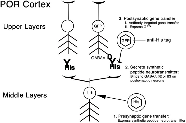
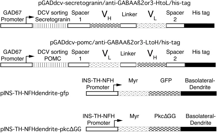
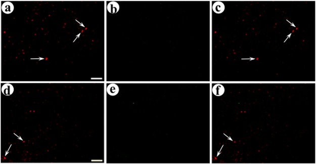
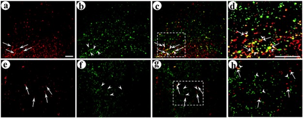
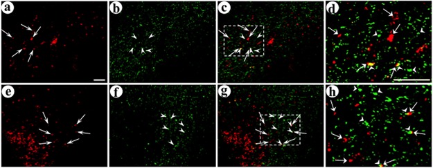
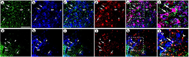
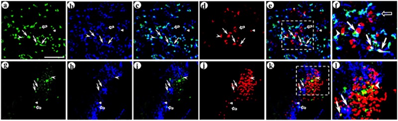
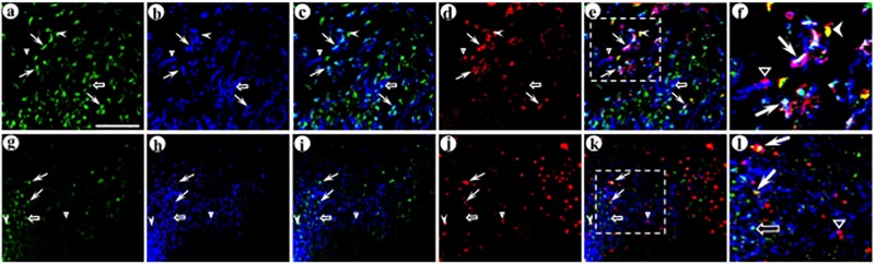
Local neocortical circuits play critical roles in information processing, including synaptic plasticity, circuit physiology, and learning, and GABAergic inhibitory interneurons have key roles in these circuits. Moreover, specific neurological disorders, including schizophrenia and autism, are associated with deficits in GABAergic transmission in these circuits. GABAergic synapses represent a small fraction of neocortical synapses, and are embedded in complex local circuits that contain many neuron and synapse types. Thus, it is challenging to study the physiological roles of GABAergic inhibitory interneurons and their synapses, and to develop treatments for the specific disorders caused by dysfunction at these GABAergic synapses. To these ends, we report a novel technology that can deliver different genes into pre- and post-synaptic neocortical interneurons connected by a GABAergic synapse: First, standard gene transfer into the presynaptic neurons delivers a synthetic peptide neurotransmitter, containing three domains, a dense core vesicle sorting domain, a GABAA receptor-binding domain, a single-chain variable fragment anti-GABAA ß2 or ß3, and the His tag. Second, upon release, this synthetic peptide neurotransmitter binds to GABAA receptors on the postsynaptic neurons. Third, as the synthetic peptide neurotransmitter contains the His tag, antibody-mediated, targeted gene transfer using anti-His tag antibodies is selective for these neurons. We established this technology by expressing the synthetic peptide neurotransmitter in GABAergic neurons in the middle layers of postrhinal cortex, and the delivering the postsynaptic vector into connected GABAergic neurons in the upper neocortical layers. Targeted gene transfer was 61% specific for the connected neurons, but untargeted gene transfer was only 21% specific for these neurons. This technology may support studies on the roles of GABAergic inhibitory interneurons in circuit physiology and learning, and support gene therapy treatments for specific disorders associated with deficits at GABAergic synapses.


Conflict of interest statement

A.I.G. has equity in Alkermes Inc. This does not alter our adherence to PLOS ONE policies on sharing data and materials.

Figures

Fig 1
Fig 1. The strategy for delivering different genes into pre- or post-synaptic neurons in POR cortex connected by GABAergic synapses that contain GABAA ß2 or ß3 subunits.
This strategy contains three steps: Step 1 is the presynaptic gene transfer: This gene transfer uses an injection site in the middle layers of POR cortex, and the gene transfer uses standard procedures. This vector expresses a synthetic peptide neurotransmitter from a GABAergic-specific promoter, the GAD67 promoter. The synthetic peptide neurotransmitter, abbreviated His within a HSV-1 vector particle (hexagon surrounded by a circle), contains three domains: i) a DCV sorting domain, ii) a ScFv anti-GABAA ß2 or ß3, and iii) the His tag. Step 2 is release of the synthetic peptide neurotransmitter (shown as His-Y, Y indicates the ScFv domain), followed by binding to GABAA receptors (shown as inverted U) on the postsynaptic neuron on the right side of the Fig. Step 3 is the postsynaptic gene transfer: This gene transfer uses an injection site is in the upper layers of POR cortex and is selective for the postsynaptic neurons that have bound the synthetic peptide neurotransmitter: The gene transfer uses antibody-mediated targeting. These vector particles contain a modified HSV-1 glycoprotein fused to the IgG binding domain from Staphylococcus A protein, and bound to anti-His tag antibody (in the top right of the Figure: HSV-1 vector particle, hexagon surrounded by a circle; modified HSV-1 glycoprotein, horizontal line from HSV-1 vector particle to anti-His tag). Complexes of these vector particles and anti-His tag antibodies are injected, and these complexes bind to the His tag on the synthetic peptide neurotransmitter, bound to GABAA ß2 or ß3 subunits on specific postsynaptic neurons. To support assays for gene transfer to connected neurons, the postsynaptic vector expresses a dendrite-targeted GFP from a pan-neuronal promoter, the INS-TH-NFH promoter. This system supports highly specific gene transfer to neurons connected by GABAA ß2 or ß3 subunit-containing synapses: As shown in the left column, axon collaterals from transduced neurons will release the synthetic peptide neurotransmitter at other synapses, but if the postsynaptic neuron lacks GABAA ß2 or ß3 subunits, that postsynaptic neuron will not be transduced.
Fig 2
Fig 2. The transcription units in the pre- or post-synaptic vectors.
These four vectors all contain a standard HSV-1 vector backbone [21]. The two presynaptic vectors use the GABAergic-specific GAD67 promoter to express a synthetic peptide neurotransmitter designed to bind to GABAA ß2 or ß3 subunits. Each synthetic peptide neurotransmitter contains a DCV sorting domain, either from Secretogranin II or POMC, a spacer (spacer 1, 42 bp), a ScFv anti-GABAA ß2 or ß3, another spacer (spacer 2, 60 bp), and the His tag. The ScFv contain either the heavy or light chain variable region, a established linker, (Gly4Ser)3 [25], and the other variable region. The two spacers separate the ScFv from the other domains, and are designed to enable each domain to function without steric hindrance from the other domains. The two postsynaptic vectors have been reported [21], and express a dendrite-targeted marker, either GFP or PkcΔGG (an enzymatically inactive PKC) from the pan-neuronal INS-TH-NFH promoter. Two dendrite-targeting signals are fused to each marker; first, a myristoylation/palmitoylation (Myr) site (from Fyn), and, second, a basolateral/ dendrite membrane-sorting domain (from the low density lipoprotein receptor) [35].
Fig 3
Fig 3. The locations of the pre- and postsynaptic injection sites in the middle or upper layers of POR cortex, and transduced neurons proximal to each injection site.
(A and B) The presynaptic vector pGADdcv-secretogranin/anti-GABAAß2or3-HtoL/his-tag, His tag-IR (Texas red-conjugated secondary antibody); (A) low power view, and (B) high power view of the box in panel A showing transduced neurons. (C and D) The postsynaptic vector pINS-TH-NFHdendrite-gfp/gC–ZZ+anti-His tag, GFP-IR (FITC-conjugated secondary antibody); (C) low power view, and (D) high power view of the box in panel C showing transduced neurons. Scale bars: (A and C) 1,200 μm; (B and D) 50 μm.
Fig 4
Fig 4. Recombinant proteins in the middle layers of POR cortex after delivery of the presynaptic vector, pGADdcv-secretogranin/anti-GABAAß2or3-HtoL/his-tag, into the middle layers of POR cortex, followed by coinjection of the two postsynaptic vectors into the upper layers POR cortex.
The presynaptic vector was injected, eight days later a 1:1 mixture of the two postsynaptic vectors was injected, and the rats were sacrificed four days later. The area proximal to the first, presynaptic gene transfer, in the middle layers of POR cortex, was analyzed for recombinant proteins by immunofluorescent costaining. Alternating sections were examined for the expression from the presynaptic vector and either the targeted or control postsynaptic vector. (A-C) Expression from pGADdcv-secretogranin/anti-GABAAß2or3-HtoL/his-tag and the targeted postsynaptic vector, pINS-TH-NFHdendrite-gfp/gC–ZZ+anti-His tag; (A) His tag-IR (Texas red-conjugated secondary antibody), (B) GFP-IR (FITC-conjugated secondary antibody), and (C) merge. Arrows, neuronal cell bodies that express the synthetic peptide neurotransmitter from the presynaptic vector. (D-F) Costaining for expression from this presynaptic vector and the untargeted postsynaptic vector pINS-TH-NFHdendrite-PkcΔGG; (D) His tag-IR, (E) flag-IR (the flag tag is fused to PkcΔGG; FITC-conjugated secondary antibody), and (F) merge. Scale bar: 50 μm.
Fig 5
Fig 5. Recombinant proteins in the middle layers of POR cortex after injection of a presynaptic vector, pGADdcv-pomc/anti-GABAAß2or3-LtoH/his-tag, into the middle layers of POR cortex followed by coinjection of the two postsynaptic vectors into the upper layers of POR cortex.
The experimental design and immunofluorescent costaining were as above. (A-C) Expression from this presynaptic vector and the targeted postsynaptic vector; (A) His tag-IR, (B) GFP-IR, and (C) merge. Arrows, cell bodies expressing the synthetic peptide neurotransmitter from the presynaptic vector. (D-F) Costaining for this presynaptic vector and the control, untargeted postsynaptic vector; (D) His tag-IR, (E) flag-IR, and (F) merge. Scale bar: 50 μm.
Fig 6
Fig 6. The synthetic peptide neurotransmitter in the presynaptic vector supports efficient gene transfer to connected neurons.
The experimental design was as detailed in Fig 3 legend. Alternating sections containing POR cortex were analyzed for expression from the presynaptic vector and either the targeted or control postsynaptic vector, using immunofluorescent costaining. The upper layers of POR cortex were examined. (A-D) Delivery of this presynaptic vector, followed by the targeted postsynaptic vector, supports efficient gene transfer to connected neurons; (A) His tag-IR (Texas red-conjugated secondary antibody), (B) GFP-IR (FITC-conjugated secondary antibody), (C) merge, and (D) merge, an enlarged area showing specific transduced axons either proximal to, or distant from, a transduced dendrite. Arrows, transduced axons; arrowheads, transduced dendrites. (E-H) Delivery of this presynaptic vector followed by the untargeted, control postsynaptic vector supported only a low level of gene transfer to connected neurons; (E) His tag-IR, (F) flag-IR (FITC-conjugated secondary antibody), (G) merge, and (H) merge, an enlarged area with His tag-IR axons and flag-IR dendrites that are distant from each other. Scale bars: (A-C and E-G) 50 μm, and (D and H) 50 μm.
Fig 7
Fig 7. The synthetic peptide neurotransmitter dcv-pomc/anti-GABAAß2or3-LtoH/his-tag supports limited gene transfer to connected neurons.
The experimental design and immunofluorescent costaining were as above. (A-D) Delivery of this presynaptic vector followed by the targeted postsynaptic vector resulted in only low levels of gene transfer to connected neurons; (A) His tag-IR, (B) GFP-IR, (C) merge, and (D) merge, with an enlarged area that contains a number of transduced axons and dendrites; some transduced axons are proximal to a transduced dendrite and other transduced axons are distant from a transduced dendrite. Arrows, transduced axons; arrowheads, transduced dendrites. (E-H) Delivery of this presynaptic vector, followed by the untargeted postsynaptic vector, supported low levels of gene transfer to connected neurons, similar to those observed using the targeted postsynaptic vector; (E) His tag-IR, (F) flag-IR, (G) merge, and (H) merge, an enlarged area that shows transduced axons that are either proximal to, or distant from, transduced dendrites. Scale bars: (A-C and E-G) 50 μm, and (D and H) 50 μm.
Fig 8
Fig 8. The synthetic peptide neurotransmitter dcv-secretogranin/anti-GABAAß2or3-HtoL/his-tag supports gene transfer to the parvalbumin-containing subtype of GABAergic neurons.
The experimental design was as above. Using immunofluorescent costaining, the upper layers of POR cortex were analyzed for expression from the presynaptic vector (His tag-IR), the targeted postsynaptic vector (GFP-IR), and the GABAergic subtype marker parvalbumin. (A-F) Image stacks show neuronal morphology for the transduced postsynaptic dendrites and cell bodies, proximal to transduced axons; (A) His tag-IR (fluorescein-conjugated secondary antibody), (B) GFP-IR (Alexa Fluor 633-conjugated secondary antibody), (C) merge of His tag-IR and GFP-IR, (D) parvalbumin-IR (TRITC-conjugated secondary antibody), (E) merge of all three IR, (F) merge, an enlarged area showing adjacent transduced axons and dendrites that contain parvalbumin-IR. Arrows, transduced axons proximal to transduced dendrites, with parvalbumin-IR. Open arrow, a transduced axon proximal to a transduced dendrite that lacks parvalbumin-IR. Arrowhead, a transduced axon distant from a transduced dendrite, but proximal to parvalbumin-IR. Open triangle, a transduced dendrite that contains parvalbumin-IR, but distant from a transduced axon. (G-L) Single confocal images in each channel show that delivery of this presynaptic vector, followed by the targeted postsynaptic vector, supported gene transfer across GABAA ß2- or ß3-containing synapses to parvalbumin-containing postsynaptic neurons; (G) His tag-IR, (H) GFP-IR, (I) merge of His tag-IR and GFP-IR, (J) parvalbumin-IR, (K) merge of all three IR, (L) merge, an enlarged area showing adjacent transduced axons and dendrites that contain parvalbumin-IR. Panels G-L show a different field than for panels A-F. Scale bar: 20 μm.
Fig 9
Fig 9. The synthetic peptide neurotransmitter dcv-secretogranin/anti-GABAAß2or3-HtoL/his-tag supports gene transfer to the calretinin-containing subtype of GABAergic neurons.
The experimental design and immunofluorescent costaining were as above, but calretinin-IR was assayed instead of parvalbumin-IR. (A-F) Image stacks show neuronal morphology for the transduced postsynaptic dendrites and cell bodies, proximal to transduced axons; (A) His tag-IR (fluorescein-conjugated secondary antibody), (B) GFP-IR (Alexa Fluor 633-conjugated secondary antibody), (C) merge of His tag-IR and GFP-IR, (D) calretinin-IR (TRITC-conjugated secondary antibody), (E) merge of all three IR, (F) merge, an enlarged area showing adjacent transduced axons and dendrites that contain calretinin-IR. Arrows, transduced axons proximal to transduced dendrites, with calretinin-IR. Open arrow, a transduced axon proximal to a transduced dendrite that lacks calretinin-IR. Arrowheads, transduced axons distant from transduced dendrites, but proximal to calretinin-IR. Open triangle, a transduced dendrite that contains calretinin-IR, but distant from a transduced axon. (G-L) Single confocal images in each channel show that delivery of this presynaptic vector, followed by the targeted postsynaptic vector, supported gene transfer across GABAA ß2- or ß3-containing synapses to calretinin-containing postsynaptic neurons; (G) His tag-IR, (H) GFP-IR, (I) merge of His tag-IR and GFP-IR, (J) calretinin-IR, (K) merge of all three IR, (L) merge, an enlarged area showing adjacent transduced axons and dendrites that contain calretinin-IR. Panels G-L show a different field than for panels A-F. Scale bar: 20 μm.
Fig 10
Fig 10. This synthetic peptide neurotransmitter supports gene transfer to the calbindin-containing subtype of GABAergic neurons.
The experimental design and immunofluorescent costaining were as above, but calbindin-IR was assayed instead of parvalbumin-IR. (A-F) Image stacks show neuronal morphology for the transduced postsynaptic dendrites and cell bodies, proximal to transduced axons; (A) His tag-IR (fluorescein-conjugated secondary antibody), (B) GFP-IR (Alexa Fluor 633-conjugated secondary antibody), (C) merge of His tag-IR and GFP-IR, (D) calbindin-IR (TRITC-conjugated secondary antibody), (E) merge of all three IR, (F) merge, an enlarged area showing adjacent transduced axons and dendrites that contain calbindin-IR. Arrow, a transduced axon proximal to a transduced dendrite, with calbindin-IR. Open arrow, a transduced axon proximal to a transduced dendrite that lacks calbindin-IR. Arrowhead, a transduced axon distant from a transduced dendrite, but proximal to calbindin-IR. Open triangle, a transduced dendrite that contains calbindin-IR, but distant from a transduced axon. (G-L) Single confocal images in each channel show that delivery of the presynaptic vector, followed by the targeted postsynaptic vector, supported gene transfer across GABAA ß2- or ß3-containing synapses to calbindin-containing postsynaptic neurons; (G) His tag-IR, (H) GFP-IR, (I) merge of His tag-IR and GFP-IR, (J) calbindin-IR, (K) merge of all three IR, (L) merge, an enlarged area showing adjacent transduced axons and dendrites that contain calbindin-IR. Panels G-L show a different field than for panels A-F. Scale bar: 20 μm.

Similar articles

Cited by

References

    1. Marin O. Interneuron dysfunction in psychiatric disorders. Nat Rev Neurosci. 2012;13(2):107–20. Epub 2012/01/19. 10.1038/nrn3155 . - DOI - PubMed
    1. Hubel DH, Wiesel TN. Ferrier lecture. Functional architecture of macaque monkey visual cortex. Proc Royal Soc London—Series B. 1977;198:1–59. - PubMed
    1. Hubel DH, Wiesel TN. Receptive fields, binocular interaction and functional architecture in the cat's visual cortex. J Physiol. 1962;160:106–54. 10.1113/jphysiol.1962.sp006837 - DOI - PMC - PubMed
    1. Dudai Y. The Neurobiology of Memory. Oxford, England: Oxford Univ. Press; 1989.
    1. Kubota Y, Karube F, Nomura M, Kawaguchi Y. The Diversity of Cortical Inhibitory Synapses. Frontiers in neural circuits. 2016;10:27 Epub 2016/05/21. 10.3389/fncir.2016.00027 . - DOI - PMC - PubMed

Publication types

MeSH terms