

Difference analysis in prevalence of incidental pancreatic cystic lesions between computed tomography and magnetic resonance imaging
- PMID: 31126251
- PMCID: PMC6534824
- DOI: 10.1186/s12880-019-0341-5
Difference analysis in prevalence of incidental pancreatic cystic lesions between computed tomography and magnetic resonance imaging
Abstract
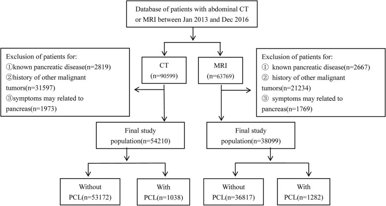
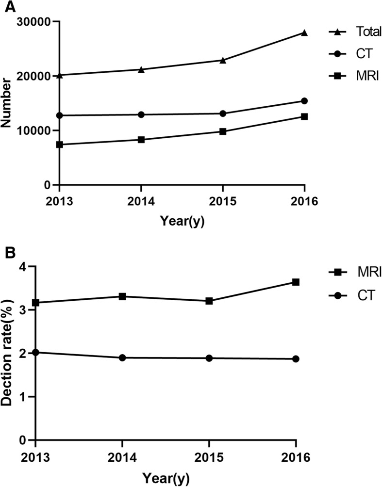
Background: The purpose was to investigate the difference of detection rate of incidental pancreatic cystic lesions (PCLs) with computed tomography (CT) and magnetic resonance imaging (MRI) and to compare the difference between CT and MRI and to explore the effect of this difference on surgical resection.
Methods: We reviewed the diagnostic reports for incidental PCLs between 2013 and 2016. Images of PCLs would be re-evaluated. Clinical and imaging data were recorded. The chi-square and independent t-test were conducted for categorical and continuous variables.
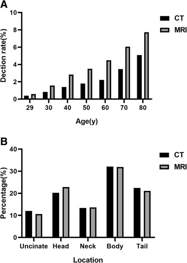
Results: The prevalence of PCLs was 1.91% (1038/54210) and 3.36% (1282/38099) on CT and MRI respectively, and increased with increasing age (P < 0.001). No significant differences were found in the annual prevalence of PCLs on CT (P = 0.796) and MRI (P = 0.213) from 2013 to 2016 while the number of examinations was increasing every year. The annual detection rate of MRI for small PCLs (< 20 mm) was significantly higher than CT (P < 0.001), but was not significantly different for large PCLs (≥20 mm). The rate of surgical resection of PCLs (≥20 mm) in MRI group was higher than CT (55.2% vs. 37.0%, P < 0.001).
Conclusions: The detection rate of PCLs on CT and MRI tended to be stable despite increasing scan volumes. Female had a slightly more frequency of PCLs than male. MRI detected more small PCLs(< 20 mm) and had higher impact on surgical resection of large PCL(≥20 mm) compared with CT.
Keywords: Computed tomography; Magnetic resonance imaging; Pancreatic cyst; Prevalence.
Conflict of interest statement
The authors declare that they have no competing interests.
Figures
Similar articles
-
Global Prevalence of Pancreatic Cystic Lesions in the General Population on Magnetic Resonance Imaging: A Systematic Review and Meta-analysis.Clin Gastroenterol Hepatol. 2024 Sep;22(9):1798-1809.e6. doi: 10.1016/j.cgh.2024.02.018. Epub 2024 Feb 28. Clin Gastroenterol Hepatol. 2024. PMID: 38423346
-
Prevalence of pancreatic cystic lesions detected by magnetic resonance imaging in the Chinese population.J Gastroenterol Hepatol. 2019 Sep;34(9):1656-1662. doi: 10.1111/jgh.14658. Epub 2019 Apr 14. J Gastroenterol Hepatol. 2019. PMID: 30883900
-
Incidental pancreatic cystic lesions: retrospective analysis of natural history and efficacy of imaging surveillance guidelines.Eur Radiol. 2025 Jul;35(7):4100-4110. doi: 10.1007/s00330-024-11307-0. Epub 2024 Dec 20. Eur Radiol. 2025. PMID: 39706924
-
Imaging-based surveillance in patients with initially detected pancreatic cystic lesions.Eur J Radiol. 2025 Jun;187:112102. doi: 10.1016/j.ejrad.2025.112102. Epub 2025 Apr 6. Eur J Radiol. 2025. PMID: 40215706
-
Diagnosis and Surveillance of Incidental Pancreatic Cystic Lesions: 2017 Consensus Recommendations of the Korean Society of Abdominal Radiology.Korean J Radiol. 2019 Apr;20(4):542-557. doi: 10.3348/kjr.2018.0640. Korean J Radiol. 2019. PMID: 30887737 Free PMC article. Review.
Cited by
-
Imaging Modalities for Early Detection of Pancreatic Cancer: Current State and Future Research Opportunities.Cancers (Basel). 2022 May 21;14(10):2539. doi: 10.3390/cancers14102539. Cancers (Basel). 2022. PMID: 35626142 Free PMC article. Review.
-
Utility of gadolinium for identifying the malignant potential of pancreatic cystic lesions.Abdom Radiol (NY). 2022 Apr;47(4):1351-1359. doi: 10.1007/s00261-022-03446-z. Epub 2022 Feb 23. Abdom Radiol (NY). 2022. PMID: 35195765
-
Opportunistic Detection for Pancreatic Cystic Lesions During Chest Multidetector CT Scans for Lung Cancer Screening.Cancer Manag Res. 2021 Oct 2;13:7559-7568. doi: 10.2147/CMAR.S327022. eCollection 2021. Cancer Manag Res. 2021. PMID: 34629902 Free PMC article.
-
Cystic Neoplasms of the Pancreas: Differential Diagnosis and Radiology Correlation.Front Oncol. 2022 Mar 1;12:860740. doi: 10.3389/fonc.2022.860740. eCollection 2022. Front Oncol. 2022. PMID: 35299739 Free PMC article. Review.
-
Global Prevalence of Pancreatic Cystic Lesions in the General Population on Magnetic Resonance Imaging: A Systematic Review and Meta-analysis.Clin Gastroenterol Hepatol. 2024 Sep;22(9):1798-1809.e6. doi: 10.1016/j.cgh.2024.02.018. Epub 2024 Feb 28. Clin Gastroenterol Hepatol. 2024. PMID: 38423346
References
-
- de Jong K, Nio CY, Hermans JJ, Dijkgraaf MG, Gouma DJ, van Eijck CH, van Heel E, Klass G, Fockens P, Bruno MJ. High prevalence of pancreatic cysts detected by screening magnetic resonance imaging examinations. Clin Gastroenterol Hepatol. 2010;8(9):806–811. doi: 10.1016/j.cgh.2010.05.017. - DOI - PubMed
Publication types
MeSH terms
LinkOut - more resources
Full Text Sources
Medical
