. 2019 Jul 18;85(15):e00988-19.
doi: 10.1128/AEM.00988-19. Print 2019 Aug 1.

Horizontal Gene Transfer as an Indispensable Driver for Evolution of Neocallimastigomycota into a Distinct Gut-Dwelling Fungal Lineage

Affiliations

Horizontal Gene Transfer as an Indispensable Driver for Evolution of Neocallimastigomycota into a Distinct Gut-Dwelling Fungal Lineage

Chelsea L Murphy et al. Appl Environ Microbiol. .

Abstract

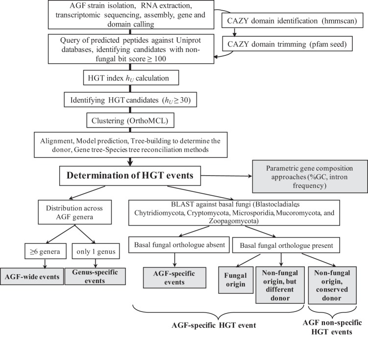
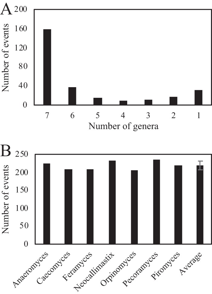
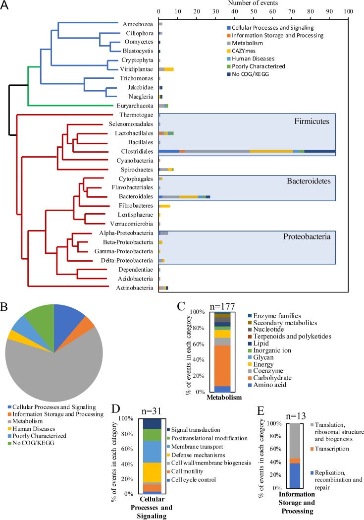
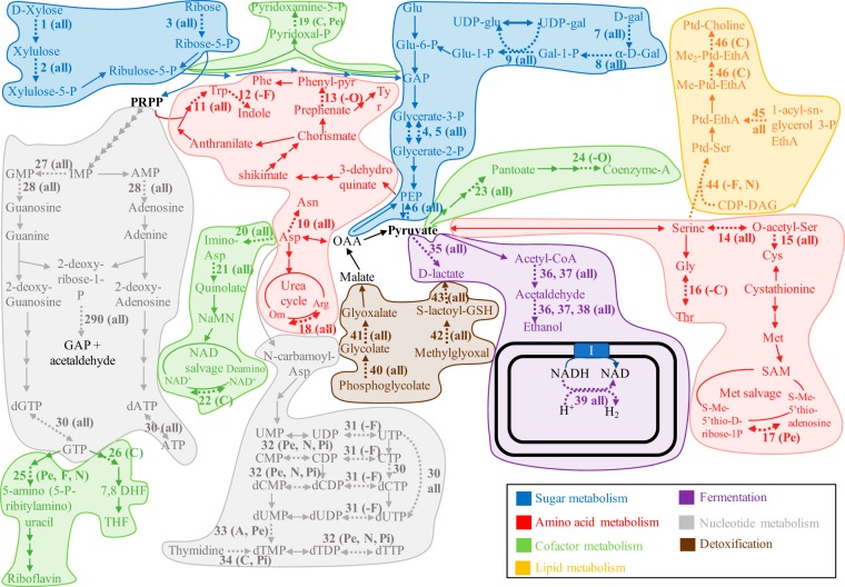
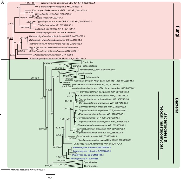
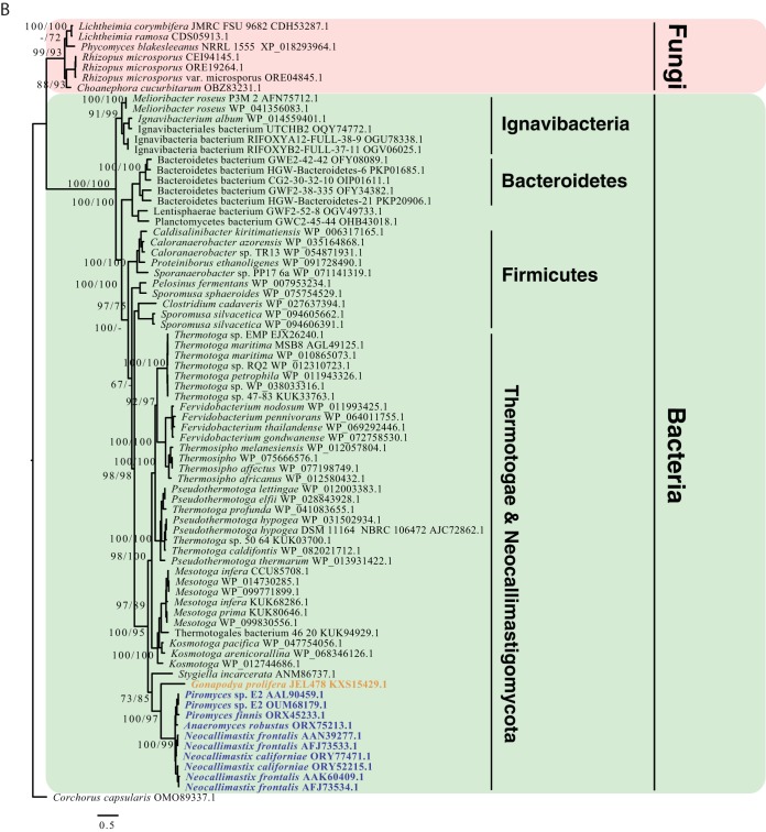
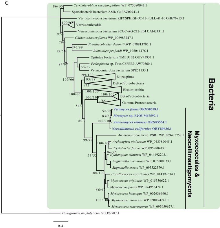
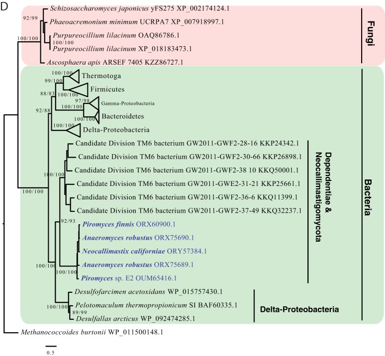
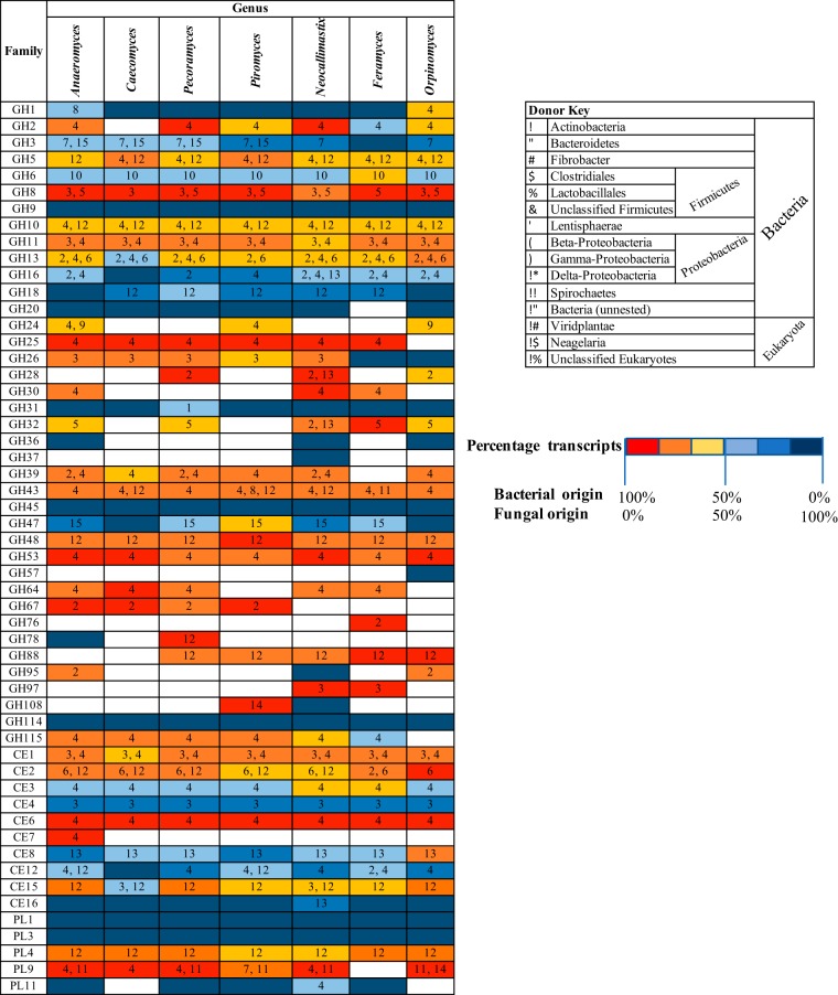
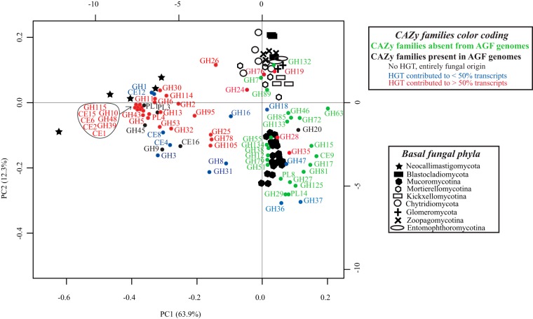
Survival and growth of the anaerobic gut fungi (AGF; Neocallimastigomycota) in the herbivorous gut necessitate the possession of multiple abilities absent in other fungal lineages. We hypothesized that horizontal gene transfer (HGT) was instrumental in forging the evolution of AGF into a phylogenetically distinct gut-dwelling fungal lineage. The patterns of HGT were evaluated in the transcriptomes of 27 AGF strains, 22 of which were isolated and sequenced in this study, and 4 AGF genomes broadly covering the breadth of AGF diversity. We identified 277 distinct incidents of HGT in AGF transcriptomes, with subsequent gene duplication resulting in an HGT frequency of 2 to 3.5% in AGF genomes. The majority of HGT events were AGF specific (91.7%) and wide (70.8%), indicating their occurrence at early stages of AGF evolution. The acquired genes allowed AGF to expand their substrate utilization range, provided new venues for electron disposal, augmented their biosynthetic capabilities, and facilitated their adaptation to anaerobiosis. The majority of donors were anaerobic fermentative bacteria prevalent in the herbivorous gut. This study strongly indicates that HGT indispensably forged the evolution of AGF as a distinct fungal phylum and provides a unique example of the role of HGT in shaping the evolution of a high-rank taxonomic eukaryotic lineage.IMPORTANCE The anaerobic gut fungi (AGF) represent a distinct basal phylum lineage (Neocallimastigomycota) commonly encountered in the rumen and alimentary tracts of herbivores. Survival and growth of anaerobic gut fungi in these anaerobic, eutrophic, and prokaryote-dominated habitats necessitates the acquisition of several traits absent in other fungal lineages. We assess here the role of horizontal gene transfer as a relatively fast mechanism for trait acquisition by the Neocallimastigomycota postsequestration in the herbivorous gut. Analysis of 27 transcriptomes that represent the broad diversity of Neocallimastigomycota identified 277 distinct HGT events, with subsequent gene duplication resulting in an HGT frequency of 2 to 3.5% in AGF genomes. These HGT events have allowed AGF to survive in the herbivorous gut by expanding their substrate utilization range, augmenting their biosynthetic pathway, providing new routes for electron disposal by expanding fermentative capacities, and facilitating their adaptation to anaerobiosis. HGT in the AGF is also shown to be mainly a cross-kingdom affair, with the majority of donors belonging to the bacteria. This study represents a unique example of the role of HGT in shaping the evolution of a high-rank taxonomic eukaryotic lineage.

Keywords: Neocallimastigomycota; anaerobic gut fungi; horizontal gene transfer.


Figures

FIG 1
FIG 1
Workflow diagram describing the procedure employed for identification HGT events in Neocallimastigomycota data sets analyzed in this study.
FIG 2
FIG 2
(A) Distribution pattern of HGT events in AGF transcriptomes demonstrating that the majority of events were Neocallimastigomycota-wide, i.e., identified in all seven AGF genera examined. (B) Total number of HGT events identified per AGF genus.
FIG 3
FIG 3
Identity of HGT donors and their contribution to the various functional classes. The x axis shows the absolute number of events belonging to each of the functional classes shown in the legend. The tree is intended to show the relationship between the donors’ taxa and is not drawn to scale. Bacterial donors are shown with red branches depicting the phylum level, with the exception of Firmicutes and Bacteroidetes donors, where the order level is shown, and Proteobacteria, where the class level is shown. Archaeal donors are shown with green branches and all belonged to the Methanobacteriales order of Euryarchaeota. Eukaryotic donors are shown with blue branches. Only the 230 events from a definitive-taxon donor are shown in the figure. The other 53 events were clearly nested within a nonfungal clade, but a definitive donor taxon could not be ascertained. Functional classification of the HGT events, determined by searching the Conserved Domain server (106) against the COG database are shown in panel B. For events with no COG classification, a search against the KEGG orthology database (107) was performed. For the major COG/KEGG categories (metabolism, cellular processes, and signaling, and information storage and processing), subclassifications are shown in panels C, D, and E, respectively.
FIG 4
FIG 4
HGT impact on AGF central metabolic abilities. Pathways for sugar metabolism are highlighted in blue, pathways for amino acid metabolism are highlighted in red, pathways for cofactor metabolism are highlighted in green, pathways for nucleotide metabolism are highlighted in gray, pathways for lipid metabolism are highlighted in orange, fermentation pathways are highlighted in purple, while pathways for detoxification are highlighted in brown. The double black lines depict the hydrogenosomal outer and inner membrane. Arrows corresponding to enzymes encoded by horizontally transferred transcripts are shown with thicker dotted lines and are given numbers 1 through 46 as follows. Sugar metabolism (1 to 9): 1, xylose isomerase; 2, xylulokinase; 3, ribokinase; 4, 2,3-bisphosphoglycerate-independent phosphoglycerate mutase; 5, 2,3-bisphosphoglycerate-dependent phosphoglycerate mutase; 6, phosphoenolpyruvate synthase; 7, aldose-1-epimerase; 8, galactokinase; 9, galactose-1-phosphate uridyltransferase. Amino acid metabolism (10 to 18): 10, aspartate-ammonia ligase; 11, tryptophan synthase (TrpB); 12, tryptophanase; 13, monofunctional prephenate dehydratase; 14, serine-o-acetyltransferase; 15, cysteine synthase; 16, low-specificity threonine aldolase; 17, 5′-methylthioadenosine nucleosidase/5′-methylthioadenosine phosphorylase (MTA phosphorylase); 18, arginase. Cofactor metabolism (19 to 26): 19, pyridoxamine 5′-phosphate oxidase; 20, l-aspartate oxidase (NadB); 21, quinolate synthase (NadA); 22, NH3-dependent NAD+ synthetase (NadE); 23, 2-dehydropantoate 2-reductase; 24, dephospho-CoA kinase; 25, dihydrofolate reductase (DHFR) family; 26, dihydropteroate synthase. Nucleotide metabolism (27 to 34): 27, GMP reductase; 28, trifunctional nucleotide phosphoesterase; 29, deoxyribose-phosphate aldolase (DeoC); 30, oxygen-sensitive ribonucleoside-triphosphate reductase class III (NrdD); 31, nucleoside/nucleotide kinase family protein; 32, cytidylate kinase-like family; 33, thymidylate synthase; 34, thymidine kinase. Pyruvate metabolism (fermentation pathways) (35 to 39): 35, d-lactate dehydrogenase; 36, bifunctional aldehyde/alcohol dehydrogenase family of Fe-alcohol dehydrogenase; 37, butanol dehydrogenase family of Fe-alcohol dehydrogenase; 38, Zn-type alcohol dehydrogenase; 39, Fe-only hydrogenase. Detoxification reactions (40 to 43): 40, phosphoglycolate phosphatase; 41, glyoxal reductase; 42, glyoxalase I; 43, glyoxalase II. Lipid metabolism (44 or 46): 44, CDP-diacylglycerol–serine O-phosphatidyltransferase; 45, lysophospholipid acyltransferase LPEAT; 46, methylene-fatty-acyl-phospholipid synthase. Following the numbers, between parentheses, the distribution of the specific event across AGF genera is shown where (all) indicates the event was detected in all seven genera, while a minus sign followed by a genus indicates that the event was detected in all but that/those genus/genera. Genera are represented by letters as follows: A, Anaeromyces; C, Caecomyces; F, Feramyces, N, Neocallimastix, O, Orpinomyces; Pe, Pecoramyces; Pi, Piromyces. Abbreviations: CDP-DAG, CDP-diacylglycerol; 7,8 DHF, 7,8-dihydrofolate; EthA, ethanolamine; Gal, galactose; GAP, glyceraldehyde-3-P; Glu, glucose; GSH, glutathione; I, complex I NADH dehydrogenase; NaMN, nicotinate d-ribonucleotide; Orn, ornithine; PEP, phosphoenol pyruvate; Phenyl-pyr, phenylpyruvate; PRPP, phosphoribosyl-pyrophosphate; Ptd, phosphatidyl; SAM; S-adenosylmethionine; THF, tetrahydrofolate.
FIG 5
FIG 5
(A) Maximum-likelihood tree showing the phylogenetic affiliation of AGF galactokinase. AGF genes highlighted in light blue clustered within the Flavobacteriales order of the Bacteroidetes phylum and were clearly nested within the bacterial domain (highlighted in green) attesting to their nonfungal origin. Fungal galactokinase representatives are highlighted in pink. (B) Maximum-likelihood tree showing the phylogenetic affiliation of AGF Fe-only hydrogenase. AGF genes highlighted in light blue clustered within the Thermotogae phylum and were clearly nested within the bacterial domain (highlighted in green) attesting to their nonfungal origin. Stygiella incarcerata (anaerobic Jakobidae) clustered with the Thermotogae as well, as has recently been suggested (55). Fe-only hydrogenases from Gonopodya prolifera (Chytridiomycota) (shown in orange text) clustered with the AGF genes. This is an example of one of the rare occasions (n = 24) where a non-AGF basal fungal representative showed an HGT pattern with the same donor affiliation as the Neocallimastigomycota. Other basal fungal Fe-only hydrogenase representatives are highlighted in pink and clustered outside the bacterial domain. (C) Maximum-likelihood tree showing the phylogenetic affiliation of AGF l-aspartate oxidase (NadB). AGF genes highlighted in light blue clustered within the Deltaproteobacteria class and were clearly nested within the bacterial domain (highlighted in green) attesting to their nonfungal origin. Since de novo NAD synthesis in fungi usually follows the five-enzyme pathway starting from tryptophan, as opposed to the two-enzyme pathway from aspartate, no NadB was found in non-AGF fungi, and hence no fungal cluster is shown in the tree. (D) Maximum-likelihood tree showing the phylogenetic affiliation of AGF oxygen-sensitive ribonucleotide reductase (NrdD). AGF genes highlighted in light blue clustered with representatives from the candidate phylum Dependentiae and were clearly nested within the bacterial domain (highlighted in green) attesting to their nonfungal origin. Fungal NrdD representatives are highlighted in pink. GenBank accession numbers are shown in parentheses. Alignment was done using the standalone MAFFT aligner (94), and trees were constructed using IQ-TREE (95).
FIG 5
FIG 5
(A) Maximum-likelihood tree showing the phylogenetic affiliation of AGF galactokinase. AGF genes highlighted in light blue clustered within the Flavobacteriales order of the Bacteroidetes phylum and were clearly nested within the bacterial domain (highlighted in green) attesting to their nonfungal origin. Fungal galactokinase representatives are highlighted in pink. (B) Maximum-likelihood tree showing the phylogenetic affiliation of AGF Fe-only hydrogenase. AGF genes highlighted in light blue clustered within the Thermotogae phylum and were clearly nested within the bacterial domain (highlighted in green) attesting to their nonfungal origin. Stygiella incarcerata (anaerobic Jakobidae) clustered with the Thermotogae as well, as has recently been suggested (55). Fe-only hydrogenases from Gonopodya prolifera (Chytridiomycota) (shown in orange text) clustered with the AGF genes. This is an example of one of the rare occasions (n = 24) where a non-AGF basal fungal representative showed an HGT pattern with the same donor affiliation as the Neocallimastigomycota. Other basal fungal Fe-only hydrogenase representatives are highlighted in pink and clustered outside the bacterial domain. (C) Maximum-likelihood tree showing the phylogenetic affiliation of AGF l-aspartate oxidase (NadB). AGF genes highlighted in light blue clustered within the Deltaproteobacteria class and were clearly nested within the bacterial domain (highlighted in green) attesting to their nonfungal origin. Since de novo NAD synthesis in fungi usually follows the five-enzyme pathway starting from tryptophan, as opposed to the two-enzyme pathway from aspartate, no NadB was found in non-AGF fungi, and hence no fungal cluster is shown in the tree. (D) Maximum-likelihood tree showing the phylogenetic affiliation of AGF oxygen-sensitive ribonucleotide reductase (NrdD). AGF genes highlighted in light blue clustered with representatives from the candidate phylum Dependentiae and were clearly nested within the bacterial domain (highlighted in green) attesting to their nonfungal origin. Fungal NrdD representatives are highlighted in pink. GenBank accession numbers are shown in parentheses. Alignment was done using the standalone MAFFT aligner (94), and trees were constructed using IQ-TREE (95).
FIG 6
FIG 6
HGT in the AGF CAZyome shown across the seven genera studied. Glycosyl hydrolase (GH), carboxyl esterase (CE), and polysaccharide lyase (PL) families are shown to the left. The color of the cells depicts the prevalence of HGT within each family. Red indicates that 100% of the CAZyme transcripts were horizontally transferred. Shades of red-orange indicate that HGT contributed to >50% of the transcripts belonging to that CAZy family. Dark blue indicates that 100% of the CAZyme transcripts were of fungal origin. Shades of blue indicate that HGT contributed to <50% of the transcripts belonging to that CAZy family. The numbers in each cell indicate the affiliation of the HGT donor as shown in the key to the right.
FIG 7
FIG 7
Principal-component analysis biplot of the distribution of CAZy families in AGF genomes (solid stars), compared to representatives of other basal fungi belonging to the Mucoromycotina (solid hexagons), Chytridiomycota (open circles), Blastocladiomycota (solid boxes), Entomophthoromycotina (ovals), Mortierellomycotina (open hexagons), Glomeromycota (“+” signs), Kickxellomycotina (open boxes), and Zoopagomycotina (“×” signs). CAZy families are shown as colored dots. The color code used was as follows: green, CAZy families that are absent from AGF genomes; black, CAZy families present in AGF genomes and with an entirely fungal origin; blue, CAZy families present in AGF genomes and for which HGT contributed to <50% of the transcripts in the examined transcriptomes; red, CAZy families present in AGF genomes and for which HGT contributed to >50% of the transcripts in the examined transcriptomes. The majority of CAZyme families defining the AGF CAZyome were predominantly of nonfungal origin (red and blue dots).

Similar articles

Cited by

References

    1. Doolittle WF. 1999. Lateral genomics. Trends Cell Biol 9:M5–M8. doi:10.1016/S0962-8924(99)01664-5. - DOI - PubMed
    1. Innan H, Kondrashov F. 2010. The evolution of gene duplications: classifying and distinguishing between models. Nat Rev Genet 11:97–10. doi:10.1038/nrg2689. - DOI - PubMed
    1. Cai J, Zhao R, Jiang H, Wang W. 2008. De novo origination of a new protein-coding gene in Saccharomyces cerevisiae. Genetics 179:487–496. doi:10.1534/genetics.107.084491. - DOI - PMC - PubMed
    1. Kaessmann H. 2010. Origins, evolution, and phenotypic impact of new genes. Genome Res 20:1313–1326. doi:10.1101/gr.101386.109. - DOI - PMC - PubMed
    1. Andersson DI, Jerlström-Hultqvist J, Näsvall J. 2015. Evolution of new functions de novo and from preexisting genes. Cold Spring Harb Perspect Biol 7:a017996. doi:10.1101/cshperspect.a017996. - DOI - PMC - PubMed

Publication types