. 2019 Jul;30(7):1174-1191.
doi: 10.1681/ASN.2018111117. Epub 2019 May 24.

Bif-1 Interacts with Prohibitin-2 to Regulate Mitochondrial Inner Membrane during Cell Stress and Apoptosis

Affiliations

Bif-1 Interacts with Prohibitin-2 to Regulate Mitochondrial Inner Membrane during Cell Stress and Apoptosis

Sung-Gyu Cho et al. J Am Soc Nephrol. 2019 Jul.

Abstract

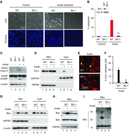
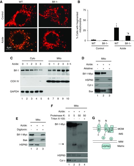
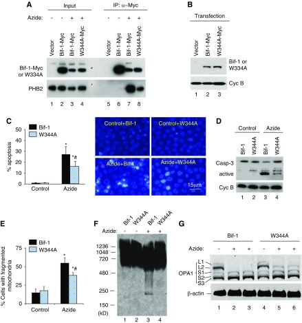
Background: Mitochondria are dynamic organelles that undergo fission and fusion. During cell stress, mitochondrial dynamics shift to fission, leading to mitochondrial fragmentation, membrane leakage, and apoptosis. Mitochondrial fragmentation requires the cleavage of both outer and inner membranes, but the mechanism of inner membrane cleavage is unclear. Bif-1 and prohibitin-2 may regulate mitochondrial dynamics.

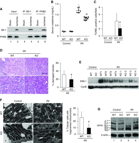
Methods: We used azide-induced ATP depletion to incite cell stress in mouse embryonic fibroblasts and renal proximal tubular cells, and renal ischemia-reperfusion to induce stress in mice. We also used knockout cells and mice to determine the role of Bif-1, and used multiple techniques to analyze the molecular interaction between Bif-1 and prohibitin-2.

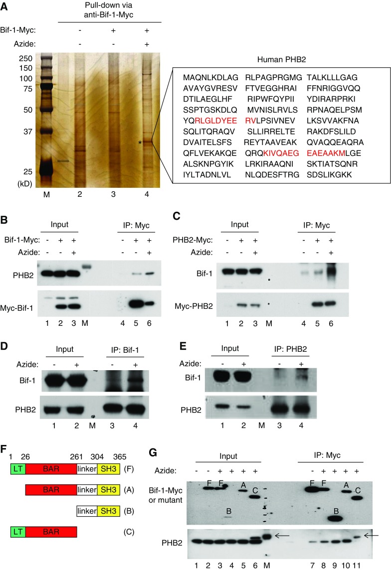
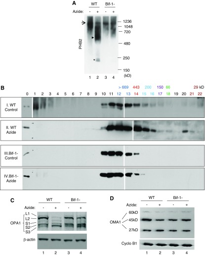
Results: Upon cell stress, Bif-1 translocated to mitochondria to bind prohibitin-2, resulting in the disruption of prohibitin complex and proteolytic inactivation of the inner membrane fusion protein OPA1. Bif-1-deficiency inhibited prohibitin complex disruption, OPA1 proteolysis, mitochondrial fragmentation, and apoptosis. Domain deletion analysis indicated that Bif-1 interacted with prohibitin-2 via its C-terminus. Notably, mutation of Bif-1 at its C-terminal tryptophan-344 not only prevented Bif-1/prohibitin-2 interaction but also reduced prohibitin complex disruption, OPA1 proteolysis, mitochondrial fragmentation, and apoptosis, supporting a pathogenic role of Bif-1/prohibitin-2 interaction. In mice, Bif-1 bound prohibitin-2 during renal ischemia/reperfusion injury, and Bif-1-deficiency protected against OPA1 proteolysis, mitochondrial fragmentation, apoptosis and kidney injury.

Conclusions: These findings suggest that during cell stress, Bif-1 regulates mitochondrial inner membrane by interacting with prohibitin-2 to disrupt prohibitin complexes and induce OPA1 proteolysis and inactivation.

Keywords: apoptosis; mitochondria; renal ischemia.


Figures

Figure 1.
Figure 1.
Azide-induced intrinsic pathway of apoptosis is blocked in Bif-1-null MEF. (A–C) Apoptosis. WT and Bif-1-null MEFs were left untreated (Control) or treated with 20 mM azide in glucose-free buffer for 3 hours, followed by 2 hours of recovery in normal culture medium. The cells were then stained with Hoechst33342 to record cellular and nuclear morphology (A) and counted to determine the percentage of cells with apoptotic morphology (B), or were lysed for immunoblot analysis of active caspase 3, PARP, and cyclophilin B (loading control) (C). Insert in (B) is an immunoblot to confirm Bif-1 expression in WT MEFs and not in Bif-1-null cells. (D–F) Cyt c release from mitochondria. WT and Bif-1-null MEFs were treated with 20 mM azide in glucose-free buffer for 3 hours or left untreated. The cells were then fractionated into membrane-bound fraction with mitochondria (Mito) and cytosolic fraction (Cyto) for immunoblot analysis of indicated proteins (D), or fixed for immunofluorescence of Cyt c to record representative images (E) and count to determine the percentage of cells with Cyt c released into cytosol (F). Arrows in (E) point to the cells with Cyt c release. (G–I) Bax accumulation, insertion, and oligomerization in mitochondria. WT and Bif-1-null MEFs were treated with 20 mM azide in glucose-free buffer for 3 hours or left untreated. The cells were then fractionated into membrane-bound fraction with mitochondria (Mito) and cytosolic fraction (Cyto) for immunoblot analysis of indicated proteins (G). In addition, the Mito fraction was subjected to alkaline treatment to remove loosely attached proteins for immunoblot analysis of membrane-inserted Bax (H) or subjected to chemical crosslinking with 1 mM DSP for immunoblot analysis of Bax oligomerization (I). Immunoblots and images (A, C–E, G–I) are representatives of at least three independent experiments. The data (B and F) represent mean±SD (n=3; 300–600 cells evaluated per condition); *P<0.05 versus control; #P<0.05 versus WT/azide.
Figure 2.
Figure 2.
Azide-induced mitochondrial fragmentation in apoptosis is attenuated in Bif-1-null MEF. (A and B) Lower mitochondrial fragmentation in Bif-1-null MEFs. WT and Bif-1-null MEFs were transfected with MitoRed to fluorescently label mitochondria, then incubated with 20 mM azide in glucose-free medium for 3 hours or left untreated. Representative images of mitochondrial morphology were recorded (A) and for quantification, the cells with fragmented mitochondria were counted to determine the percentage (B). The data in (B) represent mean±SD (n=3; >100 cells evaluated per condition); * P<0.05 versus control; #P<0.05 versus WT/azide. (C) Bif-1 translocation to mitochondria. WT MEFs were treated with 20 mM azide for 0–3 hours. The cells were then fractionated into membrane-bound fraction with mitochondria (Mito) and cytosolic fraction (Cyto) for immunoblot analysis of Bif-1. Cox IV and GAPDH were probed as cytosolic and mitochondrial markers. (D) Mitochondrial Bif-1 is resistant to alkaline stripping. HEK293 cells were transfected with Bif-1-Myc and treated with 20 mM azide for 2 hours to isolate mitochondria, which were incubated with alkaline (0.1 M Na2CO3, pH 11) buffer followed by centrifugation to collect pellet and supernatant fractions for immunoblot analysis of Bif-1-Myc, HSP60, Cyt c, and Bax. Whole mitochondria without alkaline incubation was analyzed as a control. The majority of Bif-1 remained on mitochondrial membranes after alkaline incubation. (E) Digitonin extracts Bif-1 from mitochondria. Isolated mitochondria were incubated with 0.5 mg/ml digitonin and then centrifuged to collect the soluble fraction (lane 3) and the pellet (lane 2) containing mitoplast. Nontreated mitochondria were used as a control (lane 1). Like Bak, Bif-1-Myc was solubilized by digitonin, whereas the matrix protein HSP60 remained in mitoplast. (F) Proteinase K protection assay. Isolated mitochondria were treated with 5 or 50 μg/ml proteinase K for 30 minutes on ice in the absence or presence of Triton X-100 (0.5%), and then centrifuged to collect pellet for immunoblot analysis. Without Triton X-100, 5 μg/ml proteinase K partially digested Bif-1, releasing a fragment that was detected by anti-Myc antibody (lane 2), whereas 50 μg/ml proteinase K induced more digestion, likely because of compromising the integrity of outer membrane (lane 3). In the presence of Triton X-100 (lane 4), all three proteins (Bif-1, HSP60, Cyt c) were degraded as a result of complete exposure to proteinase K. Arrow: Bif-1 fragment released after proteinase K digestion. (G) Schematic diagram of Bif-1 submitochondrial localization. Marker proteins from different compartments of mitochondria are also shown.
Figure 3.
Figure 3.
Bif-1 interacts with PHB2 via C terminus during apoptosis. (A) Identification of PHB2 as a Bif-1–interacting protein by affinity pulldown and mass spectrometry. HEK293 cells were transfected with Bif-1-Myc or empty plasmid, followed by 3 hours of 10 mM azide treatment or control incubation. Whole cell lysate was then collected with CHAPS buffer for immunoprecipitation using anti-Myc antibody-conjugated beads. The resultant immunoprecipitates were resolved by SDS-PAGE for silver staining. Azide treatment consistently induced a protein band at approximately 33 kD in the Bif-1-Myc immunoprecipitate (lane 4: *). The protein band was exercised for mass spectrometry, which identified two peptide sequences of PHB2 (red text). (B and C) Co-IP of overexpressed Bif-1 and PHB2. HEK293 cells were transfected with Bif-1-Myc (B), PHB2-Myc (C), or empty plasmid. The cells were then subjected to azide treatment or control incubation. Whole cell lysate was harvested with CHAPS buffer for IP using anti-Myc–conjugated beads. The immunoprecipitates, along with inputs, were examined by SDS-PAGE and immunoblot analysis. Bif-1/PHB2 co-IP is markedly higher in azide-treated cell lysate than that of control cell lysate (lane 6 versus 5), indicating an enhanced Bif-1/PHB2 interaction during azide treatment. (D and E) Co-IP of endogenous Bif-1 and PHB2 in azide-treated cells. Cells were subjected to azide treatment or control incubation to collect whole cell lysate with CHAPS buffer for IP using anti-Bif-1 or anti-PHB2. The immunoprecipitates, along with inputs, were examined by SDS-PAGE and immunoblot analysis. Bif-1/PHB2 co-IP was induced by azide treatment (lane 4 versus 3), indicating an enhanced Bif-1/PHB2 interaction. (F) Schematic diagram of full length Bif-1 (F) and deletion mutants (A–C). (G) Mapping of Bif-1 domains that mediate Bif-1/PHB2 interaction. HEK293 cells were transfected with Myc-tagged Bif-1 or its deletion mutants, followed by azide treatment or control incubation. Whole cell lysate was harvested with CHAPS buffer for IP using anti-Myc-conjugated beads. The immunoprecipitates, along with inputs, were examined by SDS-PAGE and immunoblot analysis with anti-Myc and anti-PHB2 antibodies. The results show PHB2 co-IP with full-length Bif-1 (F) and N-terminal deletion Bif-1 mutants (A and B), but not with C-terminal deletion mutant (C). Note: The arrows indicate protein bands in lanes 6, 11, and M (molecular weight markers) that are clearly distinguished from PHB2, but their identities are currently unclear.
Figure 4.
Figure 4.
Disruption of prohibitin complexes and proteolysis of OPA1 during apoptosis are attenuated in Bif-1-null MEF. (A) Blue native gel analysis of prohibitin complexes. WT and Bif-1-null MEFs were treated with azide for 3 hours in glucose-free buffer or left untreated. The cells were then fractionated to collect the mitochondria-enriched fraction for chemical crosslinking with 1 mM DSP. After resuspension, the samples were resolved by BN-PAGE for immunoblot analysis of PHB2. Arrow indicates a large prohibitin complex of approximately 1.2 mD; arrowhead indicates a smear of intermediate prohibitin complexes; * indicates a small prohibitin complex of approximately 200 kD. The results show that azide treatment induces a disruption of the large prohibitin complex into intermediate and small size complexes in WT MEFs, and the complex disruption is suppressed in Bif-1-null cells. (B) Size exclusion chromatography analysis of prohibitin complexes. Samples were prepared as (A) and loaded onto AKTA purifier FPLC for elution with 100 mM phosphate buffer (pH 7.0). Twenty two fractions were collected and concentrated for immunoblot analysis of PHB2. Lane “0”: HEK293 cell lysate. Sigma Gel Filtration Protein Standards were used for calibration. The molecular weight of each protein in the Standards and its elution fraction numbers are written in the same color. (C and D) OPA1 and OMA1 proteolysis. WT and Bif-1-null MEFs were treated with azide for 3 hours or left untreated. Whole cell lysate was collected for immunoblot analysis of OPA1, OMA1, and control proteins.
Figure 5.
Figure 5.
W344A mutation in Bif-1 reduces its ability of PHB2 interaction, OPA1 proteolysis, mitochondrial fragmentation, and apoptosis. (A) PHB2 interaction. HEK293 cells were transfected with Bif-1-Myc, Myc-W344A-Bif-1, or empty vector. The cells were then left untreated or treated with azide for 3 hours to collect the membrane-bound mitochondrial fraction, which was further subjected to chemical cross-linking followed by immunoprecipitation with anti-Myc. The immunoprecipitates, along with inputs, were resolved by SDS-PAGE for immunoblot analysis with anti-Myc and anti-PHB2 antibodies. The results show a much weaker co-IP of W344A-Bif-1 with PHB2 than WT Bif-1 (lane 8 versus 7), suggesting that W344A mutation diminishes Bif-1/PHB2 interaction. (B) Reconstitution of Bif-1 or W344A mutant into Bif-1-null MEFs. Bif-1-null MEFs were reconstituted with Bif-1 or W344A mutant using the lentivector system. Whole cell lysate was then collected for immunoblot analysis to confirm the expression of Bif-1 and W344A-Bif-1. (C) Apoptosis in Bif-1 or W344A-Bif-1 reconstituted MEFs. Bif-1 or W344A-Bif-1 reconstituted MEFs were left untreated or treated with azide for 3 hours followed by 2 hours of recovery in culture medium. The cells were then stained with Hoechst33342 for morphologic examination. The cells with apoptotic morphology were counted to determine the percentage of apoptosis. Representative images of nuclear staining are also presented. (D) Caspase 3 activation in Bif-1 or W344A-Bif-1 reconstituted MEFs. Cells were treated as (C) to collect cell lysate for immunoblot analysis of caspase 3 and cyclophilin B (loading control). Caspase 3 activation was indicated by the appearance of active/cleaved fragments. (E) Mitochondrial fragmentation in Bif-1 or W344A-Bif-1 reconstituted MEFs. Cells were transfected with MitoRed to fluorescently label mitochondria and then treated with azide or left untreated. The morphology of mitochondria in individual cells was examined by fluorescence microscopy to determine the percentage of cells with fragmented mitochondria. (F) Blue native gel analysis of prohibitin complexes in Bif-1 or W344A-Bif-1 reconstituted MEFs. Cells were treated with azide for 3 hours or left untreated. The cells were then fractionated to collect the mitochondria-enriched fraction for chemical crosslinking with 1 mM DSP, followed by BN-PAGE for immunoblot analysis of PHB2. (G) OPA1 proteolysis in Bif-1 or W344A-Bif-1 reconstituted MEFs. Cells were treated with azide for 3 hours or left untreated. Whole cell lysate was collected for immunoblot analysis of OPA1 and β-actin. The data in (C and D) represent mean±SD (n=3); *P<0.05 versus control; #P<0.05 versus Bif-1/azide.
Figure 6.
Figure 6.
Bif-1 binds prohibitin-2 and contributes to OPA1 proteolysis and mitochondrial fragmentation in mouse renal IRI. (A) Bif-1 and PHB2 interacts in mitochondrial fraction during renal IRI. Mice were subjected to 30 minutes of bilateral renal ischemia with brief (approximately 15 minutes) reperfusion. Kidneys were collected to isolate mitochondrial fraction for co-IP of Bif-1 and PHB2. (B–E) Partial resistance of Bif-1 knockout (KO) mice to renal IRI. Bif-1 KO and wild-type (WT) littermate mice were subjected to 30 minutes of bilateral renal ischemia with 48 hours of reperfusion to collect blood serum for creatinine measurement to indicate renal functional loss (B), and to collect kidney tissues for hematoxylin and eosin staining analysis of tubular damage (C), TUNEL assay of apoptosis (D) and immunoblot analysis of KIM1. (F) Lower mitochondrial fragmentation in Bif-1 KO mice during renal IRI. Bif-1KO and WT littermate mice were subjected to 30 minutes of bilateral renal ischemia with brief (approximately 15 minutes) reperfusion. Kidney tissues were fixed for electron microscopy to record representative mitochondrial morphology and determine percentage of the cells with mostly fragmented mitochondria. (G) Less OPA1 proteolysis in Bif-1 KO mice during renal IRI. Bif-1 KO and WT littermate mice were subjected to 30 minutes of bilateral renal ischemia with brief (approximately 15 minutes) reperfusion to collect kidney cortical tissues for immunoblot analysis of OPA1 and β-actin. The quantitative data in (B–E) represent mean±SD (n=3); *P<0.05 versus control; #P<0.05 versus WT/IRI.

Similar articles

Cited by

References

    1. Chan DC: Fusion and fission: Interlinked Processes critical for mitochondrial health. Annu Rev Genet 46: 265–287, 2012 - PubMed
    1. Nunnari J, Suomalainen A: Mitochondria: In sickness and in health. Cell 148: 1145–1159, 2012 - PMC - PubMed
    1. Youle RJ, van der Bliek AM: Mitochondrial fission, fusion, and stress. Science 337: 1062–1065, 2012 - PMC - PubMed
    1. Bhargava P, Schnellmann RG: Mitochondrial energetics in the kidney. Nat Rev Nephrol 13: 629–646, 2017 - PMC - PubMed
    1. Zhan M, Brooks C, Liu F, Sun L, Dong Z: Mitochondrial dynamics: Regulatory mechanisms and emerging role in renal Pathophysiology. Kidney Int 83: 568–581, 2013 - PMC - PubMed

Publication types

LinkOut - more resources