. 2019 Jun 25;116(26):12873-12882.
doi: 10.1073/pnas.1821945116. Epub 2019 May 24.

Mosaic origin of the eukaryotic kinetochore

Affiliations

Mosaic origin of the eukaryotic kinetochore

Eelco C Tromer et al. Proc Natl Acad Sci U S A. .

Abstract

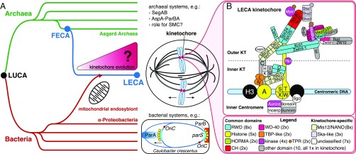
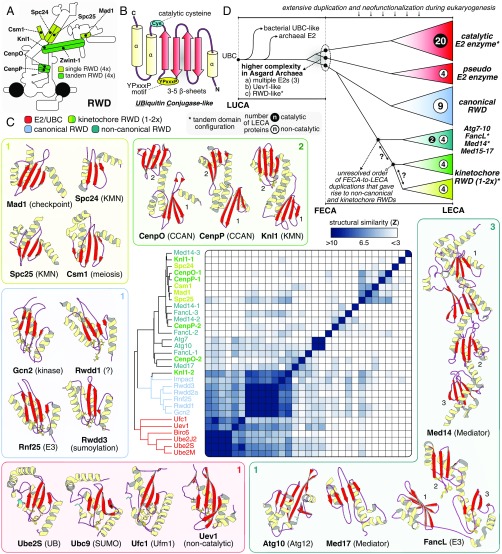
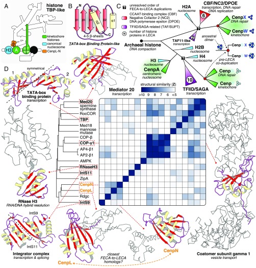
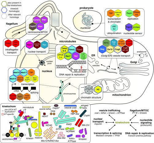
The emergence of eukaryotes from ancient prokaryotic lineages embodied a remarkable increase in cellular complexity. While prokaryotes operate simple systems to connect DNA to the segregation machinery during cell division, eukaryotes use a highly complex protein assembly known as the kinetochore. Although conceptually similar, prokaryotic segregation systems and the eukaryotic kinetochore are not homologous. Here we investigate the origins of the kinetochore before the last eukaryotic common ancestor (LECA) using phylogenetic trees, sensitive profile-versus-profile homology detection, and structural comparisons of its protein components. We show that LECA's kinetochore proteins share deep evolutionary histories with proteins involved in a few prokaryotic systems and a multitude of eukaryotic processes, including ubiquitination, transcription, and flagellar and vesicular transport systems. We find that gene duplications played a major role in shaping the kinetochore; more than half of LECA's kinetochore proteins have other kinetochore proteins as closest homologs. Some of these have no detectable homology to any other eukaryotic protein, suggesting that they arose as kinetochore-specific folds before LECA. We propose that the primordial kinetochore evolved from proteins involved in various (pre)eukaryotic systems as well as evolutionarily novel folds, after which a subset duplicated to give rise to the complex kinetochore of LECA.

Keywords: LECA; eukaryogenesis; gene duplication; kinetochore; mitosis.


Conflict of interest statement

The authors declare no conflict of interest.

Figures

Fig. 1.
Fig. 1.
The eukaryotic kinetochore and mitotic machinery originated between FECA and LECA. (A) How did the eukaryotic kinetochore originate and evolve between FECA and LECA? Eukaryotes (blue) are descended from Archaea (green) and likely are closely related to the Asgard archaeal superphylum (59). This Asgard-related lineage incorporated an Alphaproteobacterium via endosymbiosis; the latter gave rise to the eukaryotic mitochondrion. Archaea and Bacteria (red) do not separate their duplicated chromosome(s) via a mitotic spindle (–13). For example, bacteria such as Caulobacter crescentus operate the parABS partitioning system, in which parS DNA sites are recognized by the protein ParB, stimulating ParA, which in turn pulls or pushes the chromosomes apart (12). Due to these differences, the mitotic spindle and the kinetochore probably originated between the FECA and the LECA. LUCA, the last universal common ancestor. (B) The kinetochore of LECA consisted of 52 proteins that contain domains found in other, nonkinetochore eukaryotic proteins as well (“common domains”) or that are unique to the kinetochore (“kinetochore-specific”). KT, kinetochore.
Fig. 2.
Fig. 2.
Kinetochore RWDs are an expanded class of noncatalytic E2 UBCs. (A) Overview of the position of eight kinetochore proteins with a single (light green) or a tandem (dark green) RWD configuration. (B) RWD domains are part of the UBC superfamily. The secondary structure of the UBC superfamily is characterized by a “β-meander” of three to five β-sheets enclosed by ɑ-helices at both termini, a YPxxxP motif, and a catalytic cysteine residue (lost in RWDs). (C) The UBC superfamily can be subdivided into four classes: (i) E2 UBCs (E2), including noncatalytic pseudo E2s (e.g., Uev1); (ii) canonical RWDs; (iii) kinetochore RWDs; and (iv) atypical RWD/UBC-like (e.g., FancL, Med14-17). Per class, the structure of various members is depicted to show the overall structural and topological similarity, and a known molecular function is indicated between brackets. When present, YPxxxP (yellow) and the catalytic cysteine (cyan) are represented in a sticks configuration. The average linkage clustering of structural similarity scores of single UBC domains (z-scores) demonstrates the close similarity amongst E2s and canonical RWD domains. Kinetochore RWDs and noncanonical domains are more divergent and cluster together. (D) Cartoon of the evolutionary reconstruction of the UBC superfamily based on phylogenetic analyses (SI Appendix, Figs. S1E and S3) and structural comparisons (Dataset S2). Extensive duplication and neofunctionalization of an archaeal E2 UBC gave rise to a large complexity of catalytic and noncatalytic E2/RWD proteins in LECA (see numbers per class). Possibly a part of this eukaryotic complexity was already present in FECA, since Asgard Archaea contain multiple E2 conjugases, an Uev-like homolog, and an RWD-like domain (SI Appendix, Fig. S3). Kinetochore RWDs might have a monophyletic origin, although a structural affiliation with other divergent proteins signify a more complex evolutionary scenario (see question marks).
Fig. 3.
Fig. 3.
A common origin of kinetochore histones and TBP-like proteins with complexes involved in DNA repair and transcription. (A) Overview of the position of CenpA and CenpS-X-T-W (histones, green) and CenpL-N (TBP-like, orange) in the kinetochore. (B) The TBP-like fold is a set of curved β-strands that form an interaction surface for substrates (RNA/DNA, amino acid motifs) and potential dimer interfaces. (C) A cartoon of the evolutionary reconstruction of kinetochore-related histone proteins CenpA and CenpS-T-X-W (based on SI Appendix, Fig. S1I). A histone of archaeal descent duplicated and subfunctionalized many times, giving rise to a large diversity of histone proteins in eukaryotes, including those involved in the kinetochore, chromatin structure (nucleosome), transcription (TAF/SUPT/NC2/CBF), and DNA repair (DPOE). CenpA is the closest homolog of the nucleosomal histone H3. CenpS-T and CenpX-W are likely each other’s closest paralogs, signifying a coduplication of an ancient dimer to form the tetramer CenpS-X-T-W. The CenpS-X dimer also plays a role in the Fanconi anemia pathway (DNA repair). (D) Yellow (helices) and red (sheets) show the location of a TBP-like domain in a subset of available TBP-like protein structures. The gray ribbon representation indicates the nonhomologous parts of the proteins; their cellular function is indicated between brackets. CenpL and CenpN contain a TBP-like fold. Average linkage clustering of similarity scores (z-scores) indicates that CenpN and CenpL could be each other’s closest homologs.
Fig. 4.
Fig. 4.
The Mis12 and NANO complex have a common ancestry. (A) Overview of the position of the Mis12 complex and NANO tetramers in the LECA kinetochore. (B) Cartoon of the consensus topology of all eight Mis12/NANO subunits, illustrating disordered and globular regions. (C) Profile-versus-profile hits with HHsearch (dark blue) and PRC (light blue) indicate that Mis12, Nnf1, Nsl1, Nkp1 and Nkp2 are homologous (SI Appendix, Text and Dataset S1). No sequence similarity between CenpU, Dsn1, and CenpQ with any of the other Mis12/NANO subunits was detected. (D) The subunits of the Mis12 and NANO display a high degree of similarity with respect to the (i) size and orientation of the head domains, (ii) length of the coiled coils, and (iii) presence of disordered N-terminal tails. Based on these three criteria, we defined a ‘Mis12’ and ‘Dsn1’ subtype. We propose that the Mis12 and NANO complex are the result of an ancient whole complex duplication, which was preceded by two rounds of Mis12/Dsn1 subtype duplication. Distances in the tree do not reflect measured distances but indicate a higher degree of sequence and structural variation for the Dsn1 type compared with the Mis12 type.
Fig. 5.
Fig. 5.
The HORMA-Trip13 module is of prokaryotic origin. Shown are phylogenetic trees of HORMA domain proteins and AAA+ ATPases. In eukaryotes, HORMAD, Mad2, and p31comet are structurally modified by a Trip13 hexamer (Upper, right side). In prokaryotes, HORMA (types 1 and 2) and Trip13 are present in a single operon, strongly suggesting that they also interact in these species, and thus that this interaction is ancient. The phylogenetic trees indeed suggest that the eukaryotic HORMA domain and Trip13 were derived from prokaryotes. In addition, the prokaryotic operons include proteins involved in nucleotide signaling [yellow, nucleotide transferase (SMODS); red, transposase-related; black, unknown] (34). The uncollapsed trees are shown in SI Appendix, Figs. S1 F and G. Asterisks indicate the species for which we discovered a HORMA-Trip13 operon (annotation in Dataset S5 and SI Appendix, Fig. S5).
Fig. 6.
Fig. 6.
Mosaic origin of the eukaryotic kinetochore. Overview of the eukaryotic and prokaryotic close(st) homologs of LECA kinetochore proteins, which play roles in a wide variety of cellular processes, signifying the mosaic origin of the eukaryotic kinetochore. Relevant eukaryotic and prokaryotic homologs (hexagons) of LECA kinetochore proteins are colored based on the presence of a common domain (Bottom Left: overview of kinetochore parts), and projected onto the location(s) in the eukaryotic cell at which they operate (SI Appendix, Table S1). The hexagons of homologs are lined with different colors indicate a LECA kinetochore protein with a nonkinetochore function (green), the closest homolog to a LECA kinetochore protein (blue), and other close homologs of LECA kinetochore proteins (black). In addition, distantly related homologs of TBP-like, histones, UBC/RWD, and HORMA domain-containing kinetochore proteins were already present in prokaryotes (Top Right). (Bottom Left) Overview of the different number and types of domains in the LECA kinetochore. The Mis12/NANO and Ska domains are kinetochore-specific and thus are not found in other systems. The dotted lines indicate a potential intrakinetochore duplication during eukaryogenesis leading to the formation of various heteromeric (sub)complexes within the kinetochore. (Bottom Right) summary of the evolutionary links between the kinetochore and selected prokaryotic/eukaryotic molecular systems.

Comment in

References

    1. Makarova M., Oliferenko S., Mixing and matching nuclear envelope remodeling and spindle assembly strategies in the evolution of mitosis. Curr. Opin. Cell Biol. 41, 43–50 (2016). - PMC - PubMed
    1. De Souza C. P. C., Osmani S. A., Mitosis, not just open or closed. Eukaryot. Cell 6, 1521–1527 (2007). - PMC - PubMed
    1. Drechsler H., McAinsh A. D., Exotic mitotic mechanisms. Open Biol. 2, 120140 (2012). - PMC - PubMed
    1. Sazer S., Lynch M., Needleman D., Deciphering the evolutionary history of open and closed mitosis. Curr. Biol. 24, R1099–R1103 (2014). - PMC - PubMed
    1. Henikoff S., Ahmad K., Malik H. S., The centromere paradox: Stable inheritance with rapidly evolving DNA. Science 293, 1098–1102 (2001). - PubMed

Publication types

LinkOut - more resources