

NIH Initiative to Improve Understanding of the Pancreas, Islet, and Autoimmunity in Type 1 Diabetes: The Human Pancreas Analysis Program (HPAP)
- PMID: 31127054
- PMCID: PMC6609987
- DOI: 10.2337/db19-0058
NIH Initiative to Improve Understanding of the Pancreas, Islet, and Autoimmunity in Type 1 Diabetes: The Human Pancreas Analysis Program (HPAP)
Abstract
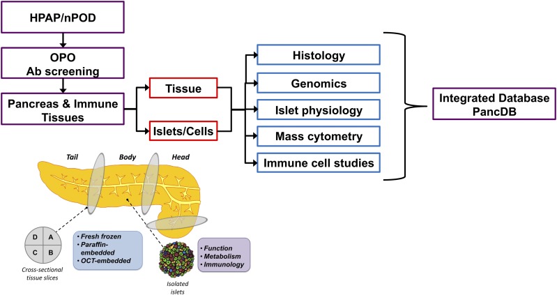
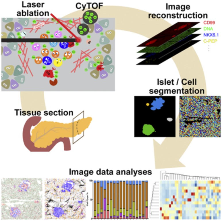
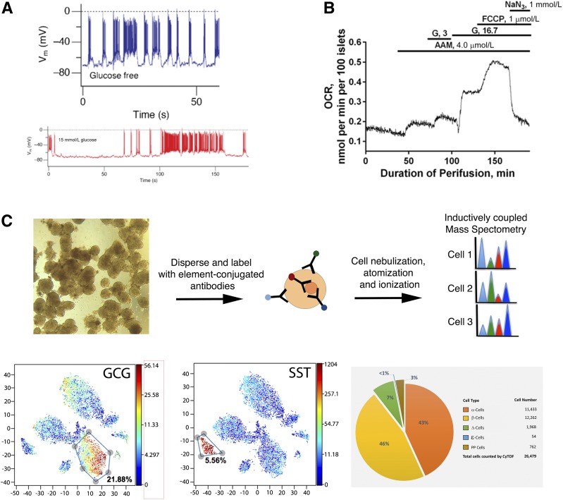
Type 1 diabetes risk can reliably be predicted by markers of autoimmunity, but approaches to prevent or modify the underlying disease process are needed. We posit this void fundamentally results from a limited understanding of immune-islet cell interactions within the pancreas and relevant immune organs, contributions of β-cells to their own demise, and epigenetic predispositions affecting both immune and islet cells. Because biopsy of the human pancreas and pancreatic lymph nodes carries risk and the pancreas begins to autodigest soon after death, detailed cellular and molecular phenotyping of the human type 1 diabetes pancreas is lacking, limiting our understanding of the mechanisms of β-cell loss. To address these challenges, the National Institutes of Health/National Institute of Diabetes and Digestive and Kidney Diseases established the Human Pancreas Analysis Program (HPAP) to procure human type 1 diabetes pancreata for an extensive array of tissue-based, cellular, and epigenetic assays aimed at critical knowledge gaps in our understanding of the local immune attack and loss of β-cells. In this Methodology Review, we describe how HPAP is performing detailed islet and immune cell phenotyping and creating publicly available data sets with the goals of an improved understanding of type 1 diabetes and the development of more effective treatments to prevent or reverse the disease.
© 2019 by the American Diabetes Association.
Figures
References
-
- Katsarou A, Gudbjörnsdottir S, Rawshani A, et al. . Type 1 diabetes mellitus. Nat Rev Dis Primers 2017;3:17016. - PubMed
-
- Gepts W. Islet morphology in type I diabetes. Behring Inst Mitt 1984;75:39–41 - PubMed
-
- Gepts W, Lecompte PM. The pancreatic islets in diabetes. Am J Med 1981;70:105–115 - PubMed
-
- LeCompte PM, Gepts W. The Pathology of Juvenile Diabetes. Volk BWWKF, Ed. New York, Plenum Press, 1977
-
- Gepts W. Islet changes suggesting a possible immune aetiology of human diabetes mellitus. Acta Endocrinol Suppl (Copenh) 1976;205:95–106 - PubMed
Publication types
MeSH terms
Substances
Grants and funding
LinkOut - more resources
Full Text Sources
Other Literature Sources
Medical
Molecular Biology Databases
