. 2019 May 24;10(1):2297.
doi: 10.1038/s41467-019-09915-2.

Candidalysin activates innate epithelial immune responses via epidermal growth factor receptor

Affiliations

Candidalysin activates innate epithelial immune responses via epidermal growth factor receptor

Jemima Ho et al. Nat Commun. .

Abstract

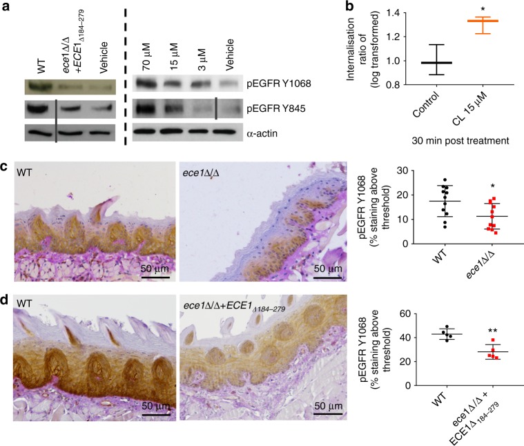
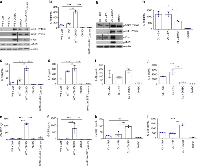
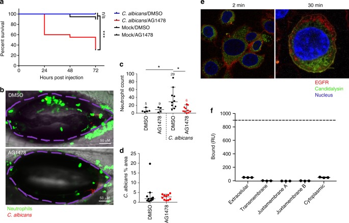
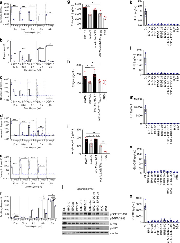
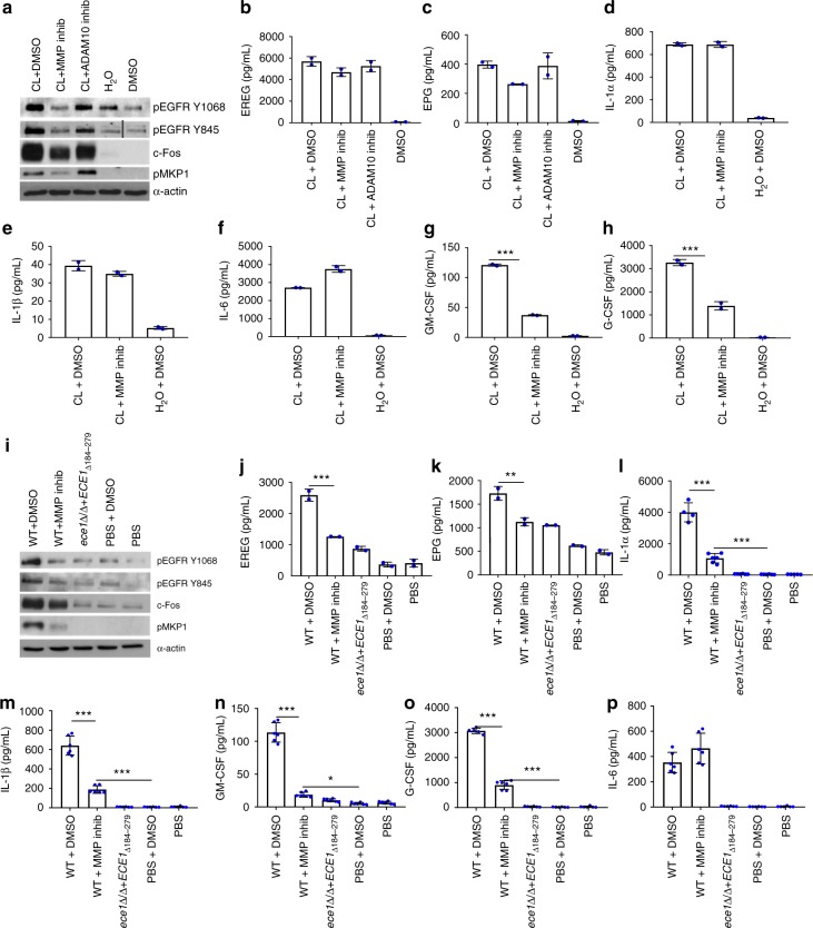
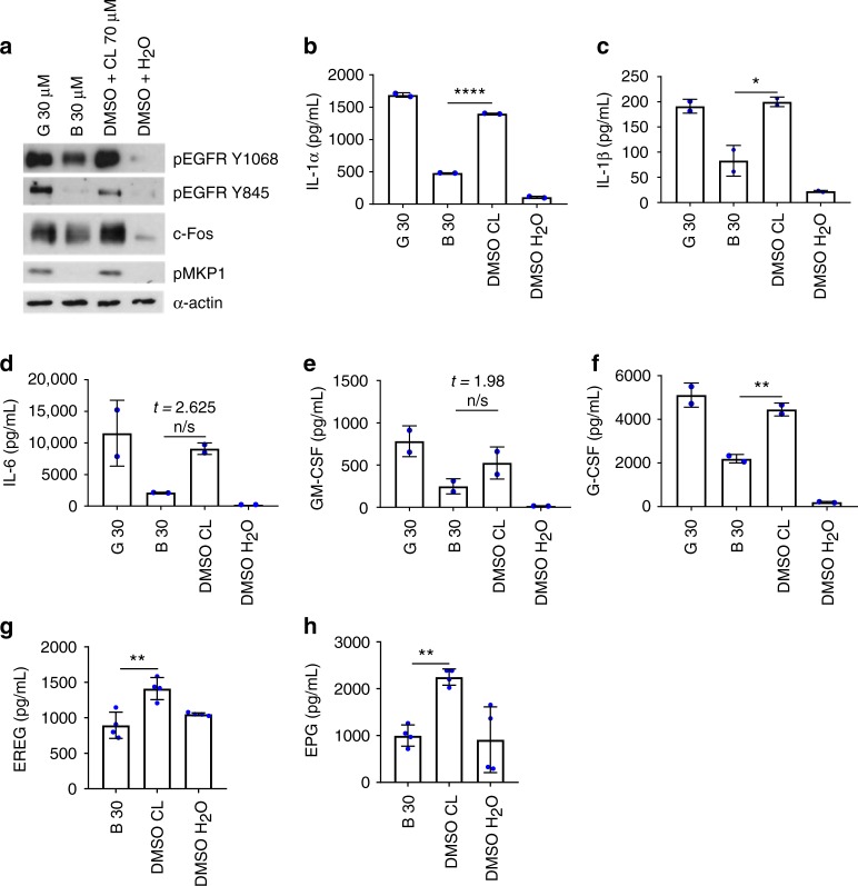
Candida albicans is a fungal pathobiont, able to cause epithelial cell damage and immune activation. These functions have been attributed to its secreted toxin, candidalysin, though the molecular mechanisms are poorly understood. Here, we identify epidermal growth factor receptor (EGFR) as a critical component of candidalysin-triggered immune responses. We find that both C. albicans and candidalysin activate human epithelial EGFR receptors and candidalysin-deficient fungal mutants poorly induce EGFR phosphorylation during murine oropharyngeal candidiasis. Furthermore, inhibition of EGFR impairs candidalysin-triggered MAPK signalling and release of neutrophil activating chemokines in vitro, and diminishes neutrophil recruitment, causing significant mortality in an EGFR-inhibited zebrafish swimbladder model of infection. Investigation into the mechanism of EGFR activation revealed the requirement of matrix metalloproteinases (MMPs), EGFR ligands and calcium. We thus identify a PAMP-independent mechanism of immune stimulation and highlight candidalysin and EGFR signalling components as potential targets for prophylactic and therapeutic intervention of mucosal candidiasis.


Conflict of interest statement

The authors declare no competing interests.

Figures

Fig. 1
Fig. 1
Candidalysin (CL) activates EGFR. a Phosphorylation: Infection of TR146 cells with WT C. albicans induces EGFR phosphorylation at two distinct tyrosine sites, Y1068 and Y845, while a strain lacking the candidalysin-encoding region (ece1Δ/Δ+ECE1Δ184–279) does not (left panel). Phosphorylation of EGFR at both sites was also induced following direct candidalysin treatment in a dose-dependent manner (right panel). Data are representative of three biological repeats. Protein lysates were taken at 2 h p.i. for western blot analysis. Solid vertical lines indicate omitted, extraneous portions of blot images. b Internalisation: At 30 min post CL treatment, EGFR is significantly internalised. Data collected from a total of 120,000 cells per group over three independent experiments. Median value indicated, error bars represent min-max data points. Values are shown as a Log transformed ratio of EGFR staining intensity inside the cell to the total cell intensity of EGFR staining. c Murine pEGFR staining: Immune compromised mice were infected with a C. albicans ece1Δ/Δ mutant (lacking ECE1 which encodes the parent protein from which candidalysin is derived) and tongues were harvested at day 1 p.i. Paraffin-embedded, 3 mm tissue sections were prepared and stained for pEGFR (Y1068). The ece1Δ/Δ mutant exhibits reduced capacity to phosphorylate EGFR above a given threshold (threshold set using Image J software), when compared to WT C. albicans infections. Data from 9 (WT) and 11 (ece1Δ/Δ) individual mice obtained over two independent experiments. d Murine pEGFR staining: Immune competent mice were infected with the candidalysin-deficient ece1Δ/Δ+ECE1Δ184–279 mutant which also exhibited a reduced ability to phosphorylate EGFR at Y1068, when compared to WT infection. Data obtained from one experiment, 5 mice per group. Unpaired T-tests were used to determine statistical significance in (b, c, d). Error bars from (c, d) represent Standard deviation (SD). *p < 0.05, **p < 0.01
Fig. 2
Fig. 2
Inhibition of EGFR activity suppresses C. albicans- and Candidalysin (CL)-induced protein activation and expression. Use of either Gefitinib or PD153035 EGFR inhibitor suppresses WT C. albicans-induced (af) and CL-induced (gl) pEGFR (Y1068 and Y845), c-Fos and pMKP1 (a, g), as well as secretion of GM-CSF (e, k) and G-CSF (f, l). Both EGFR inhibitors suppressed C. albicans-induced IL-1α, IL-1β, IL-6 (bd) but not CL-induced IL-1α, IL-1β, IL-6 (hj). The CL-deficient ece1Δ/Δ+ECE1Δ184–279 strain poorly induced all proteins investigated. The data are representative images (a, g) or combined averages of three independent experiments (bf, hl). Protein lysates taken at 2 h post infection for western blot analysis, cytokines assessed at 24 h post infection via luminex. Solid vertical lines indicate omitted, extraneous portions of blot images. Unmatched, one-way ANOVA with Bonferroni multiple comparison’s test was used to assess statistical significance. Error bars represent SD, *p < 0.05, **p < 0.01, ***p < 0.001
Fig. 3
Fig. 3
Inhibition of EGFR suppresses neutrophil recruitment and enhances mortality in a zebrafish swimbladder model of candidiasis. Candidalysin does not bind EGFR. a Mortality: In the presence of AG1478 EGFR inhibitor, C. albicans-infected fish exhibited enhanced mortality with 70% death by day 3 p.i. (red), whereas all the infected vehicle treated fish survived (blue). b, c Neutrophil recruitment: EGFR inhibition resulted in significantly fewer neutrophils (green) recruited into the infected swimbladder (outlined in purple). Numbers in graph c provide the median neutrophil count per group. d Fungal load: No significant change in fungal burden was observed in the presence of AG1478 inhibitor, as quantified by the number of red pixels, normalised to the area of the swimbladder. Log-rank with a Bonferroni correction was used for (a), Kruskal-Wallis with Dunn’s post-test correction for (c) and Mann-Whitney for (d). Median and interquartile range are plotted in (c, d). *p < 0.05, ***p < 0.001, n.s. p > 0.05. e Candidalysin does not co-localise with EGFR: Confocal images of TR146 cells show that at 2 min post exposure, fluorescently labelled candidalysin-488 (green) can be seen within the cell cytoplasm whilst EGFR (red) remains at the cell surface. At 30 min post toxin exposure, although both EGFR and candidalysin are found within cells, no co-localisation was observed, and staining patterns are distinct for each. Confocal point images have a 91.36 µm x 90.79 µm (left image) and 35.9 µm x 35.9 µm (right image) field of view. Images are representative of three individual experiments. f Surface plasmon resonance response of candidalysin and EGFR domains : SPR signal response for the different domains of EGFR did not reach above 50 RU when interacting with biotinylated-candidalysin (180 s injection of 500 nM of titrant at 10 µL/min flow, 25 °C). Approximate response expected for binding of extracellular or cytoplasmic domains is 900 RU (dotted line) at a Kd of 20 mM
Fig. 4
Fig. 4
Candidalysin-induced release of EREG and EPG contribute to subsequent EGFR-mediated signalling. Release of EREG (a), EPG (b) and NRGs 2, 3 and 4 (ce) was observed following treatment of candidalysin to TR146 cells, in a dose-dependent manner with rapid onset (within 15 min). AREG was also detected in candidalysin-treated cell supernatants, but the release was gradual and accumulated over 6 h (f). At 24 h p.i. epiregulin (EREG) (g) and epigen (EPG) (h) were induced by candidalysin-expressing (WT and ece1Δ/Δ+ECE1 (black bars)) but not candidalysin-deficient (ece1Δ/Δ and ece1Δ/Δ+ECE1Δ184–279 (grey bars)) C. albicans strains. While amphiregulin (AREG) was induced by all fungal strains tested, diminished potency was observed by those unable to express candidalysin (i). Increasing concentrations of EREG, EPG or EREG + EPG, were used to stimulate TR146 oral epithelial cells. j Phosphorylation of EGFR (at Y1068 and Y845 sites) and MKP1 proteins occurred in a dose-dependent manner in response to EREG and EREG + EPG, but not EPG alone. Induction of c-Fos by ligand stimulation was not dose-dependent (j). The effects of ligand exposure are not comparable to that of 70 µM candidalysin. While a dose-dependent trend of IL-6 (m), GM-CSF (n) and G-CSF (o) induction is observed in response to increasing concentrations of all ligand combinations, the changes are not statistically significant. IL-1α (k) and IL-1β (l) are not induced by ligand exposure. One-way ANOVA followed by a Bonferroni multiple comparison’s test was used to calculate statistical significance between groups. Graphs are an average of 3 (ag, io) or 2 (h) independent experiments; one-way ANOVA with Bonferroni multiple comparison’s test used to assess statistical significance between samples from the same timepoint. Error bars represent SD. *p < 0.05, **p < 0.01, ***p < 0.001
Fig. 5
Fig. 5
MMP inhibition suppresses the candidalysin and C. albicans-induced response pathway. Pre-treatment of TR146 cells with Marimastat (MMP inhibitor, 10 µM) but not GI-253023X (ADAM10 inhibitor, 5 µM), significantly suppressed candidalysin-induced expression of pEGFR (Y1068 and Y845), c-Fos and pMKP1 (a). MMP inhibition had no suppressive effects on candidalysin-induced EREG (b) EPG (c), IL-1α (d), IL-1β (e) or IL-6 (f) but did suppress GM-CSF (g) and G-CSF (h) cytokine release. In Marimastat-inhibited, WT C. albicans-infected cells, significant suppression of all investigated proteins (i–o) except IL-6 (p), was observed. The candidalysin-deficient ece1Δ/Δ+ECE1Δ184–279 strain also failed to induce phosphorylation of EGFR or MKP1, c-Fos expression (i) or our panel of cytokines (lp). Protein lysates were taken at 2 h post infection for western blot analysis, cytokines assessed at 24 h post infection via luminex. Solid vertical lines indicate omitted, extraneous portions of blot images. Unmatched, one-way ANOVA with Bonferroni multiple comparison’s test used to assess statistical significance. All images and graphs are representative of three independent experiments. One-way ANOVA with Bonferroni multiple comparison’s test was used to assess statistical significance. Error bars represent SD. *p < 0.05, **p < 0.01, ***p < 0.001
Fig. 6
Fig. 6
Calcium is required for candidalysin-triggered immune responses and lies upstream of EGFR. Pre-treatment of TR146 cells with a calcium chelator (Bapta-AM 30 µM) reduced levels of pEGFR Y1068 and Y845, c-Fos, pMKP1 (a), IL-1α (b), IL-1β (c), G-CSF (f), EREG (g) and EPG (h), following candidalysin exposure. This is not observed when using the potassium inhibitor glibenclamide (30 µM). IL-6 (d) and GM-CSF (e) are not significantly suppressed by either inhibitor, though a reduction is observed. Protein lysates were taken at 2 h post infection for western blot analysis, cytokines assessed at 24 h post infection via luminex. Unmatched, one-way ANOVA with Bonferroni multiple comparison’s test was used to assess statistical significance. Images and graphs are representative of 2 (a, g, h) or 3 (bf) independent experiments, respectively. Error bars represent SD. *p < 0.05, **p < 0.01, ***p < 0.001, n.s. p > 0.05

References

    1. Brown, G. D., Denning, D. W. & Levitz, S. M. Tackling human fungal infections. Science336, 647 (2012). - PubMed
    1. Moyes DL, et al. Candidalysin is a fungal peptide toxin critical for mucosal infection. Nature. 2016;532:64–68. doi: 10.1038/nature17625. - DOI - PMC - PubMed
    1. Richardson JP, et al. Processing of Candida albicans Ece1p Is Critical for Candidalysin maturation and fungal virulence. MBio. 2018;9:e02178–17. doi: 10.1128/mBio.02178-17. - DOI - PMC - PubMed
    1. Richardson JP, et al. Candidalysin drives epithelial signaling, neutrophil recruitment, and immunopathology at the vaginal mucosa. Infect. Immun. 2017;86:e00645–17. doi: 10.1128/IAI.00645-17. - DOI - PMC - PubMed
    1. Moyes DL, et al. Activation of MAPK/c-Fos induced responses in oral epithelial cells is specific to Candida albicans and Candida dubliniensis hyphae. Med. Microbiol. Immunol. 2012;201:93–101. doi: 10.1007/s00430-011-0209-y. - DOI - PMC - PubMed

Publication types

MeSH terms

LinkOut - more resources