. 2019 May 24;10(1):2314.
doi: 10.1038/s41467-019-10324-8.

Selective binding of the PHD6 finger of MLL4 to histone H4K16ac links MLL4 and MOF

Affiliations

Selective binding of the PHD6 finger of MLL4 to histone H4K16ac links MLL4 and MOF

Yi Zhang et al. Nat Commun. .

Abstract

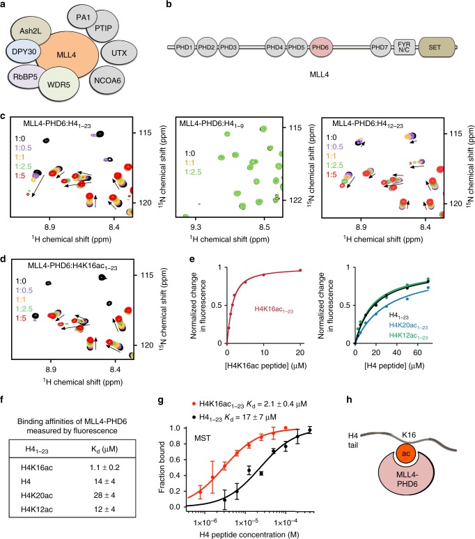
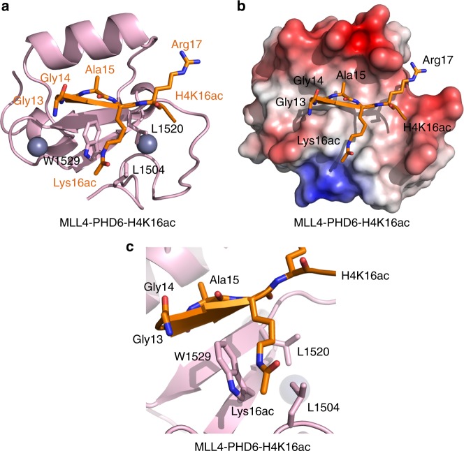
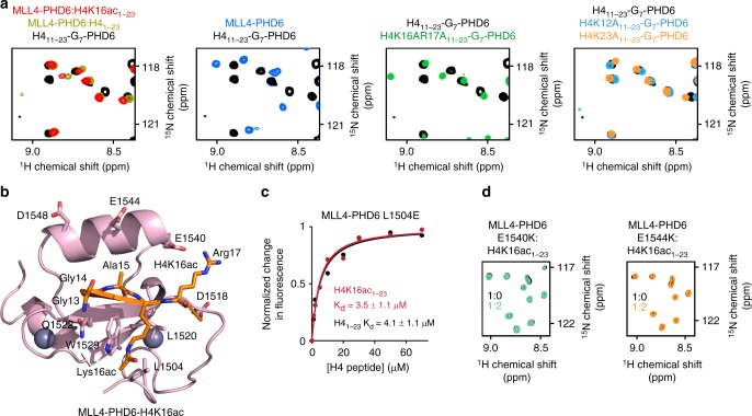
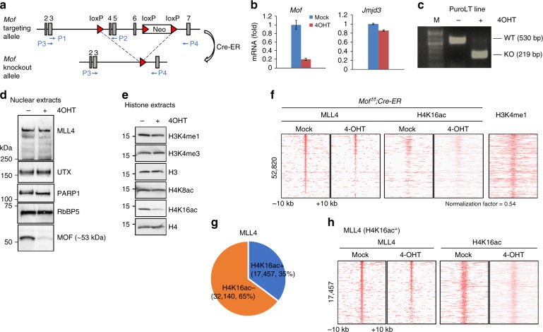
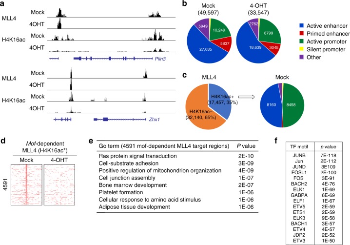
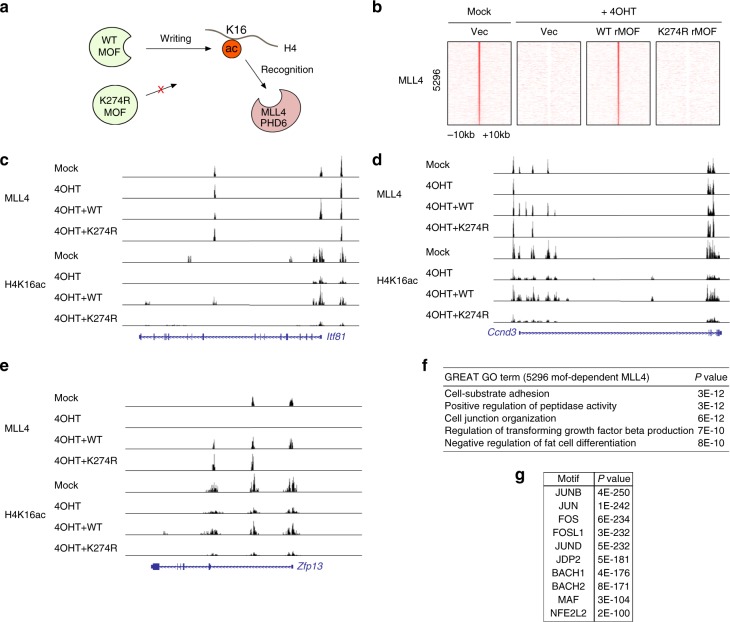
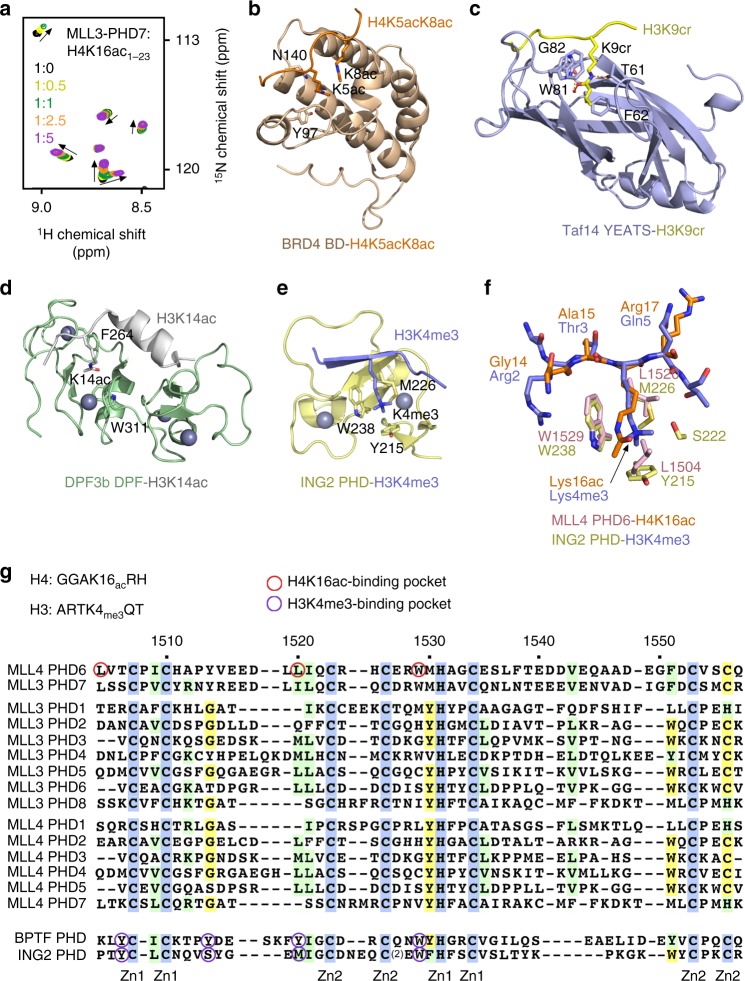
Histone methyltransferase MLL4 is centrally involved in transcriptional regulation and is often mutated in human diseases, including cancer and developmental disorders. MLL4 contains a catalytic SET domain that mono-methylates histone H3K4 and seven PHD fingers of unclear function. Here, we identify the PHD6 finger of MLL4 (MLL4-PHD6) as a selective reader of the epigenetic modification H4K16ac. The solution NMR structure of MLL4-PHD6 in complex with a H4K16ac peptide along with binding and mutational analyses reveal unique mechanistic features underlying recognition of H4K16ac. Genomic studies show that one third of MLL4 chromatin binding sites overlap with H4K16ac-enriched regions in vivo and that MLL4 occupancy in a set of genomic targets depends on the acetyltransferase activity of MOF, a H4K16ac-specific acetyltransferase. The recognition of H4K16ac is conserved in the PHD7 finger of paralogous MLL3. Together, our findings reveal a previously uncharacterized acetyllysine reader and suggest that selective targeting of H4K16ac by MLL4 provides a direct functional link between MLL4, MOF and H4K16 acetylation.


Conflict of interest statement

The authors declare no competing interests.

Figures

Fig. 1
Fig. 1
MLL4-PHD6 recognizes the histone mark H4K16ac. a Schematic of the MLL4 complex. b MLL4 domain architecture. c Superimposed 1H,15N heteronuclear single quantum coherence (HSQC) spectra of MLL4-PHD61503–1562 collected upon titration with H41–23, H41–9, and H412–23 peptides. Spectra are color coded according to the protein:peptide molar ratio. See also Supplementary Fig. 1, first panel. d Superimposed 1H,15N HSQC spectra of MLL4-PHD61503–1562 collected upon titration with H4K16ac1–23 peptide. Spectra are color coded according to the protein:peptide molar ratio. e Representative binding curves used to determine the Kd values by fluorescence spectroscopy (also see Supplementary Fig. 5). f Binding affinities of wild-type MLL4-PHD6 for the indicated histone peptides measured by tryptophan fluorescence. Error represents s.d. in triplicate measurements. Source data are provided as a Source Data file. g Binding curves used to determine the Kd values by microscale thermophoresis. Error represents s.d. in triplicate measurements. h A schematic showing specific reading of the H4K16ac mark by MLL4-PHD6
Fig. 2
Fig. 2
Structure of MLL4-PHD6 in complex with H4K16ac peptide. a A ribbon diagram of the solution nuclear magnetic resonance structure of MLL4-PHD6 (pink) in complex with the H4K16ac peptide (orange). b Electrostatic surface potential of MLL4-PHD6 colored blue and red for positive and negative charges, respectively. The bound H4K16ac peptide is shown in a stick model. c A zoom-in view of the H4K16ac-binding pocket
Fig. 3
Fig. 3
Molecular basis for the specific targeting of H4K16ac by MLL4-PHD6. a Superimposed 1H,15N heteronuclear single quantum coherence (HSQC) spectra of the linked H411–23-G7-PHD6 construct, wild-type (black) and indicated mutants (green, light blue, and orange), the isolated MLL4-PHD6 domain (blue), isolated H4-bound MLL4-PHD6 (dark yellow), and isolated H4K16ac-bound MLL4-PHD6 (red). b The structure of the H4K16ac-bound MLL4-PHD6 finger. c Representative binding curves used to determine the Kd values for the L1504E mutant by fluorescence spectroscopy (also see Supplementary Fig. 5). Source data are provided as a Source Data file. d Superimposed 1H,15N HSQC spectra of MLL4-PHD6 mutants collected upon titration with H4K16ac peptide. Spectra are color coded according to the protein:peptide molar ratio
Fig. 4
Fig. 4
A subset of MLL4-bound genomic regions overlaps with H4K16ac. SV40T-immortalized Moff/f;Cre-ER brown preadipocytes were treated with 4-hydroxytamoxifen to delete Mof. Cells were collected for reverse transcriptase PCR (RT-PCR), western blot and chromatin immunoprecipitation–sequencing (ChIP-Seq) analysis. a Schematics of targeting allele and knockout allele of Moff/f mice. In the targeting allele, a single loxP site was inserted in the intron before exon 4 and a neomycin selection cassette flanked by loxP sites was inserted in the intron after exon 6. The locations of RT-PCR primers P1–P2 (used in Fig. 4b) and P3–P4 (used in Fig. 4c) are indicated by arrows. b Quantitative RT-PCR confirmation of Mof deletion in preadipocytes. The data are presented as mean ± s.d. Two technical replicates from a single experiment were performed. Source data are provided as a Source Data file. c RT-PCR confirmation of wild-type and truncated Mof mRNA. d Western blot analysis of MLL4, UTX, and MOF. PARP1 and RbBP5 were used as loading controls. e Western blot analysis of histone modifications (also see Supplementary Fig. 15). f Heat maps around MLL4-binding sites. H4K16ac ChIP-Seq data were normalized with global H4K16ac levels measured by western blot. Published H3K4me1 data were obtained from GEO database (GSE74189). g Pie chart illustrating that 35% of MLL4-binding sites overlap with H4K16ac. h Heat maps of MLL4 and H4K16ac around 17,457 H4K16ac+ MLL4-binding sites
Fig. 5
Fig. 5
Targeting of MLL4 to active promoters is dependent on H4K16ac mark. Moff/f;Cre-ER brown preadipocytes were treated with 4-hydroxytamoxifen and collected for chromatin immunoprecipitation–sequencing (ChIP-Seq) analysis. a ChIP-Seq profiles of MLL4 and H4K16ac on Plin3 and Zhx1 gene loci. b Deletion of Mof decreases MLL4 binding to all sites. c H4K16ac-enriched (H4K16ac+) MLL4-binding regions are mainly located on active enhancers and active promoters. d Heat maps of 4591 Mof-dependent H4K16ac+ MLL4-binding sites. e Gene ontology analysis of the genes associated with 4591 Mof-dependent H4K16ac+ MLL4-binding regions identified in d. f Motif analysis of 4591 Mof-dependent H4K16ac+ MLL4-binding regions identified in d
Fig. 6
Fig. 6
MOF (Males absent On the First) catalytic activity is necessary for Mof-dependent chromatin targeting of MLL4. Moff/f;Cre-ER brown preadipocytes were infected with lentiviruses expressing wild-type (WT) or enzyme dead mutant (K274R) form of rMOF, followed by 4-hydroxytamoxifen treatment. Cells were collected for chromatin immunoprecipitation–sequencing (ChIP-Seq) analysis of MLL4 and H4K16ac. a Schematic representation of WT and enzyme dead MOF. b Heat maps of 5296 MOF enzyme activity-dependent MLL4-binding sites. ce ChIP-Seq profiles of MLL4 and H4K16ac on Zfp13 (c), Ccnd3 (d), and Itf81 (e) gene loci. f Gene ontology analysis of the genes associated with 5296 MOF catalytic activity-dependent MLL4-binding regions. g AP-1 family transcription factor motifs are enriched at MOF catalytic activity-dependent MLL4-binding regions. Top 3000 MLL4-binding regions were used for motif analysis
Fig. 7
Fig. 7
Comparative analysis of the H3K4me3 and H4K16ac recognition by histone readers. a Superimposed 1H,15N heteronuclear single quantum coherence spectra of MLL3-PHD71083–1143 collected upon titration with H4K16ac peptide. Spectra are color coded according to the protein:peptide molar ratio. bd Structures of the BRD4 BD, Taf14 YEATS, and DPF3b DPF readers in complex with the indicated histone peptides,,. PDB IDs: 3UVW (b), 5IOK (c), and 5I3l (d). e Structure of the ING2 PHD finger in complex with H3K4me3. PDB ID: 2G6Q. f A zoom-in view of the overlaid histone tail-binding sites of the ING2 PHD-H3K4me3 and MLL4 PHD6-H4K16ac complexes. g Alignment of amino acid sequences of the PHD fingers from human MLL4, MLL3, BPTF, and ING2. Absolutely, moderately, and weakly conserved residues are colored blue, green, and yellow, respectively. Zinc-coordinating cysteine/histidine residues are labeled

Similar articles

Cited by

References

    1. Froimchuk E, Jang Y, Ge K. Histone H3 lysine 4 methyltransferase KMT2D. Gene. 2017;627:337–342. doi: 10.1016/j.gene.2017.06.056. - DOI - PMC - PubMed
    1. Lee JE, et al. H3K4 mono- and di-methyltransferase MLL4 is required for enhancer activation during cell differentiation. eLife. 2013;2:e01503. doi: 10.7554/eLife.01503. - DOI - PMC - PubMed
    1. Wang C, et al. Enhancer priming by H3K4 methyltransferase MLL4 controls cell fate transition. Proc. Natl. Acad. Sci. USA. 2016;113:11871–11876. doi: 10.1073/pnas.1606857113. - DOI - PMC - PubMed
    1. Lai B, et al. MLL3/MLL4 are required for CBP/p300 binding on enhancers and super-enhancer formation in brown adipogenesis. Nucleic acids Res. 2017;45:6388–6403. doi: 10.1093/nar/gkx234. - DOI - PMC - PubMed
    1. Jin Q, et al. Distinct roles of GCN5/PCAF-mediated H3K9ac and CBP/p300-mediated H3K18/27ac in nuclear receptor transactivation. EMBO J. 2011;30:249–262. doi: 10.1038/emboj.2010.318. - DOI - PMC - PubMed

Publication types

MeSH terms