

Absorbable hemostatic hydrogels comprising composites of sacrificial templates and honeycomb-like nanofibrous mats of chitosan
- PMID: 31127114
- PMCID: PMC6534699
- DOI: 10.1038/s41467-019-10290-1
Absorbable hemostatic hydrogels comprising composites of sacrificial templates and honeycomb-like nanofibrous mats of chitosan
Erratum in
-
Author Correction: Absorbable hemostatic hydrogels comprising composites of sacrificial templates and honeycomb-like nanofibrous mats of chitosan.Nat Commun. 2025 Aug 13;16(1):7530. doi: 10.1038/s41467-025-62683-0. Nat Commun. 2025. PMID: 40804059 Free PMC article. No abstract available.
Abstract
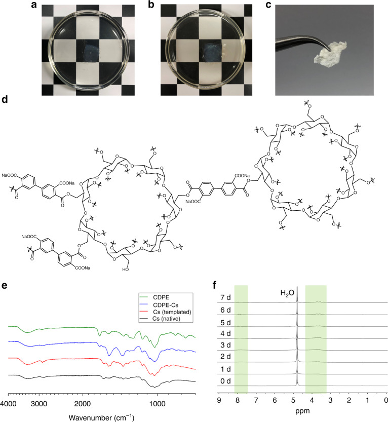
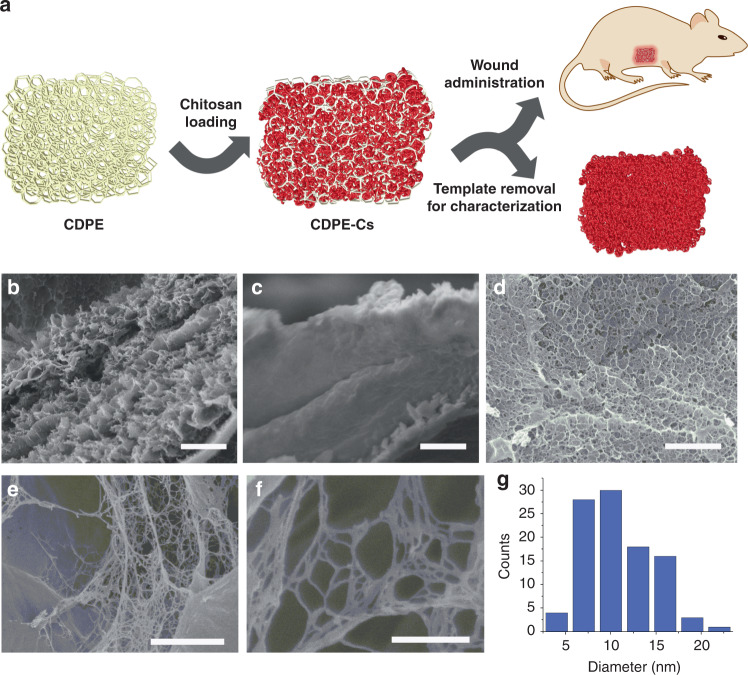
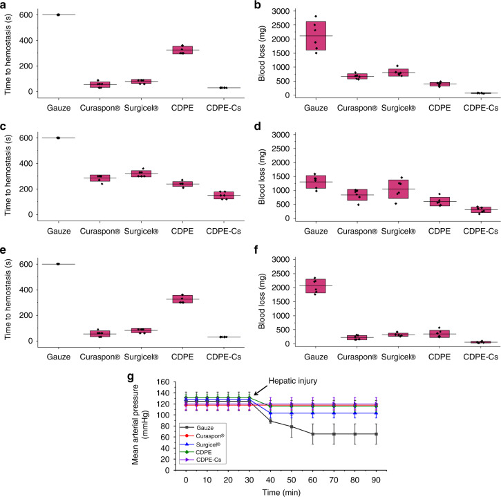
The development of hemostatic technologies that suit a diverse range of emergency scenarios is a critical initiative, and there is an increasing interest in the development of absorbable dressings that can be left in the injury site and degrade to reduce the duration of interventional procedures. In the current study, β-cyclodextrin polyester (CDPE) hydrogels serve as sacrificial macroporous carriers, capable of degradation under physiological conditions. The CDPE template enables the assembly of imprinted chitosan honeycomb-like monolithic mats, containing highly entangled nanofibers with diameters of 9.2 ± 3.7 nm, thereby achieving an increase in the surface area of chitosan to improve hemostatic efficiency. In vivo, chitosan-loaded cyclodextrin (CDPE-Cs) hydrogels yield significantly lower amounts of blood loss and shorter times to hemostasis compared with commercially available absorbable hemostatic dressings, and are highly biocompatible. The designed hydrogels demonstrate promising hemostatic efficiency, as a physiologically-benign approach to mitigating blood loss in tissue-injury scenarios.
Conflict of interest statement
The authors declare no competing interests.
Figures
References
-
- Florence, C., Haegerich, T., Simon, T., Zhou, C. & Luo, F. Estimated lifetime medical and work-loss costs of emergency department-treated nonfatal injuries—United States, 2013. MMWR64, 1078–1082 (2015). - PubMed
-
- Wedmore, I., McManus, J. G., Pusateri, A. E. & Holcomb, J. B. A special report on the chitosan-based hemostatic dressing. J. Trauma Acute Care Surg.60, 655–658 (2006). - PubMed
-
- Jayakumar, R., Prabaharan, M., Sudheesh Kumar, P. T., Nair, S. V. & Tamura, H. Biomaterials based on chitin and chitosan in wound dressing applications. Biotechnol. Adv.29, 322–337 (2011). - PubMed
-
- Brown, M. A., Daya, M. R. & Worley, J. A. Experience with Chitosan dressings in a civilian EMS system. J. Emerg. Med.37, 1–7 (2009). - PubMed
Publication types
MeSH terms
Substances
Grants and funding
LinkOut - more resources
Full Text Sources
Medical
