. 2020 Jan;27(1):176-191.
doi: 10.1038/s41418-019-0351-4. Epub 2019 May 24.

LncRNA H19 initiates microglial pyroptosis and neuronal death in retinal ischemia/reperfusion injury

Affiliations

LncRNA H19 initiates microglial pyroptosis and neuronal death in retinal ischemia/reperfusion injury

Peixing Wan et al. Cell Death Differ. 2020 Jan.

Erratum in

Abstract

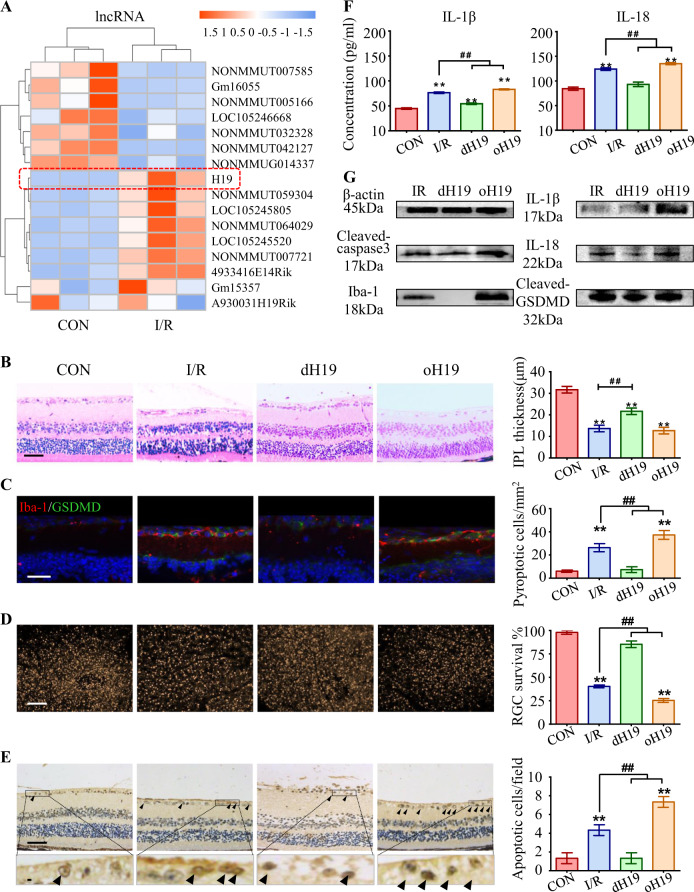
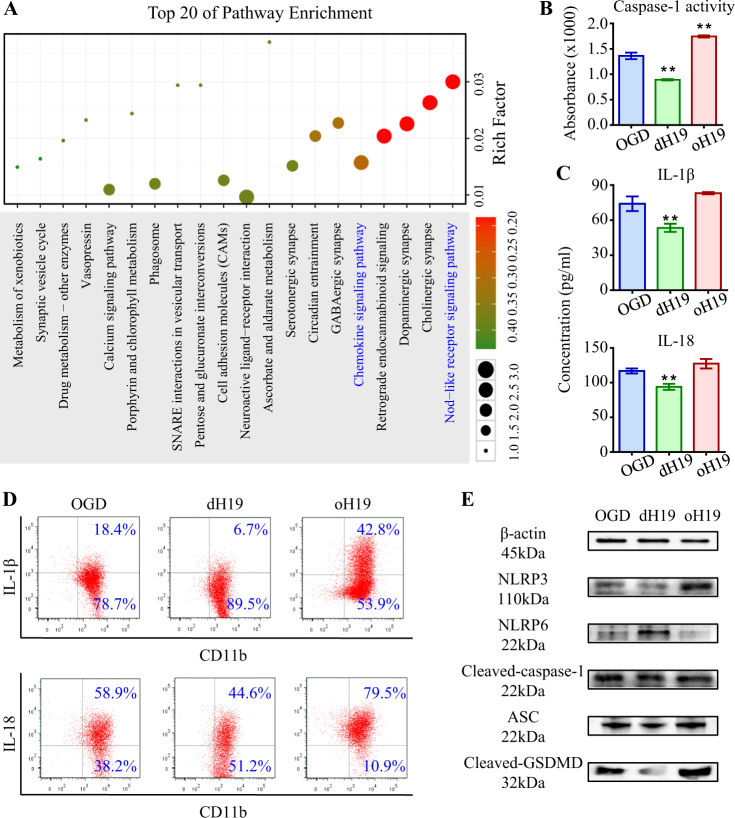
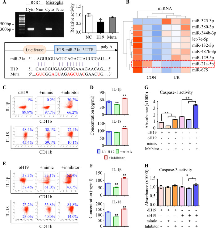
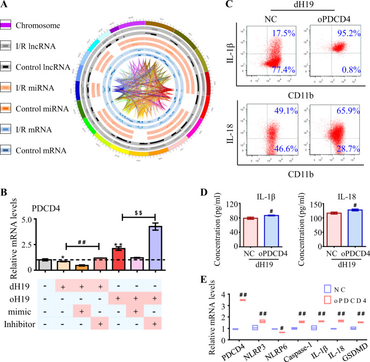
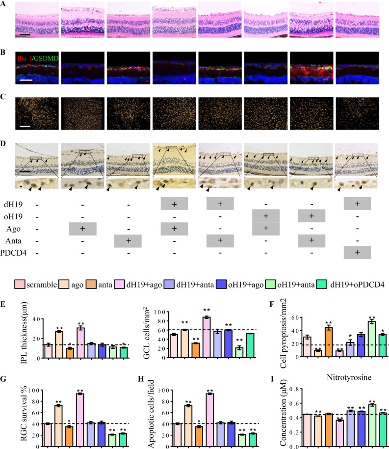
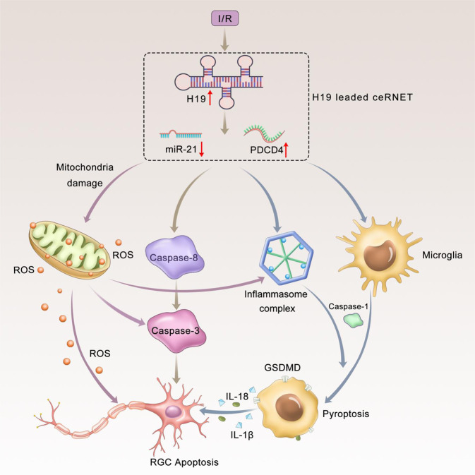
Ischemia-reperfusion (I/R) is a common pathology when the blood supply to an organ was disrupted and then restored. During the reperfusion process, inflammation and tissue injury were triggered, which were mediated by immunocytes and cytokines. However, the mechanisms initiating I/R-induced inflammation and driving immunocytes activation remained largely unknown. In this study, we identified long non-coding RNA (lncRNA)-H19 as the key onset of I/R-induced inflammation. We found that I/R increased lncRNA-H19 expression to significantly promote NLRP3/6 inflammasome imbalance and resulted in microglial pyroptosis, cytokines overproduction, and neuronal death. These damages were effectively inhibited by lncRNA-H19 knockout. Specifically, lncRNA-H19 functioned via sponging miR-21 to facilitate PDCD4 expression and formed a competing endogenous RNA network (ceRNET) in ischemic cascade. LncRNA H19/miR-21/PDCD4 ceRNET can directly regulate I/R-induced sterile inflammation and neuronal lesion in vivo. We thus propose that lncRNA-H19 is a previously unknown danger signals in the molecular and immunological pathways of I/R injury, and pharmacological approaches to inhibit H19 seem likely to become treatment modalities for patients in the near future based on these mechanistic findings.


Conflict of interest statement

The authors declare that they have no conflict of interest.

Figures

Fig. 1
Fig. 1
Predominant role of H19 in microglial pyroptosis and neuronal apoptosis. a Heat map listed the top 16 dysregulated lncRNAs in response to I/R injury. LncRNA H19 (red box) was the most upregulated lncRNA in the I/R retina. b Retinas exposed to I/R injury displayed a significant decrease in IPL thickness. This I/R-induced IPL attenuation was significantly ameliorated in dH19 mice and aggravated by H19 overexpression in retinas. c Iba-1+ microglia (red) had increased GSDMD-N (green) localized in the plasma membrane of I/R-treated mice compared with that of control retinas. Of note, less co-expression of Iba-1 and GSDMD-N was observed in the retina of dH19 mice. H19 overexpression promoted GSDMD-N augmentation in activated microglia (Iba-1+). d As measured by FG retro-labeling, viable RGC was presented as gold dots in flat-mounted retina. The RGC survival rate was noticeably decreased in response to I/R injury. H19 overexpression accentuated RGC apoptosis with less viable RGCs in retinas, which was prevented by H19 knockout. e Compared with the wild type counterparts, the dH19 retina had less TUNEL-positive cells (black arrows) in response to I/R injury, indicating that apoptosis was inhibited by H19 knockout. However, H19 overexpression markedly increased TUNEL-positive cells in GCL. f In retinal homogenates, H19 excision effectively inhibited the I/R-mediated overproduction of IL-1β and IL-18. This anti-inflammatory effect was abolished by H19 overexpression as measured by ELISA. g H19 knockout effectively prevented I/R-induced caspase-3 cleavage, indicating the pro-apoptotic effect of H19. Also, the retinas of H19-null mice exhibited lower protein levels of Iba-1, cleaved-GSDMD, IL-1β, and IL-18. The relative level of each target protein was normalized to β-actin from the same sample (Fig. S2F). Scale bar = 100 μm. Data were represented as means ± SD (n = 6). Compared with the normal control (CON): **P < 0.01. Compared with the I/R retina: ##P < 0.01. I/R, ischemia and reperfusion; dH19, H19 knockout; oH19, H19 overexpression; GCL, ganglion cell layer; IPL, inner plexiform layer; FG, flurogold
Fig. 2
Fig. 2
H19 regulates neuro-inflammation by rebalancing NLRP3/NLRP6 inflammasomes. a GO analysis annotated biological function to dysregulated mRNAs in I/R retina. Canonical pathways related to Nod-like receptor signaling and chemokine production acquired vital importance in I/R injury. b In primary microglia, H19 knockout significantly prevented OGD/R-induced caspase-1 activation, which was exacerbated by H19 overexpression. c As measured by ELISA, OGD/R-induced overproduction of IL-1β and IL-18 was suppressed by H19 knockout and aggravated by H19 overexpression in cultured microglia. d As measured by flow cytometry, H19 excision effectively decreased IL-1β or IL-18 overproduction in microglia. And H19 overexpression increased production of these two pro-inflammatory cytokines. e Reciprocal activation of NLRP3/NLRP6 inflammasomes was observed in microglia underwent OGD/R. Imbalance activation of inflammasomes resulted in increased ASC and cleavage of caspase-1 and GSDMD. The upregulation of these pro-inflammatory proteins was aggravated in oH19 microglia as attenuated by H19 knockout as measured by immunoblot. The relative level of each target protein was normalized to β-actin from the same sample (Fig. S2H). Data were represented as means ± SD (n = 6). Compared with the OGD/R group (OGD): **P < 0.01. OGD/R, oxygen-glucose deprivation and reperfusion; dH19, H19 knockout; oH19, H19 overexpression; ASC, apoptosis-associated speck-like protein containing a CARD
Fig. 3
Fig. 3
H19 sponges miR-21 to regulate neuro-inflammation. a As measured by qPCR, H19 was specifically expressed in the cytoplasm instead of the nucleus in both RGCs and microglia. Compared to NC RNA, miR-21 reduced luciferase activity by 43.4% ± 1.4% through binding to H19. The miR-21-dependent inhibition of luciferase activity was abolished after mutations (red) in the binding site. Data were represented as means ± SD (n = 3). Compared with NC RNA: *P < 0.05. b The top nine dysregulated miRNAs in I/R retinas. c, d Anti-inflammatory effect of H19 excision was further promoted by the miR-21 mimic with less IL-1β and IL-18 production in supernatant of retinal microglia. As measured by flow cytometry and ELISA, the miR-21 inhibitor greatly antagonized the anti-inflammation effect of dH19 with more production of IL-1β and IL-18. e, f In H19 overexpressed microglia, miR-21 downregulation further aggravated the production of IL-1β and IL-18, which was ameliorated by miR-21 upregulation as measured by flow cytometry and ELISA. g, h Caspase-1 activity was further restrained in the microglia deficient of both H19 and miR-21 compared with the single H19 excision. The miR-21 inhibitor further augmented caspase-1 activity in oH19 microglia. Either H19 or miR-21 had no significant effect on apoptotic activity of caspase-3 in microglia. Data were represented as means ± SD (n = 6). Compared with the dH19 group: **P < 0.01. Compared with the oH19 group: ##P < 0.01. Cyto, cytoplasm; Nuc, nucleus; NC, normal control; Muta, mutation; dH19, H19 knockout; oH19, H19 overexpression
Fig. 4
Fig. 4
H19 competed with PDCD4 for miR-21 to form ceRNET. a Sketch of the dysregulated transcriptome and their correlations. From outside in: chromosome position, lncRNAs in the I/R retina, lncRNAs in the healthy control, miRNAs in the I/R group, miRNAs in the control group, mRNAs in the I/R retina, mRNAs in the healthy control, and interactions among these three types of RNAs in the dysregulated transcriptome. b In cultured microglia, PDCD4 expression was up-regulated by oH19 and the miR-21 inhibitor, and down-regulated by dH19 and the miR-21 mimic. PDCD4 dysregulation was aggravated by simultaneous H19 and miR-21 treatment at transcriptional level. c, d As measured by flow cytometry and ELISA, H19 excision-mediated prevention of IL-1β and IL-18 overproduction was abolished by PDCD4 overexpression in microglia. e As measured by real-time PCR, PDCD4 competed with H19 in regulating the NLRP3/NLRP6 inflammasome balance in microglia. And the expression of caspase-1, GSDMD, and the pro-inflammatory cytokines was increased by PDCD4 overexpression. Data were represented as means ± SD (n = 6). Compared with the OGD/R group: *P < 0.05, **P < 0.01. Compared with dH19 microglia: #P < 0.05, ##P < 0.01. Compared with oH19 microglia: $$P < 0.01. NC, normal control lentivirus; dH19, H19 knockout; oH19, H19 overexpression; oPDCD4, PDCD4 overexpression
Fig. 5
Fig. 5
H19 functions in ceRNET during I/R-induced retinal damage. a, e Neuroprotective effect of H19 excision was enhanced by miR-21 up-regulation via inhibiting IPL attenuation and cell death in GCL. miR-21 downregulation or oPDCD4 eliminated the protective effect of H19 knockout. H19 overexpression and miR-21 antagomir worked both singly and together to increase I/R-induced retinal damage by augmenting IPL attenuation and cell death in GCL. b, f Microglial pyroptosis was suppressed by dH19 and miR-21 upregulation alone or together as shown by less cells co-labeled with Iba-1 (red) and GSDMD-N (green). H19 overexpression, miR-21 knockdown, and oPDCD4 augmented I/R-induced microglial pyroptosis with more GSDMD-N anchored in the plasma membrane of Iba-1+ cells. c, g H19/miR-21/PDCD4 worked in ceRNET to regulate RGC survival (gold dots) in I/R retinas. Both H19 knockout and miR-21 upregulation protected RGCs from I/R-induced apoptosis. By contrast, H19 overexpression and PDCD4 increased RGC apoptosis in response to I/R injury as shown by fewer gold dots in retinal flat mounts. d, h Compared with the wild type retina, dH19 excision and miR-21 upregulation exhibited fewer TUNEL-positive cells (black arrows) in response to I/R injury. This protective effect was eliminated by H19 overexpression, miR-21 down-regulation, or PDCD4 with markedly increased TUNEL+ cells in GCL. i Nitrotyrosine (ROS derivative) accumulation in retinas was significantly decreased by dH19 and miR-21 agomir. H19 overexpression, miR-21 antagomir, and oPDCD4 increased nitrotyrosine accumulation in response to I/R. Scale bar = 100 μm. Data were represented as means ± SD (n = 6). Compared with the I/R group: *P < 0.05, **P < 0.01. dH19, H19 knockout; oH19, H19 overexpression; oPDCD4, PDCD4 overexpression; ago, agomir of miR-21; anta, antagomir of miR-21
Fig. 6
Fig. 6
Graphical summary of H19/miR-21/PDCD4 ceRNET in I/R injury. H19 responded to ischemic insults with increased expression level. Upregulated H19 then competed with PDCD4 for miR-21 to form the ceRNET. The ceRNET is capable of processing several pathological signals into a unitary and coherent adaptive response. H19-mediated ceRNET triggered reciprocal activation of NLRP3/NLRP6 inflammasomes. Thereafter, activated caspase-1 was recruited to cleave GSDMD, which characterized microglial pyroptosis. Meanwhile, on the inflammasome platform, mature forms of IL-1β and IL-18 were managed, which mediated pyroptosis to exert neurotoxicity. Besides inflammatory caspases, the H19/miR-21/PDCD4 ceRNET also activated apoptotic caspases (caspase-3 and caspase-8) to directly regulate RGC apoptosis in I/R injury. Mitochondrial dysfunction (marked by MMP collapse) and consequent ROS overproduction were identified as independent activators of inflammasomes and apoptotic signals. H19 knockout effectively relieved I/R-induced neuro-inflammation, neuronal death and mitochondrial failure. Simultaneous regulation of the H19/miR-21/PDCD4 ceRNET further blocked the ischemic cascade to improve the protective effect. I/R, ischemia/reperfusion injury; RGC, retinal ganglion cell; MMP, mitochondrial membrane potential; ROS, reactive oxygen species

References

    1. Eltzschig HK, Eckle T. Ischemia and reperfusion—from mechanism to translation. Nat Med. 2011;17:1391–401. doi: 10.1038/nm.2507. - DOI - PMC - PubMed
    1. Kawaguchi M, Takahashi M, Hata T, Kashima Y, Usui F, Morimoto H, et al. Inflammasome activation of cardiac fibroblasts is essential for myocardial ischemia/reperfusion injury. Circulation. 2011;123:594–604. - PubMed
    1. Yang Y, Salayandia VM, Thompson JF, Yang LY, Estrada EY, Yang Y. Attenuation of acute stroke injury in rat brain by minocycline promotes blood-brain barrier remodeling and alternative microglia/macrophage activation during recovery. J Neuroinflamm. 2015;12:26. doi: 10.1186/s12974-015-0245-4. - DOI - PMC - PubMed
    1. Hu G, Tang Q, Sharma S, Yu F, Escobar TM, Muljo SA, et al. Expression and regulation of intergenic long noncoding RNAs during T cell development and differentiation. Nat Immunol 2013. 2013;14:1190–8. - PMC - PubMed
    1. Schmitt AM, Garcia JT, Hung T, Flynn RA, Shen Y, Qu K, et al. An inducible long noncoding RNA amplifies DNA damage signaling. Nat Genet 2016. 2016;48:1370–6. - PMC - PubMed

Publication types

MeSH terms