. 2019 Jul 9;47(12):6029-6044.
doi: 10.1093/nar/gkz354.

Fatty acid conjugation enhances potency of antisense oligonucleotides in muscle

Affiliations

Fatty acid conjugation enhances potency of antisense oligonucleotides in muscle

Thazha P Prakash et al. Nucleic Acids Res. .

Abstract

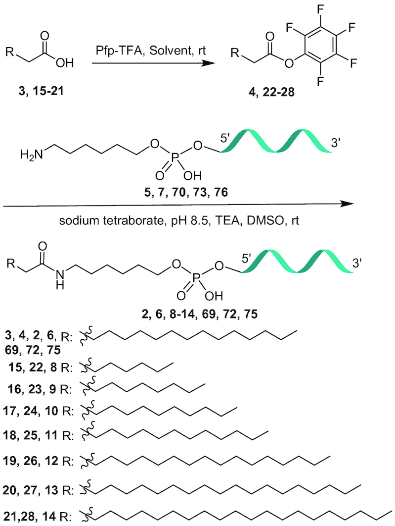
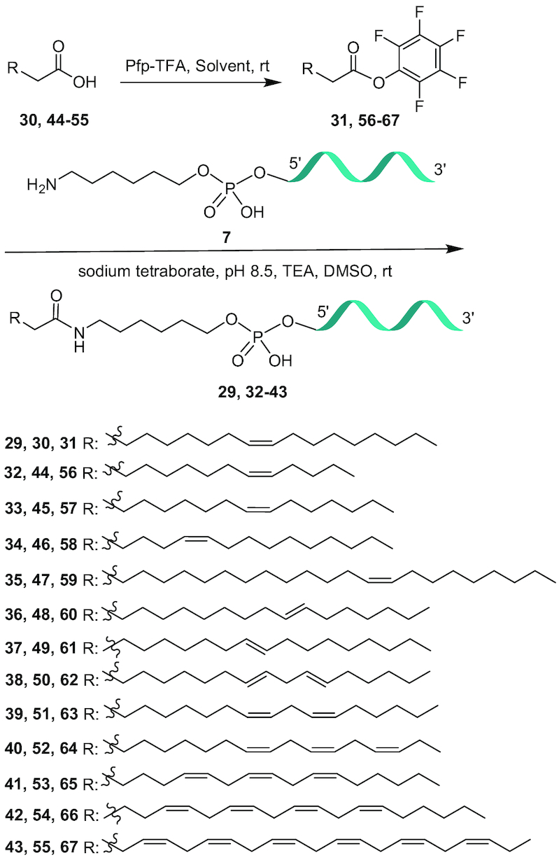
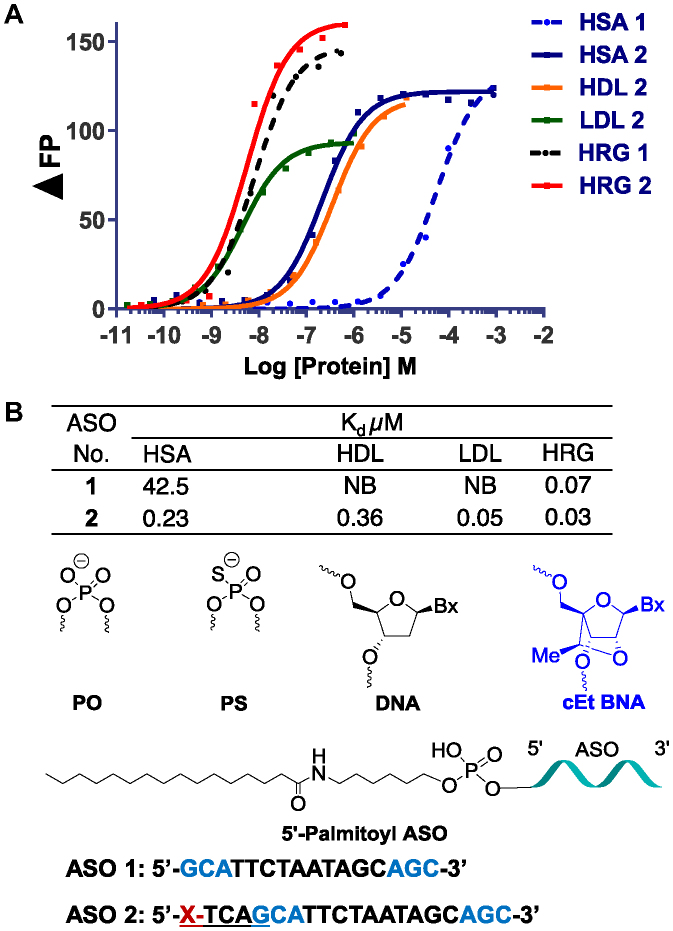
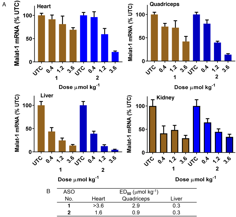
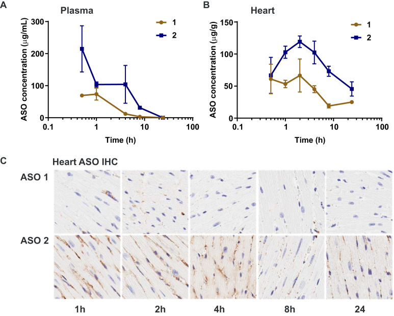
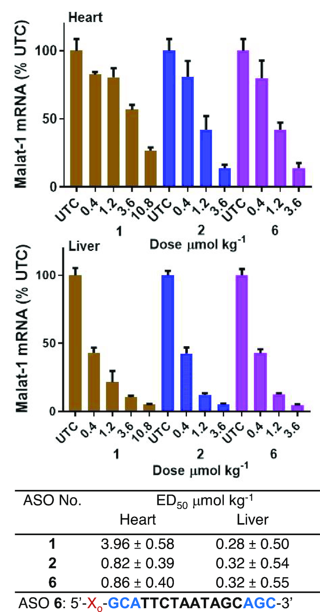
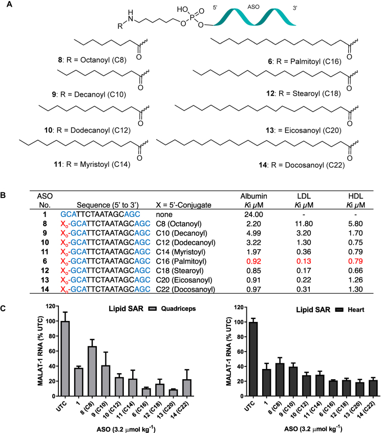
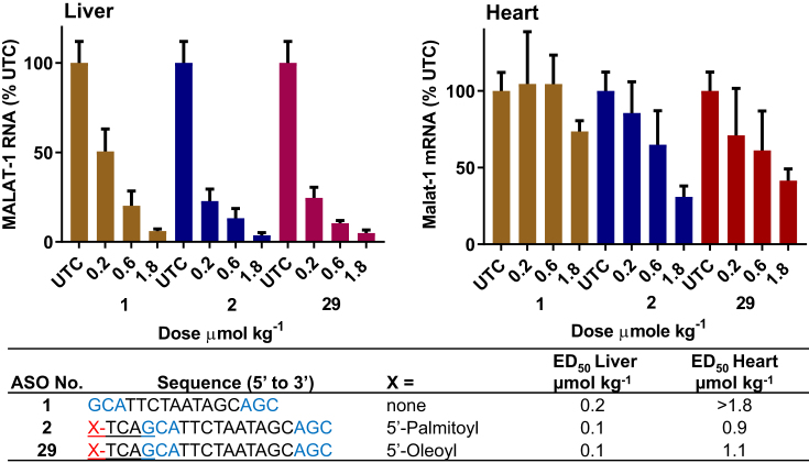
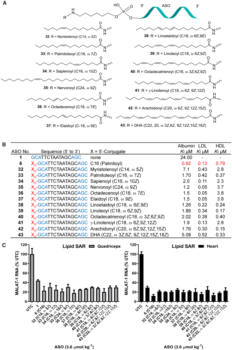
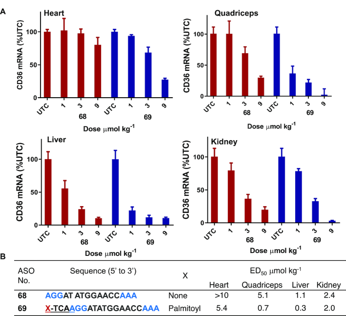
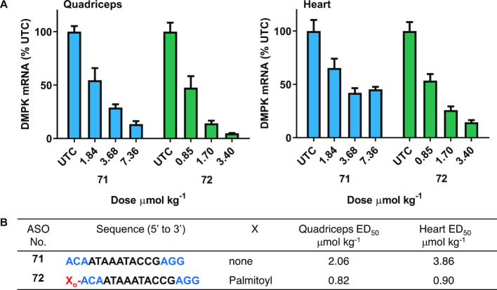
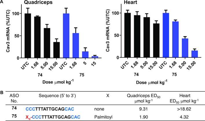
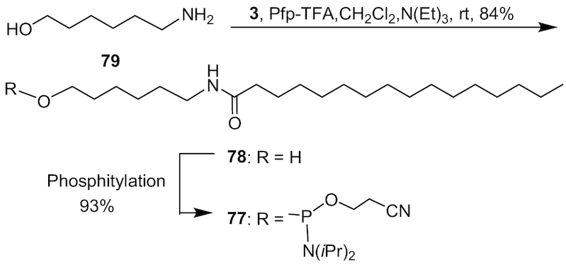
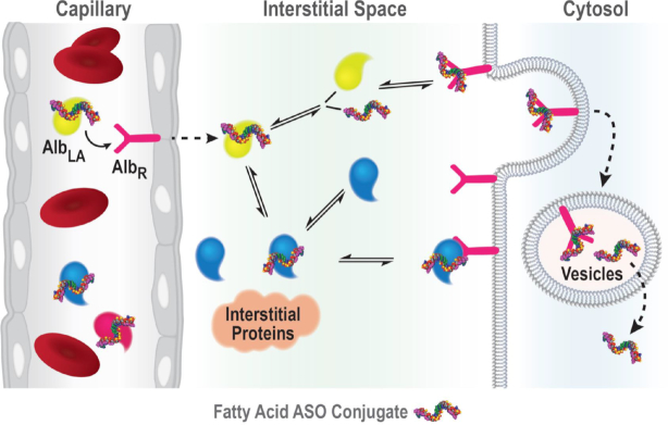
Enhancing the functional uptake of antisense oligonucleotide (ASO) in the muscle will be beneficial for developing ASO therapeutics targeting genes expressed in the muscle. We hypothesized that improving albumin binding will facilitate traversal of ASO from the blood compartment to the interstitium of the muscle tissues to enhance ASO functional uptake. We synthesized structurally diverse saturated and unsaturated fatty acid conjugated ASOs with a range of hydrophobicity. The binding affinity of ASO fatty acid conjugates to plasma proteins improved with fatty acid chain length and highest binding affinity was observed with ASO conjugates containing fatty acid chain length from 16 to 22 carbons. The degree of unsaturation or conformation of double bond appears to have no influence on protein binding or activity of ASO fatty acid conjugates. Activity of fatty acid ASO conjugates correlated with the affinity to albumin and the tightest albumin binder exhibited the highest activity improvement in muscle. Palmitic acid conjugation increases ASO plasma Cmax and improved delivery of ASO to interstitial space of mouse muscle. Conjugation of palmitic acid improved potency of DMPK, Cav3, CD36 and Malat-1 ASOs (3- to 7-fold) in mouse muscle. Our approach provides a foundation for developing more effective therapeutic ASOs for muscle disorders.


Figures

Scheme 1.
Scheme 1.
Synthesis of 5′-fatty acid conjugated ASO 2, 6, 8–14, 69, 72 and 75; ASO 5: 5′-TmCAGkmCkAk TdTdmCd TdAdAdTdAd GdmCd AkGkmCk 3′, ASO 7: GkmCk AkTdTdmCd TdAdAdTdAd GdmCd AkGkmCk, ASO 70: 5′- TdmCdAdAkGkGkAdTd AdTd GdGdAdAdmCdmCd AkAkAk-3′, ASO 73: 5′-AkmCkAk AdTd AdAd AdTdAdmCdmCdGd AkGkGk-3′, ASO 76: 5′-mCkmCkmCk Td TdTdAd TdTd GdmCdAdGdmCkAkmCk, k: cEt BNA, d: DNA, mC: 5-methyl cytidine, Backbone all PS, underline PO.
Scheme 2.
Scheme 2.
Synthesisof 5’-unsaturated fatty acid conjugated ASO 29, 32-43.
Figure 1.
Figure 1.
Characterizing the effect of palmitoyl conjugation on binding to selected plasma protein of Gapmer ASOs. (A) Binding curves showing differences between binding of unconjugated Malat-1 ASO 1 and palmitic acid conjugated ASO 2. (B) Binding constants for ASO 1 and 2 to selected plasma proteins; NB: no binding; HSA: human serum albumin; HDL: high-density lipoprotein; LDL: low-density lipoprotein; HRG: histidine rich glycoprotein; ASO sequence blue: cEt BNA, black: DNA, C: 5-methylcytidine, backbone all PS; underline PO.
Figure 2.
Figure 2.
Palmitic acid conjugation enhances potency of Malat-1 ASO in mouse heart and quadriceps. Mice (C57BL/6, n = 4/group) were injected subcutaneously Malat-1 ASO 1 and 5′-palmitoyl conjugated Malat-1 ASO 2 at 0.4, 1.2 and 3.6 μmol/kg once a week for 3 weeks for a total of three doses and sacrificed after 5 days. (A) Malat-1 RNA expression analyzed in mice heart, quadriceps, liver and kidney using qRT-PCR. All data are expressed as mean ± standard deviation. (B) ED50 (μmol/kg/week) for reducing Malat-1 RNA in mouse heart, quadriceps and liver.
Figure 3.
Figure 3.
Pharmacokinetics of Malat-1 ASO 1 and 5′-palmitoyl conjugated Malat-1 ASO 2; mice plasma total ASO concentration (A) and mice heart tissue total ASO concentration (B) after 0.5, 1, 2, 4, 8 and 24 h of subcutaneous administration of ASOs 12 (7.5 μmol kg−1); (C) ASO distribution in mice heart tissue after 1, 2, 4, 8 and 24 h of subcutaneous administration ASOs 12 (7.5 μmol kg−1) analyzed for ASOs by immunohistochemistry (IHC) using a PS-oligonucleotide antibody. Shown is a representative example of one of the four animals per group.
Figure 4.
Figure 4.
Potency of palmitic acid conjugated Malat-1 ASO with and without PO d(TCA) linker is similar in mouse heart and liver. Mice (C57BL/6, n = 4/group) were injected subcutaneously ASO 1 at 0.4, 1.2, 3.6, 10.8 and ASO 2 with PO d(TCA), 6 without PO d(TCA) at 0.4, 1.2 and 3.6 μmol/kg for 3 weeks for a total of 3 doses then sacrificed after 5 days. Malat-1 RNA expression analyzed in mice heart and liver by qRT-PCR. All data are expressed as mean ± standard deviation. ED50 (μmol/kg/wk) for reducing Malat-1 RNA in mouse heart and ASO sequence: blue = cEt BNA, black = DNA, C = 5-methylcytidine, backbone all PS; o = PO, X = palmitoyl.
Figure 5.
Figure 5.
(A) Structures of ASO fatty acid conjugates 6, 814; (B) protein binding affinity of fatty acid ASO conjugates 6, 814; (C) Malat-1 RNA expression in mice heart and quadriceps after subcutaneous administration of ASO 6, 8–14 at 3.6 μmol/kg once a week for three weeks for a total of three doses; ASO sequence: X = lipid, blue: cEt BNA, black: DNA, C: 5-methylcytidine, Backbone all PS, o: PO.
Figure 6.
Figure 6.
5′-Oleoyl ASO 29 and 5′-palmitoyl Malat-1 ASO 2 exhibited similar potency in mice heart and liver. Mice (C57BL/6, n = 4/group) were injected subcutaneously with Malat-1 ASO 1, 5′-plamitoyl Malat-1 ASO 2 and 5′-oleyol Malat-1 ASO 29 at 0.2, 0.6 and 1.8 μmol/kg once a week for 3 weeks, sacrificed after 5 days. (A) Malat-1 RNA expression analyzed in mice heart and liver by qRT-PCR. All data are expressed as mean ± standard deviation. (B) ED50 (μmol/kg/week) for reducing Malat-1 RNA in mouse heart and liver. ASO sequence: X = lipid, blue: cEt BNA, black: DNA, C: 5-methylcytidine, Backbone all PS, Underline PO. (C) structure of 5′-oleic acid conjugated Malat-1 ASO 29.
Figure 7.
Figure 7.
(A) Structure of ASO conjugates 3243; (B) protein binding affinity of lipid conjugates ASOs 3243; (C) Malat-1 RNA expression from hearts and quadriceps of ASOs 32–43 administered mice.; ASO sequence: X = lipid, blue: cEt BNA, black: DNA, C: 5-methylcytidine, Backbone all PS, o: PO.
Figure 8.
Figure 8.
Palmitic acid conjugation enhances potency of CD36 ASO in mice heart, quadriceps and, liver. Mice (C57BL/6, n = 4/group) were injected intravenously CD36 ASO 68, 5′-plamitoyl CD36 ASO 68 at 1, 3 and 9 μmol/kg once a week for 3 weeks, sacrificed after 5 days. (A) CD36 mRNA expression analyzed in mice hearts quadriceps, liver and kidney by qRT-PCR. All data are expressed as mean ± standard deviation. (B) ED50 (μmol/kg/week) for reducing CD36 mRNA in mouse heart, quadriceps, liver and kidney. ASO sequence: X = lipid, blue: cEt BNA, black: DNA, C: 5-methylcytidine, backbone all PS, underline PO.
Figure 9.
Figure 9.
Palmitic acid conjugation enhances potency of DMPK ASO in mice heart and quadriceps. Mice (Bal/c, n = 4/group) were administered subcutaneously DMPK ASO 71 at 1.84, 3.68 and 7.36 and 5′-palmitoyl DMPK ASO 72 at 0.85, 1.70, 3.40, μmol/kg once a week for 3.5 weeks, sacrificed after 2 days. (A) DMPK mRNA expression analyzed in mice heart and quadriceps by qRT-PCR. All data are expressed as mean ± standard deviation. (B) ED50 (μmol/kg/week) for reducing DMPK mRNA in mouse heart and quadriceps. ASO sequence: X = lipid, blue: cEt BNA, black: DNA, C: 5-methylcytidine, backbone all PS, o = PO.
Figure 10.
Figure 10.
Activity of Cav3 ASO 74 and 5′-palmitoyl Cav3 ASO 75 in mice heart and quadriceps; Mice (C57BL/6, n = 4/group) were injected subcutaneously Cav3 ASO 74, 5′-palmitoyl Cav3 ASO 75 at 1.68, 5 and 15 μmol/kg once a week for 2 weeks, sacrificed after 4 days. (A) Cav3 mRNA expression analyzed in mice heart and quadriceps by qRT-PCR. All data are expressed as mean ± standard deviation. (B) ED50 (μmol/kg/week) for reducing Cav3 mRNA in mouse heart and quadriceps. ASO sequence: X = lipid, blue: cEt BNA, black: DNA, C: 5-methylcytidine, backbone all PS, o = PO.
Scheme 3.
Scheme 3.
Synthesis of 6-palmitamidohexyl phosphoramidite 77; Pfp-TFA: pentaflurophenyl trifluoroacetate.
Figure 11.
Figure 11.
Schematic representation of the likely route taken by lipid conjugated ASO from the capillary to cytosol in muscle. AlbLA: albumin bound lipid ASO; AlbR: albumin receptor.

Similar articles

Cited by

References

    1. Khvorova A., Watts J.K.. The chemical evolution of oligonucleotide therapies of clinical utility. Nat. Biotech. 2017; 35:238–248. - PMC - PubMed
    1. Stein C.A., Castanotto D.. FDA-Approved Oligonucleotide Therapies in 2017. Mol. Ther. 2017; 25:1069–1075. - PMC - PubMed
    1. Crooke S.T., Witztum J.L., Bennett C.F., Baker B.F.. RNA-targeted therapeutics. Cell Metab. 2018; 27:714–739. - PubMed
    1. Prakash T.P., Graham M.J., Yu J., Carty R., Low A., Chappell A., Schmidt K., Zhao C., Aghajan M., Murray H.F. et al. .. Targeted delivery of antisense oligonucleotides to hepatocytes using triantennary N-acetyl galactosamine improves potency 10-fold in mice. Nucleic Acids Res. 2014; 42:8796–8807. - PMC - PubMed
    1. Ammala C., Drury W.J. 3rd, Knerr L., Ahlstedt I., Stillemark-Billton P., Wennberg-Huldt C., Andersson E.M., Valeur E., Jansson-Lofmark R., Janzen D. et al. .. Targeted delivery of antisense oligonucleotides to pancreatic β-cells. Sci Adv. 2018; 4:eaat3386. - PMC - PubMed

Publication types

MeSH terms