. 2019 Nov;56(11):7872-7887.
doi: 10.1007/s12035-019-01649-y. Epub 2019 May 24.

Activation of Peroxisome Proliferator-Activated Receptor-α Increases the Expression of Nuclear Receptor Related 1 Protein (Nurr1) in Dopaminergic Neurons

Affiliations

Activation of Peroxisome Proliferator-Activated Receptor-α Increases the Expression of Nuclear Receptor Related 1 Protein (Nurr1) in Dopaminergic Neurons

Carl G Gottschalk et al. Mol Neurobiol. 2019 Nov.

Abstract

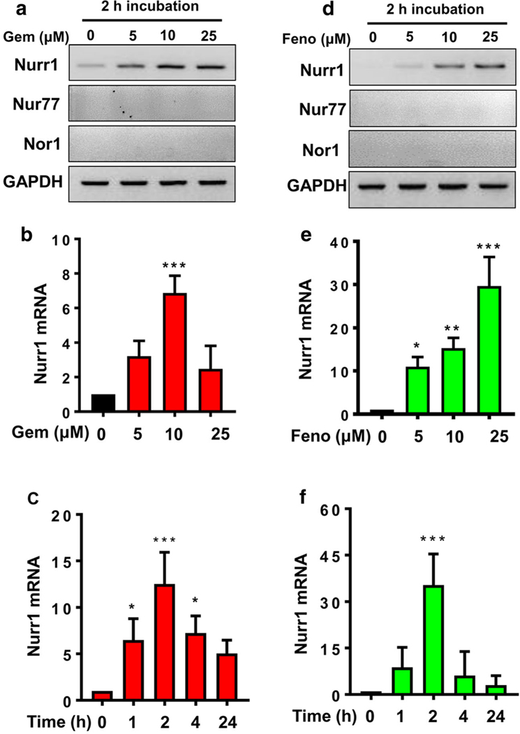
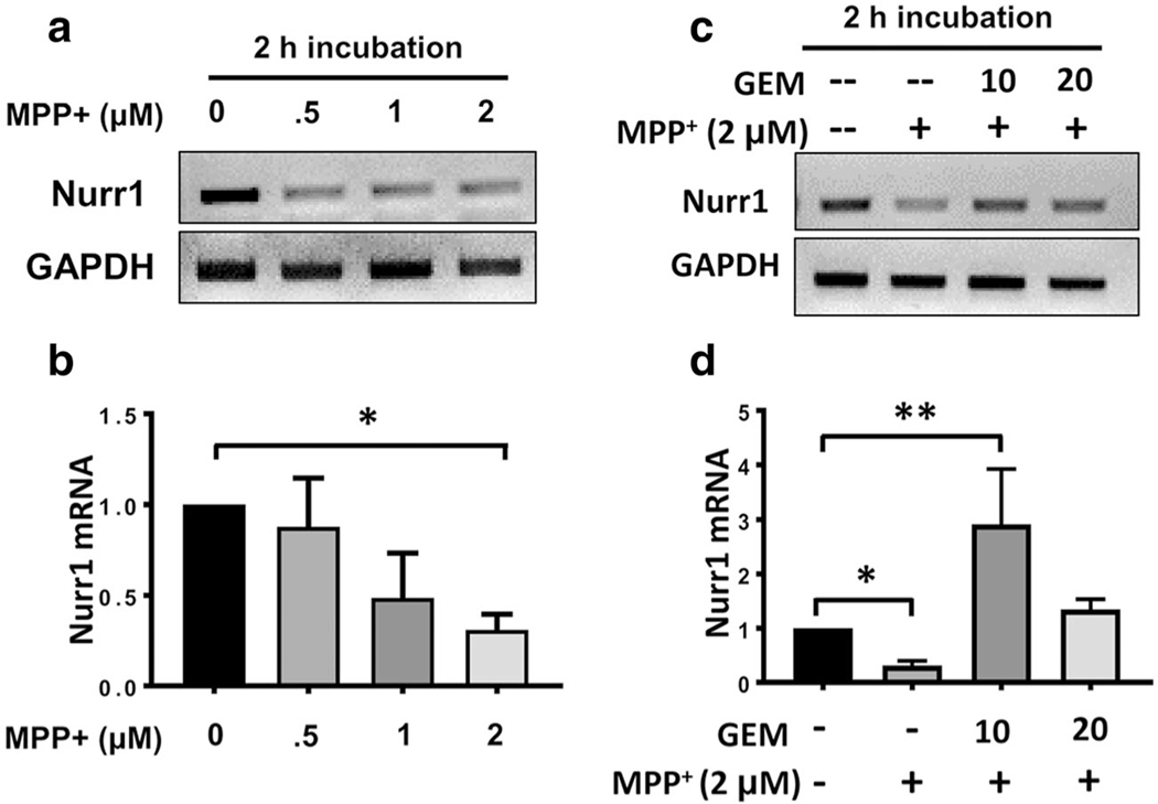
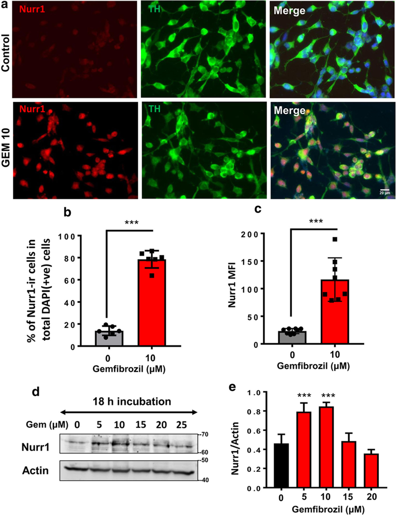
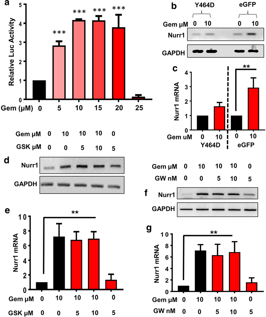
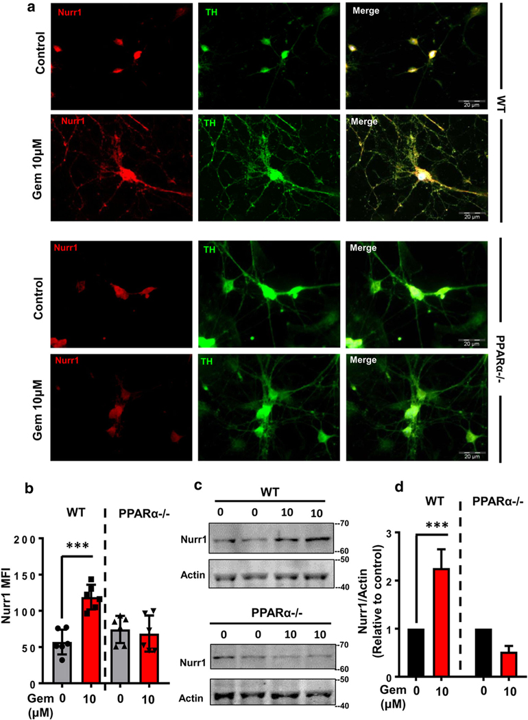
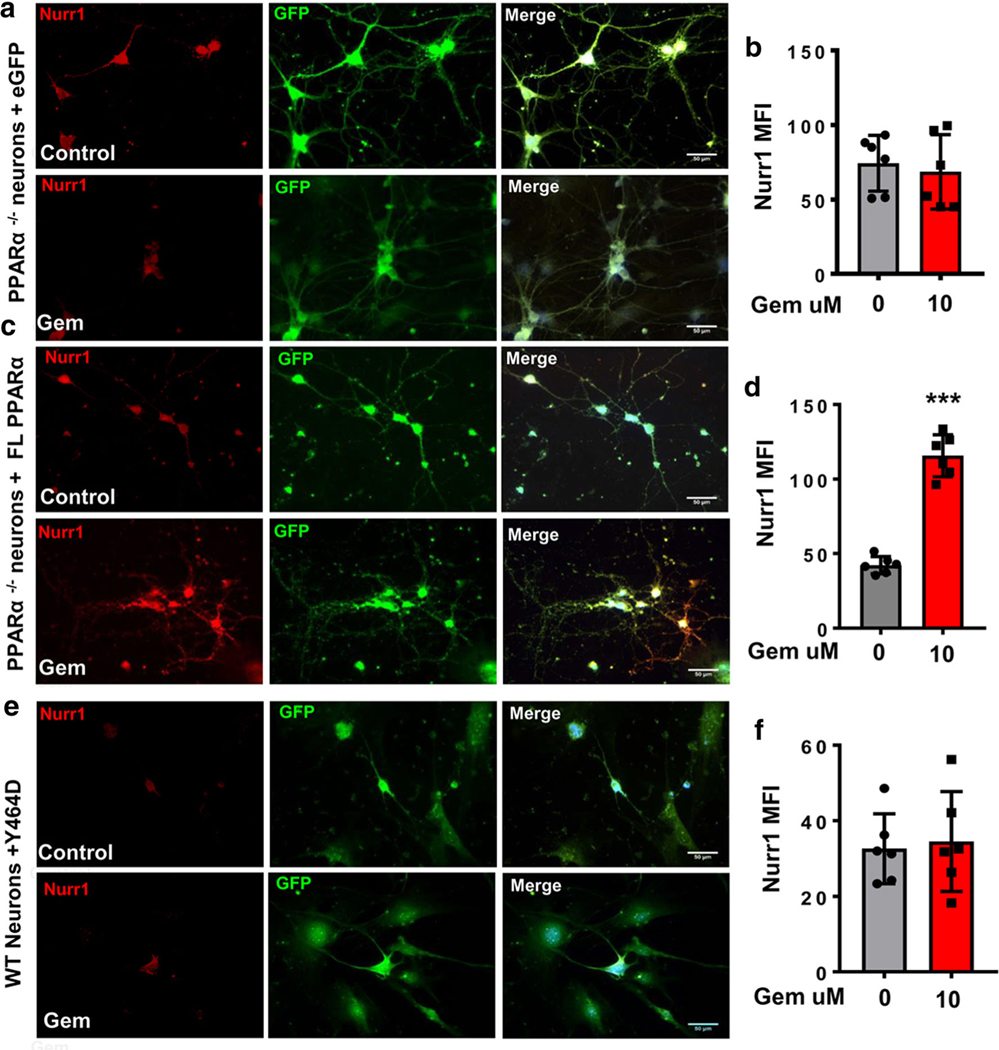
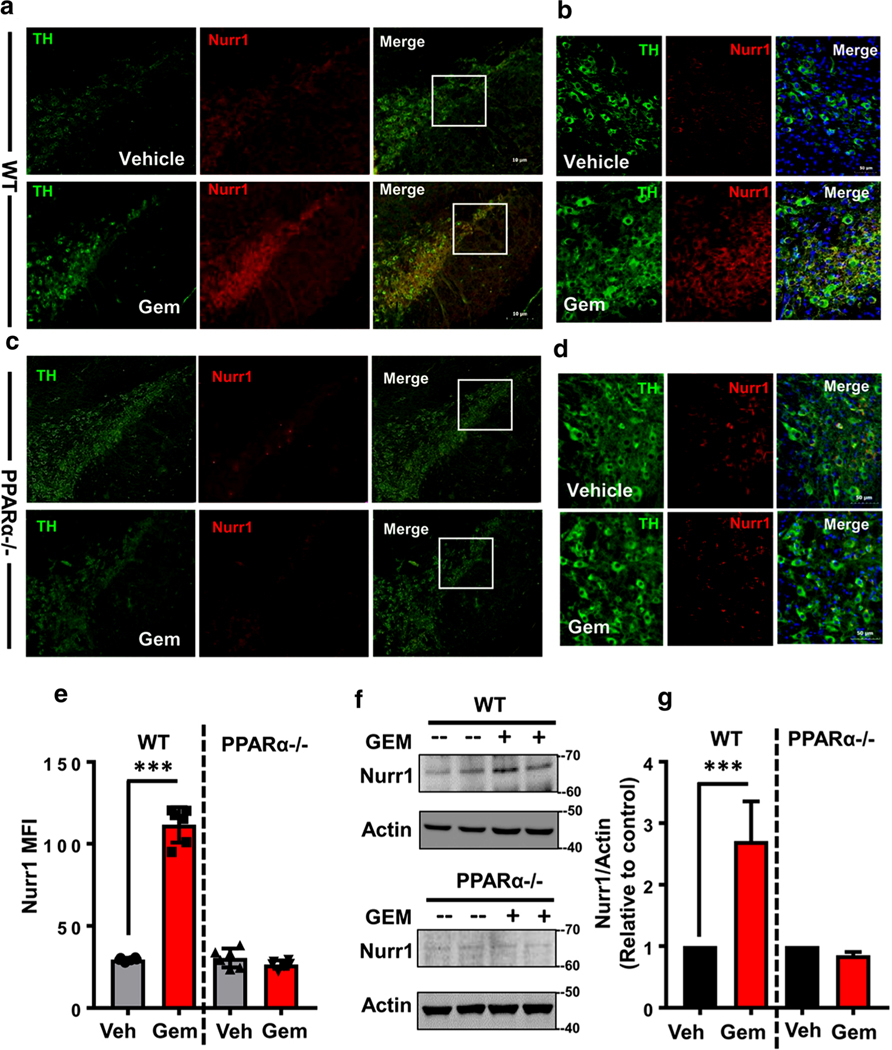
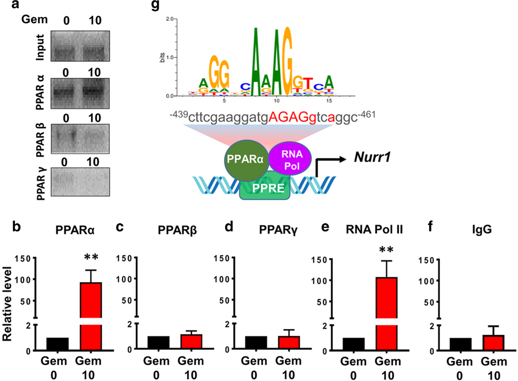
Nuclear receptor related 1 protein (Nurr1) is an important transcription factor required for differentiation and maintenance of midbrain dopaminergic (DA) neurons. Since decrease in Nurr1 function either due to diminished expression or rare mutation is associated with Parkinson's disease (PD), upregulation of Nurr1 may be beneficial for PD. However, such mechanisms are poorly understood. This study underlines the importance of peroxisome proliferator-activated receptor (PPAR)α in controlling the transcription of Nurr1. Our mRNA analyses followed by different immunoassays clearly indicated that PPARα agonist gemfibrozil strongly upregulated the expression of Nurr1 in wild-type, but not PPARα-/-, DA neurons. Moreover, identification of conserved PPRE in the promoter of Nurr1 gene followed by chromatin immunoprecipitation analysis, PPRE luciferase assay, and manipulation of Nurr1 gene by viral transduction of different PPARα plasmids confirmed that PPARα was indeed involved in the expression of Nurr1. Finally, oral administration of gemfibrozil increased Nurr1 expression in vivo in nigra of wild-type, but not PPARα-/-, mice identifying PPARα as a novel regulator of Nurr1 expression and associated protection of DA neurons.

Keywords: Dopaminergic neurons; Gemfibrozil; Nurr1; PPARα; Parkinson’s disease.


Figures

Fig. 1
Fig. 1
Both gemfibrozil and fenofibrate increase the expression of Nurr1 mRNA in MN9D neuronal cells. a, b Differentiated MN9D cells were treated with increasing doses of gemfibrozil for 2 h under serum-free condition followed by semi-quantitative and real-time mRNA expression of Nurr1. A one way ANOVA, followed by post hoc Tukey, determined significant differences between the groups [(F(3,8) = 9.59 > Fc = 3.47) *** = p < 0.0003]. c MN9D cells were treated with 10 μM of gemfibrozil for different time points. Significance of the mean was tested by ANOVA followed by post hoc Tukey to determine significant differences between the groups [(F(4,10) = 13.9 > Fc = 3.57) *** = p < 0.0003; * = p < 0.03]. The effect of fenofibrate on the expression of Nurr1 mRNA was evaluated with a dose-dependent study in MN9D cells as shown by d semi-quantitative and e real-time PCR analyses. Significance of the mean was tested by ANOVA followed by post hoc Tukey to determine significant differences between the groups [(F(3,8) = 13.9 > Fc = 3.57); *** = p < 0.0003; **= p < 0.003; * = p < 0.03]. f Time-sensitive expression of Nurr1 mRNA was measured after treating MN9D cells with 25 μM of fenofibrate. Significance of the mean was tested by ANOVA followed by post hoc Tukey to determine significant differences between the groups [(F(4,10) = 13.9 > Fc = 3.7; *** = p < 0.0003]. Results are mean ± SEM of three independent experiments
Fig. 2
Fig. 2
Gemfibrozil rescues Nurr1 mRNA expression in MPP+-insulted MN9D neuronal cells. a Semi-quantitative and b real-time PCR analyses showing a dose-dependent reduction in Nurr1 mRNA expression in MN9D cells upon MPP+ challenge. Significance of the mean was tested by ANOVA followed by a post hoc Tukey to determine differences between the groups [(F(4,10) = 9.06; > Fc = 3.47 * = p < 0.03]. c Semi-quantitative and d real-time PCR studies indicate that the addition of 10 μM of gemfibrozil significantly upregulates Nurr1 mRNA in MN9D cells pre-treated with 2 μM of MPP+. Significance of the mean was tested by ANOVA by a post hoc Tukey [(F(4,10) = 13.7> Fc = 3.47; **= p < 0.003; * = p < 0.03]
Fig. 3
Fig. 3
Gemfibrozil upregulates the expression of Nurr1 Protein in MN9D neuronal cells. a Double-labeling immunofluorescence studies were adopted to test the effect of 10 μM gemfibrozil on the expression of Nurr1 (anti-rabbit; red or cy5) in TH–ir MN9D cells (anti-mouse; green or cy2). Results were confirmed after three independent experiments. b Percentage of Nurr1 (red) in total number of cells stained DAPI (blue) was quantified in 10 independent fields. Significance of mean tested with unpaired t test that results in (t = 19.2812; p < 0.0001). c Differentiated MN9D cells were treated with 10 μM of gemfibrozil followed by immunolabeling with Nurr1 (scale bar = 20 μm). Mean fluorescence intensities (MFI) of Nurr1 (cy-5)–ir cells were quantified after normalizing signal with background and total area of the cell. An average of 800 cells were analyzed for MFI per treatment group and averaged to generate resultant MFI. An unpaired t test was done to test the significance of mean (t = 6.6814; p < 0.0001). d Representative immunoblot indicates in the expression of Nurr1 protein with increasing concentrations of gemfibrozil. e Densitometric analysis of three independent blots quantifying Nurr1 expression. Results are mean ± SD of three independent experiments. A one-way ANOVA was adopted to test the significance of mean between groups that results in [F(4,14) = 25.53 > Fc = 3.47; *** = p < 0.0003]
Fig. 4
Fig. 4
Involvement of PPARα in gemfibrozil-mediated upregulation of Nurr1 in MN9D neuronal cells. a MN9D cells were transfected with tk-PPREx3-Luc for 24 h followed by the treatment with increasing doses of gemfibrozil (gem) for another 5 h. Then cells were analyzed for PPRE luciferase activity. Results are mean ± SD of three independent experiments with significance of mean tested by one-way ANOVA, which generates [F(5,12) = 101.5 > Fc =3.11; *** = p < 0.0003)]. Semi-quantitative (b) and real-time PCR (c) analyses of Nurr1 gene in MN9D cells transduced with either Y464D-e-GFP (dominant negative mutant of PPARα) or control e-GFP lentiviral construct and treated with 10 μM of gemfibrozil. Results are mean ± SD of three experiments and tested with one-way ANOVA for significance of mean [(F3, 8 = 21.98 > Fc = 4.06); ** = p < 0.003)]. MN9D cells pretreated with different doses of GSK0660 for 1 h were treated with 10 μM of gem for 4 h followed by semi-quantitative (d) and real-time (e) PCR analyses to verify Nurr1 mRNA expression. One-way ANOVA was adopted to test the significance of mean between groups [(F(1,8) = 21.98 > Fc = 4.066); ** = p < 0.003)]. Similar experiments were performed in MN9Dcells with different doses of GW9662. The nurr1 mRNA expression was monitored by semi-quantitative (f) and real-time (g) PCR analyses. Results are mean ± SD of three experiments and tested with one-way ANOVA for significance of mean [F(1,8) = 20.78 > Fc = 3.47); ** = p < 0.003)]
Fig. 5
Fig. 5
Gemfibrozil increases Nurr1 expression in primary DA neurons via PPARα. a Mouse primary DA neurons isolated from WT and PPARα−/−mice were treated with 10 μM gemfibrozil for 18 h followed by doublelabeling for Nurr1 (green) and TH (red). Scale bar = 20 μm. b Nurr1 MFI measurements were performed from an average of 800 cells per group [(F(5,15) = 233.6 > Fc = 3.09); *** = p < 0.0003] from two-way ANOVA].c Representative immunoblots display the effect of gemfibrozil on the expression of Nurr1 in mouse primary DA neuron isolates from both WT (top) and PPARα−/− (bottom) fetuses. d Densitometric analyses of Nurr1 were performed from immunoblots, normalized with β-actin and then plotted. Results are mean ± SD of three independent immunoblots. A two-wayANOVA was adopted to justify the significance of mean in Nurr1 expressionbetween WT neurons and PPARα−/− neurons, [F(1,8) = 6.68 > Fc =5.31); *** = p < 0.0003; * = p < 0.03] for treatment; and [F(1,8) = 73.76>Fc = 5.31); *** = p < 0.0003] for genotype
Fig. 6
Fig. 6
Over-expression of PPARα restores Nurr1-upregulating efficacy of gemfibrozil in PPARα−/− DA neurons. Primary DA neurons harvested from PPARα−/− mice were transduced with lenti-empty vector e-GFP (a, b), lenti-full-length PPARα (c, d), or lenti-Y464D PPARα (e, f) for 24 h. Then cells were treated with 10 μM of gemfibrozil for 18 h followed by immunolabeling for Nurr1 (Cy5; red). MFI of Nurr1-ir was quantified for an average of 800 cells per group (b lenti-eGFP; d lenti-FL PPARα; f lenti-Y464D PPARα). A paired t test was utilized to compare significant differences from the means of these two groups resulting in t = 0.448710; p = 0.66 (b). An unpaired t test analysis was performed to show significant differences between the means (t = 11.8110; = p < 0.0003) (d). Unpaired t test shows t = 0.2910; p = 0.77 (f). All results are mean ± SD of three independent experiments
Fig. 7
Fig. 7
Oral administration of gemfibrozil increases Nurr1 expression in vivo in nigra via PPARα. WT (a, b) and PPARα−/− (c, d) animals were gavage fed 7.5 mg/kg gemfibrozil or 0.5% methyl cellulose (Veh) for 14 days via oral route. After that, nigral sections were double-labeled with anti-mouse TH (cy2) and anti-rabbit Nurr1 (cy5) antibodies. Total n = 5 animals were used per group. e MFI analysis of nigral sections indicates a significant difference between genotypes [(F(3,23) = 233.55> Fc = 3.287); *** = p < 0.0003]. f Representative immunoblots using isolated nigral sections from both WT and PPARα−/− animals in this experimental paradigm. g Densitometric analysis of f indicates a significant increase in Nurr1 protein expression from WT, but not PPARα−/− animals fed GEM as shown by a two-way ANOVA [(F(3, 11) = 20.01 > Fc = 4.757); ***p < 0.0003]
Fig. 8
Fig. 8
Gemfibrozil treatment induces the recruitment of PPARα, but neither PPARβ nor PPARγ, to the Nurr1 gene promoter in MN9D neuronal cells. MN9D cells were treated with 10 μM gemfibrozil for 2 h. After that, we performed chromatin immunoprecipitation (ChIP) followed by semi-quantitative (a) and real-time (b–f) PCR of promoter DNA as described in the “Materials and Methods” section. An ANOVA analysis was used to test the significance of the mean differences between groups and shows [(F(9,29) = 23.13 > Fc = 2.40) ** = p < 0.003)]. Post hoc unpaired t tests indicate a significant difference in samples incubated with antibodies to both PPARα (t = 5.654; p = 0.004) and RNA polymerase II (t = 4.794; p = 0.008). g A schema depicts a detailed map of promoter analysis of nurr1 gene. The map reveals a conserved PPAR-responsive element (PPRE) in the promoter of Nurr1 gene at − 439 to − 461 upstream of the Nurr1 start site on chromosome II

Similar articles

Cited by

References

    1. Roy A, Pahan K (2011) Prospects of statins in Parkinson disease. Neuroscientist 17(3):244–255. 10.1177/1073858410385006 - DOI - PMC - PubMed
    1. Sanyal J, Chakraborty DP, Sarkar B, Banerjee TK, Mukherjee SC, Ray BC, Rao VR (2010) Environmental and familial risk factors of Parkinsons disease: case-control study. Can J Neurol Sci Le journal canadien des sciences neurologiques 37(5):637–642 - PubMed
    1. Pinto F, Mazza S (1971) Psychic symptoms in levodopa treatment of Parkinsons’ disease. II. Riv Neurobiol 17(2):155–159 - PubMed
    1. Smith ML, King J, Dent L, Mackey V, Muthian G, Griffin B, Charlton CG (2014) Effects of acute and sub-chronic L-dopa therapy on striatal L-dopa methylation and dopamine oxidation in an MPTP mouse model of Parkinsons disease. Life Sci 110(1):1–7. 10.1016/j.lfs.2014.05.014 - DOI - PubMed
    1. Dong J, Li S, Mo JL, Cai HB, Le WD (2016) Nurr1-based therapies for Parkinson’s disease. CNS Neurosci Ther 22(5):351–359. 10.1111/cns.12536 - DOI - PMC - PubMed

MeSH terms

LinkOut - more resources