Review
. 2019 Oct 1;40(5):1187-1206.
doi: 10.1210/er.2018-00138.

Bone Marrow Adiposity: Basic and Clinical Implications

Affiliations
Review

Bone Marrow Adiposity: Basic and Clinical Implications

Zachary L Sebo et al. Endocr Rev. .

Abstract

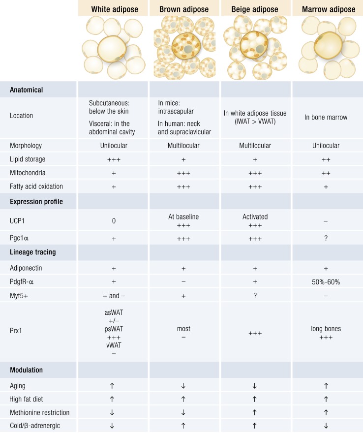
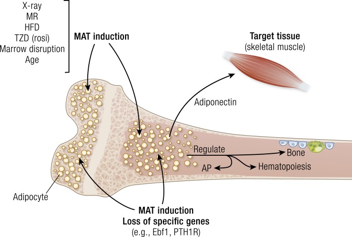
The presence of adipocytes in mammalian bone marrow (BM) has been recognized histologically for decades, yet, until recently, these cells have received little attention from the research community. Advancements in mouse transgenics and imaging methods, particularly in the last 10 years, have permitted more detailed examinations of marrow adipocytes than ever before and yielded data that show these cells are critical regulators of the BM microenvironment and whole-body metabolism. Indeed, marrow adipocytes are anatomically and functionally separate from brown, beige, and classic white adipocytes. Thus, areas of BM space populated by adipocytes can be considered distinct fat depots and are collectively referred to as marrow adipose tissue (MAT) in this review. In the proceeding text, we focus on the developmental origin and physiologic functions of MAT. We also discuss the signals that cause the accumulation and loss of marrow adipocytes and the ability of these cells to regulate other cell lineages in the BM. Last, we consider roles for MAT in human physiology and disease.


Figures

Figure 1.
Figure 1.
Phenotypes of white, brown, beige, and marrow adipocytes. A plus sign indicates that the gene/protein is expressed; a minus sign indicates that the gene/protein is not expressed; a plus/minus sign indicates a mixed population of cells; a question mark indicates that expression is unknown; an up arrow indicates increased numbers of cells; and a down arrow indicates decreased numbers of cells. Note that the VWAT includes perigonadal, retroperitoneal, and mesenteric WAT. asWAT, anterior subcutaneous WAT; psWAT, posterior subcutaneous WAT; vWAT, visceral WAT.
Figure 2.
Figure 2.
(Left) The tibia from B6 or C3H mice. The tibia was isolated, fixed in 10% formalin, and decalcified for 20 d in 4% EDTA. The bones were washed and stained with osmium tetroxide for 48 h, washed, and MAT was imaged by micro-CT. The tibia from B6 mice had low MAT in the proximal tibia with significant MAT visible in the distal tibia and above the growth plate. In contrast, the tibia from C3H mice had extensive MAT throughout the medullary canal. Even the fibula can be imaged in the C3H mice because of MAT filling the marrow space. (Right) A 5-μm thin histologic section of osmium-stained distal tibia (below the tibia–fibular junction) is shown. Marrow adipocytes fill the BM space from endosteum to endosteum.
Figure 3.
Figure 3.
(Top) Schematic of the mT/mG double reporter system. (Bottom) Adiponectin-cre:mT/mG double reporter mice were fed a rosiglitazone-containing diet for 6 wk. The femur was isolated and the femoral head was removed. A 20-gauge needle was slipped down the medullary canal and punched out through the distal epiphysis. The needle was attached to a 5-cc syringe, and the BM was extruded onto a microscope slide. The BM was stained with LipidTox (a lipophilic dye), washed, immersed in Fluoromount, and coverslipped, and fluorescence was viewed by confocal microscopy. LipidTox (left) was used to identify marrow adipocytes. All of the marrow adipocytes were traced with eGFP, (center left), whereas none was with dTomato+ (center right). These data indicate that all marrow adipocytes and their progenitors express adiponectin.
Figure 4.
Figure 4.
Marrow adipogenesis can be increased in long bones of B6 mice by a variety of external inducers, including x-irradiation, feeding mice a diet containing rosiglitazone, a methionine-restricted diet, or a high-fat diet, or physically disrupting the BM. Alternatively, loss of certain genes [e.g., early B cell factor 1 (Ebf1), PTH/PTHrP receptor (PTH1R)] can also result in increased marrow adipogenesis; however, this occurs in the absence of any exogenous induction. This increased marrow adipogenesis occurs above the growth plate, in the secondary center of ossification, and just below the growth plate, in the intratrabecular spaces. Adipocytes can extend down the medullary canal through the metaphysis and into the diaphysis. Nerves that intercalate the marrow channel can contact these adipocytes directly. Marrow adipocytes may regulate bone cells (osteoblast and osteoclast precursors) as well as hematopoietic cells, either by direct cell contact or the secretion of adipokines in a paracrine manner. It is also possible that mature adipocytes regulate the development of adipocyte precursors in the marrow. Lastly, this ability of marrow adipocytes to regulate other cells is not restricted to BM. Secretion of adipokines, such as adiponectin, can regulate cells outside the BM in an endocrine manner. AP, adipocyte progenitor; HFD, high-fat diet; MR, methionine-restricted diet; rosi, rosiglitazone; TZD, thiazolidinedione; X-ray, x-irradiation.

Similar articles

Cited by

References

    1. Gregory CA, Prockop DJ, Spees JL. Non-hematopoietic bone marrow stem cells: molecular control of expansion and differentiation. Exp Cell Res. 2005;306(2):330–335. - PubMed
    1. Bruder SP, Fink DJ, Caplan AI. Mesenchymal stem cells in bone development, bone repair, and skeletal regeneration therapy. J Cell Biochem. 1994;56(3):283–294. - PMC - PubMed
    1. Gimble JM, Robinson CE, Wu X, Kelly KA. The function of adipocytes in the bone marrow stroma: an update. Bone. 1996;19(5):421–428. - PubMed
    1. Cawthorn WP, Scheller EL, Learman BS, Parlee SD, Simon BR, Mori H, Ning X, Bree AJ, Schell B, Broome DT, Soliman SS, DelProposto JL, Lumeng CN, Mitra A, Pandit SV, Gallagher KA, Miller JD, Krishnan V, Hui SK, Bredella MA, Fazeli PK, Klibanski A, Horowitz MC, Rosen CJ, MacDougald OA. Bone marrow adipose tissue is an endocrine organ that contributes to increased circulating adiponectin during caloric restriction. Cell Metab. 2014;20(2):368–375. - PMC - PubMed
    1. Veldhuis-Vlug AG, Rosen CJ. Clinical implications of bone marrow adiposity. J Intern Med. 2018;283(2):121–139. - PMC - PubMed

Publication types