

Distorted gaze direction input to attentional priority map in spatial neglect
- PMID: 31128129
- PMCID: PMC6667735
- DOI: 10.1016/j.neuropsychologia.2019.05.017
Distorted gaze direction input to attentional priority map in spatial neglect
Abstract
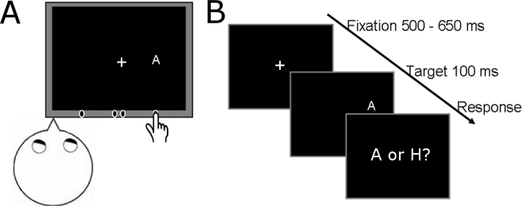
A contribution of the gaze signals to the attention imbalance in spatial neglect is presumed. Direct evidence however, is still lacking. Theoretical models for spatial attention posit an internal representation of locations that are selected in the competition for neural processing resources - an attentional priority map. Following up on our recent research showing an imbalance in the allocation of attention after an oculoproprioceptive perturbation in healthy volunteers, we investigated here whether the lesion in spatial neglect distorts the gaze direction input to this representation. Information about one's own direction of gaze is critical for the coordinate transformation between retinotopic and hand proprioceptive locations. To assess the gaze direction input to the attentional priority map, patients with left spatial neglect performed a cross-modal attention task in their normal, right hemispace. They discriminated visual targets whose location was cued by the patient's right index finger hidden from view. The locus of attention in response to the cue was defined as the location with the largest decrease in reaction time for visual discrimination in the presence vs. absence of the cue. In two control groups consisting of healthy elderly and patients with a right hemisphere lesion without neglect, the loci of attention were at the exact location of the cues. In contrast, neglect patients allocated attention at 0.5⁰-2⁰ rightward of the finger for all tested locations. A control task using reaching to visual targets in the absence of visual hand feedback ruled out a general error in visual localization. These findings demonstrate that in spatial neglect the gaze direction input to the attentional priority map is distorted. This observation supports the emerging view that attention and gaze are coupled and suggests that interventions that target gaze signals could alleviate spatial neglect.
Keywords: Attention; Coordinate transformation; Spatial neglect; Stroke.
Copyright © 2019 The Authors. Published by Elsevier Ltd.. All rights reserved.
Figures


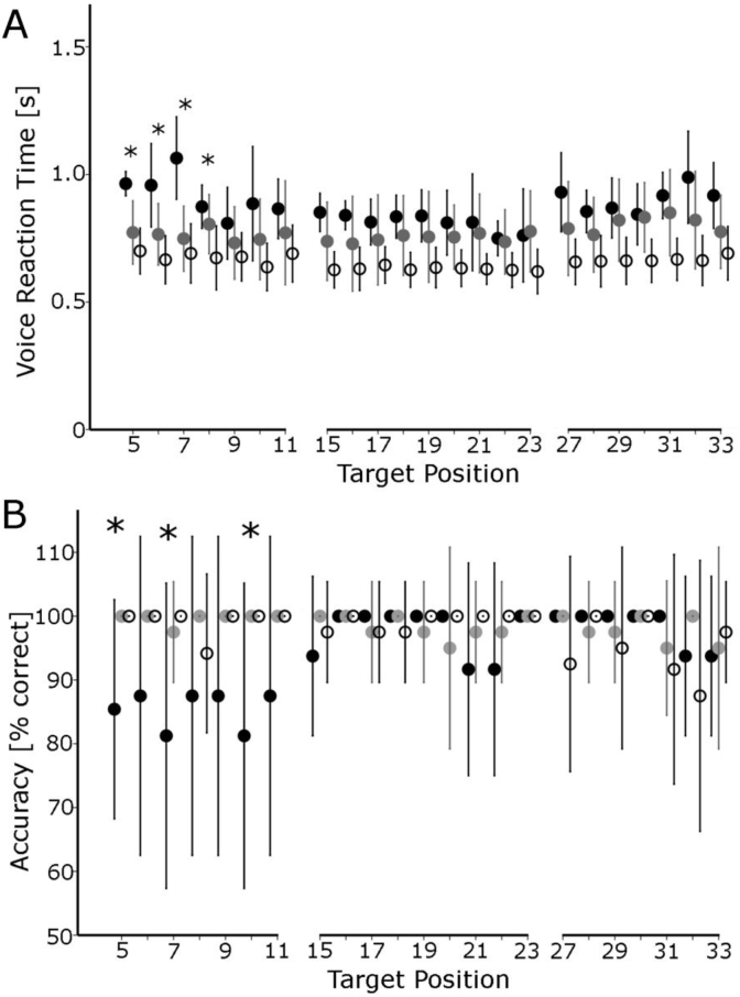
 ) and healthy controls (◯) for targets located further than 11° to the right of the body midline (error bars show one standard deviation, * denotes p< 0.05, independent samples t-tests, neglect vs. control, for either control group).
 ) and the absence of the finger position cue (
 ). The arrow indicates the location of the cue in trials when the cue was present. NEG – patients with spatial neglect, HEC – healthy elderly controls, PCG – patient control group. Error bars show one standard deviation.
 , ASG-
 , SG-
, SG- , UH-
, UH- ; PCG - patient control group; HEC – healthy elderly controls.
 , ASG-, SG-, UH-; PCG-patient control group; HEC – healthy elderly controls.
Similar articles
-
Value-driven attentional capture in neglect.Cortex. 2018 Dec;109:260-271. doi: 10.1016/j.cortex.2018.09.015. Epub 2018 Oct 5. Cortex. 2018. PMID: 30391880
-
Acute visual neglect and extinction: distinct functional state of the visuospatial attention system.Brain. 2011 Nov;134(Pt 11):3310-25. doi: 10.1093/brain/awr220. Epub 2011 Sep 23. Brain. 2011. PMID: 21948940
-
Rightward exogenous attentional shifts impair perceptual memory of spatial locations in patients with left unilateral spatial neglect.Cortex. 2020 Jan;122:187-197. doi: 10.1016/j.cortex.2019.10.002. Epub 2019 Oct 30. Cortex. 2020. PMID: 31761271
-
The contribution of spatial remapping impairments to unilateral visual neglect.Neurosci Biobehav Rev. 2004 Apr;28(2):181-200. doi: 10.1016/j.neubiorev.2004.03.003. Neurosci Biobehav Rev. 2004. PMID: 15172763 Review.
-
[Unilteral Spatial Neglect: Clinical Manifestations and Neural Correlates].Brain Nerve. 2024 Jun;76(6):749-754. doi: 10.11477/mf.1416202673. Brain Nerve. 2024. PMID: 38853504 Review. Japanese.
Cited by
-
Seeing without a Scene: Neurological Observations on the Origin and Function of the Dorsal Visual Stream.J Intell. 2024 May 11;12(5):50. doi: 10.3390/jintelligence12050050. J Intell. 2024. PMID: 38786652 Free PMC article.
-
Visual-Spatial Search in Neglect Syndrome as a Function of the Number of Stimuli in the Hemifields.Healthcare (Basel). 2024 Nov 28;12(23):2387. doi: 10.3390/healthcare12232387. Healthcare (Basel). 2024. PMID: 39685009 Free PMC article.
-
Prism adaptation combined with eye movement training for unilateral spatial neglect after stroke: Study protocol for a single-blind prospective, randomized controlled trial.Front Neurol. 2023 Jan 5;13:1081895. doi: 10.3389/fneur.2022.1081895. eCollection 2022. Front Neurol. 2023. PMID: 36686538 Free PMC article.
References
-
- Albert M. A simple test of visual neglect. Neurology. 1973;23:658–664. - PubMed
Publication types
MeSH terms
LinkOut - more resources
Full Text Sources
