. 2019 Oct:100:101651.
doi: 10.1016/j.jchemneu.2019.101651. Epub 2019 May 22.

Noradrenergic terminal density varies among different groups of hypoglossal premotor neurons

Affiliations

Noradrenergic terminal density varies among different groups of hypoglossal premotor neurons

Caroline E Boyle et al. J Chem Neuroanat. 2019 Oct.

Abstract

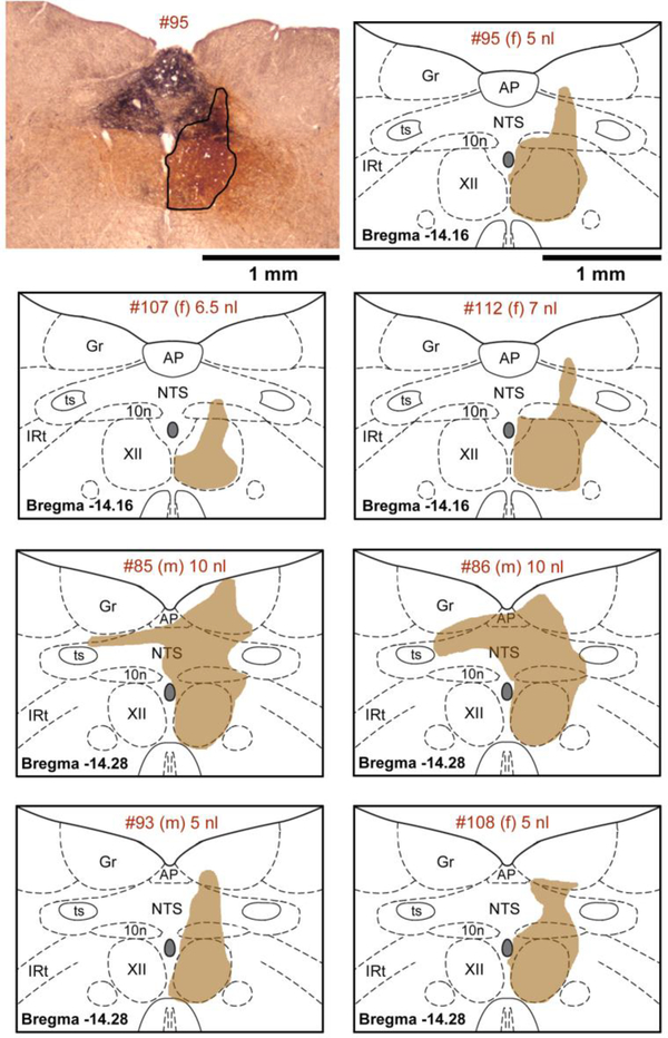
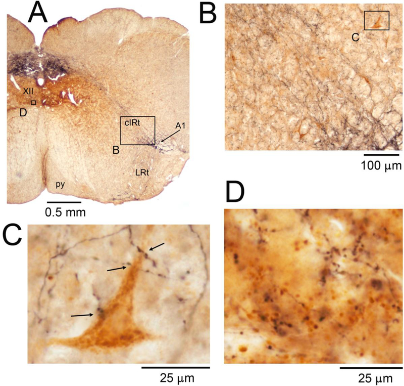
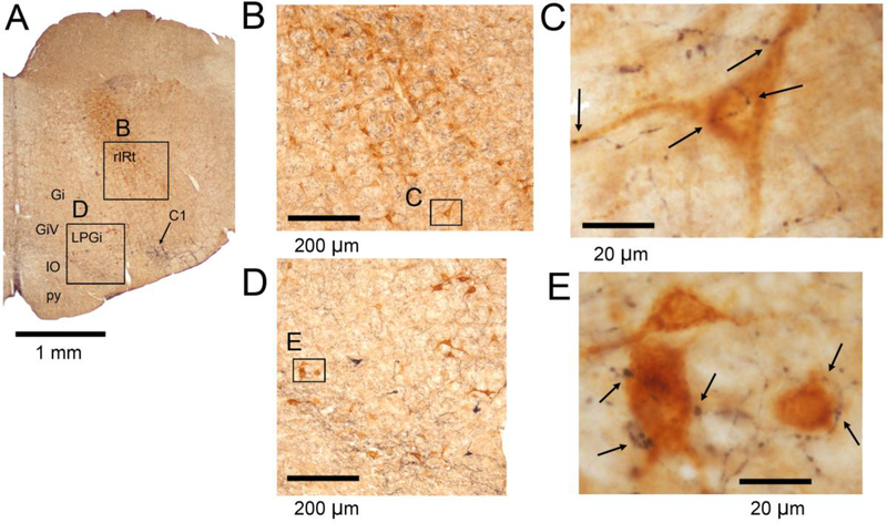
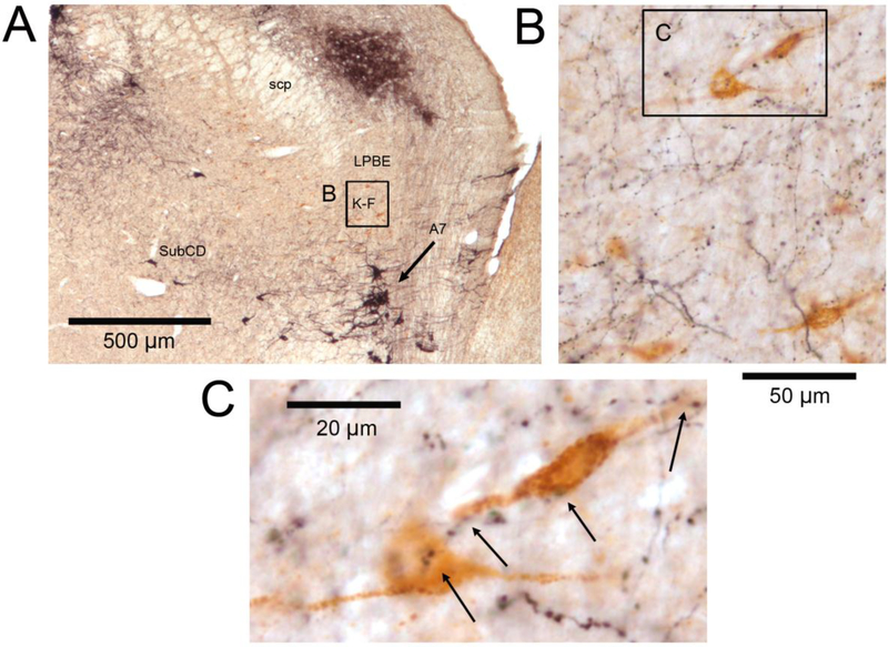
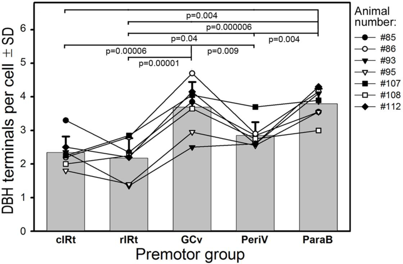
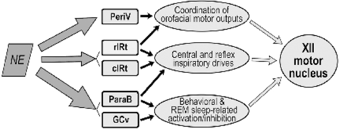
In obstructive sleep apnea (OSA) patients, contraction of the muscles of the tongue is needed to protect the upper airway from collapse. During wakefulness, norepinephrine directly excites motoneurons that innervate the tongue and other upper airway muscles but its excitatory effects decline during sleep, thus contributing to OSA. In addition to motoneurons, NE may regulate activity in premotor pathways but little is known about these upstream effects. To start filling this void, we injected a retrograde tracer (beta-subunit of cholera toxin-CTb; 5-10 nl, 1%) into the hypoglossal (XII) motor nucleus in 7 rats. We then used dual immunohistochemistry and brightfield microscopy to count dopamine beta-hydroxylase (DBH)-positive axon terminals closely apposed to CTb cells located in five anatomically distinct XII premotor regions. In different premotor groups, we found on the average 2.2-4.3 closely apposed DBH terminals per cell, with ˜60% more terminals on XII premotor neurons located in the ventrolateral pontine parabrachial region and ventral medullary gigantocellular region than on XII premotor cells of the rostral or caudal intermediate medullary reticular regions. This difference suggests stronger control by norepinephrine of the interneurons that mediate complex behavioral effects than of those mediating reflexes or respiratory drive to XII motoneurons.

Keywords: Pons; Reticular formation; Sleep apnea; Swallowing; Tongue; motor control.


Conflict of interest statement

Declaration of conflict of interest: none.

Figures

Figure 1:
Figure 1:
Location of the tracer injection sites. The upper left panel shows a microphotograph of staining (outlined) following unilateral tracer injection into the center of the right Xii nucleus (5 nl of 1 % beta subunit of cholera toxin - CTb). The tracer covers the entire XII nucleus on one side with limited diffusion ventral and lateral to the nucleus and a moderate spill into the dorsal nucleus of the vagus (10n) and nucleus of the solitary tract (NTS). The dense black dopamine beta hydroxylase (DBH) staining marks the characteristic aggregation of noradrenergic and adrenergic cells and fibers dorsal to the XII nucleus. The top panel on the right shows CTb spread in the same rat (#95) redrawn and superimposed onto the closest standard medullary cross-sections derived from a rat brain atlas [Paxinos & Watson, 2007]. The six panels below the top row show in the same format CTb distribution in the remaining 6 rats of this study. The identifying number for each rat, its sex (female (f) or male (m)) and the volume of tracer injected are specified at the top of each panel. Abbreviations: AP – area postrema; Gr – gracile nucleus; IRt – intermediate medullary reticular region; ts – solitary tract.
Figure 2:
Figure 2:
DBH terminals on and near retrogradely labeled cIRt XII PMNs and, for comparison, DBH terminals in the ventromedial region of the XII motor nucleus. A: low magnification image of the brain section containing the cIRt region. Arrow points to a cluster of DBH-stained cells representing NE neurons of the A1 group. Other abbreviations for local anatomical landmarks: LRt – lateral reticular nucleus, py – pyramidal tract. Boxes mark the locations of the enlarged details shown in panels B-D. B: the cIRt region ipsilateral to the tracer injection site shown at an intermediate magnification suitable for visualization of labeled XII PMNs (brown). C: one of the labeled XII PMNs of panel B illustrated at the high magnification used to identify DBH-stained terminals closely apposed to the cell bodies and proximal dendrites of retrogradely labeled XII PMNs; arrows show the sites identified as putative points of close appositions. D: DBH terminals in the ventromedial region of the XII nucleus. DBH fibers and terminals are shown in black; the brown background represents residual diffusion of the retrograde tracer into the side opposite to the injection (see the small box in the left XII nucleus in A). Comparison of panels C and D, both shown at the same magnification, illustrates that DBH terminal density is much higher in the ventromedial XII nucleus than in the cIRt region.
Figure 3:
Figure 3:
DBH terminals in the rIRt and GCvXII premotor regions. A: low magnification image of the brain section from which the specific examples were derived. Arrow points to a cluster of DBH-stained cells which at this level mainly represent adrenergic neurons of the C1 group. Other abbreviations for local anatomical landmarks: Gi – gigantocellular reticular nucleus, GiV – ventral gigantocellular reticular nucleus, IO – inferior olive, LPGi – lateral paragigantocellular nucleus, py – pyramidal tract. B and C: rIRt XII PMNs (brown) shown at increasing magnifications. The image in C was obtained at the magnification used to identify DBH-stained terminals closely apposed to cell bodies and proximal dendrites of identified XII PMNs; arrows point to such a putative synaptic contacts. D and E: GCv XII PMNs shown at increasing magnifications using the same format as in B and C.
Figure 4:
Figure 4:
DBH terminals on PeriV XII PMNs. A: PeriV XII PMNs are characteristically distributed along the perimeter of the trigeminal motor nucleus (Mo5 – dashed line). The black-stained area in the upper left corner represents the locus coeruleus (LC). Other abbreviations for local anatomical landmarks: SubCD – dorsal part of the subcoeruleus region, SubCV – ventral part of the subcoeruleus region. B and C: selected PeriV XII PMNs (brown) shown at progressively higher magnification. The relatively dense black-stained axonal swellings seen in the lower left portion of panel B represent DBH-stained fibers and terminals located within the Mo5 proper. Arrow in C shows a site of multiple close appositions between a dendrite of a retrogradely labeled PeriV XII premotor neuron and DBH-stained boutons.
Figure 5:
Figure 5:
DBH terminals on ParaB XII PMNs. A: low-magnification image of the section through the dorsolateral pons. DBH-staining (black) marks a plexus of fibers in the dorsolateral parabrachial region and a dense cluster of cells belonging to the pontine A7 group (arrow). Visible are also scattered DBH-positive cells of the dorsal subcoeruleus region (SubCD). Other abbreviations for local anatomical landmarks: K-F – Kölliker-Fuse nucleus, LPBE – external portion of the lateral parabrachial region, scp – superior cerebellar peduncle. The box marked B and the corresponding panel B contain numerous XII PMNs retrogradely labeled with CTb (brown). C: enlarged image of a pair of XII PMNs of the K-F nucleus enclosed in the box in B. Arrows point to DBH-stained axonal swellings closely apposed to cell bodies and proximal dendrites of these two XII PMNs.
Figure 6:
Figure 6:
Average numbers of closely apposed DBH-stained boutons on XII PMNs belonging to different premotor groups. Bars show the mean and standard deviation values for the 7 rats of the present study. The superimposed lines represent individual means obtained from 20 retrogradely labeled XII PMNs selected from each premotor group in individual rats; data from the same animal are marked by the same symbol and are connected by lines. The overall order of closely apposed terminal counts was: cIRt ≈ rIRt < PeriV < GCv ≈ ParaB, and the means for individual rats well approximated this order. Pairwise comparisons among the premotor groups conducted with correction for multiple comparisons revealed many highly significant differences.
Figure 7:
Figure 7:
Scheme of NE innervation of different groups of XII PMNs in relation to their prevailing functions in the control of XII motoneurons. Thickness of the arrows on the left side is proportional to the relative density of DBH-stained presynaptic boutons found in our study to be closely apposed to XII PMNs (cf. Fig. 6). We suggest that NE plays a particularly prominent role in the regulation of excitability in those XII PMNs that mediate behavioral effects related to sleep-wake states and various forebrain-originating commands during wakefulness (GCv and ParaB). In contrast, NE-dependent modulation of the premotor pathways that mediate central and reflex inspiratory drives is modest (rIRt and cIRt).

Similar articles

Cited by

References

    1. Agnati LF, Zoli M, Stromberg I, & Fuxe K (1995). Intercellular communication in the brain: wiring versus volume transmission. Neuroscience 69, 711–726. - PubMed
    1. Aldes LD & Boone TB (1985). Organization of projections from the principal sensory trigeminal nucleus to the hypoglossal nucleus in the rat: an experimental light and electron microscopic study with axonal tracer techniques. Exp Brain Res 59, 16–29. - PubMed
    1. Aldes LD, Chapman ME, Chronister RB, & Haycock JW (1992). Sources of noradrenergic afferents to the hypoglossal nucleus in the rat. Brain Res Bull 29, 931–942. - PubMed
    1. Aston-Jones G & Bloom FE (1981). Activity of norepinephrine-containing locus coeruleus neurons in behaving rats anticipates fluctuations in the sleep-waking cycle. J Neurosci 1, 876–886. - PMC - PubMed
    1. Aston-Jones G, Rajkowski J, Kubiak P, Valentino RJ, & Shipley MT (1996). Role of the locus coeruleus in emotional activation. Progr Brain Res 107, 379–402. - PubMed

Publication types

LinkOut - more resources