Comparative Study
. 2019 May 24;11(10):2968-2997.
doi: 10.18632/aging.101947.

The small non-coding RNA profile of mouse oocytes is modified during aging

Affiliations
Comparative Study

The small non-coding RNA profile of mouse oocytes is modified during aging

Bettina P Mihalas et al. Aging (Albany NY). .

Abstract

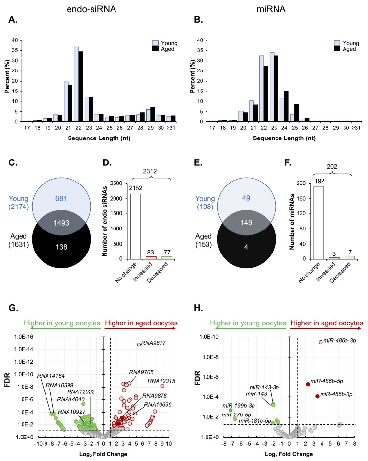
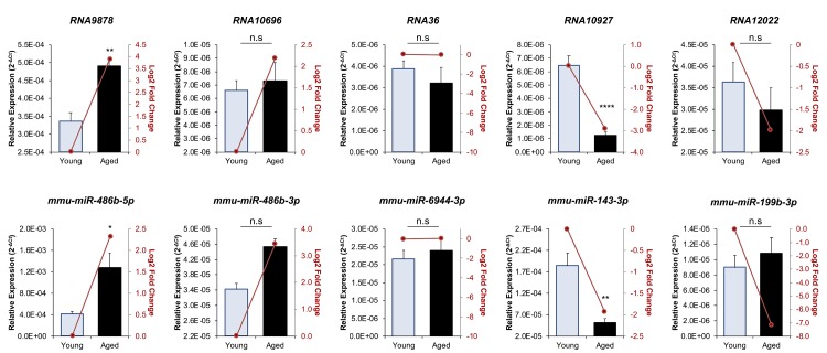
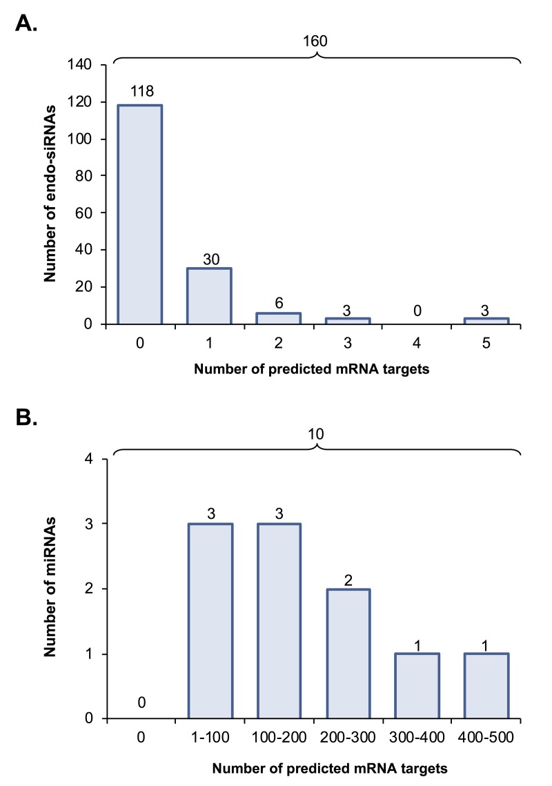
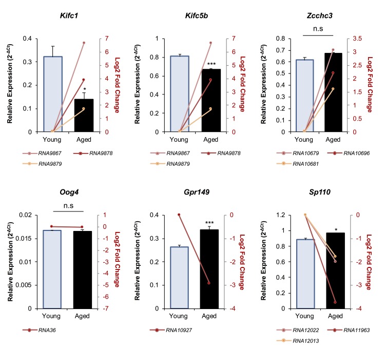
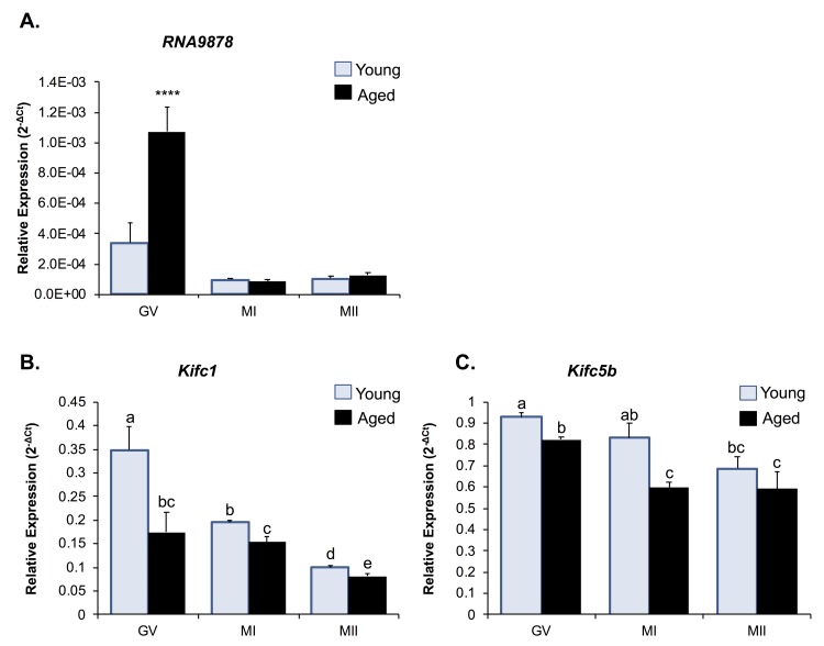
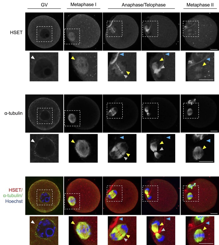
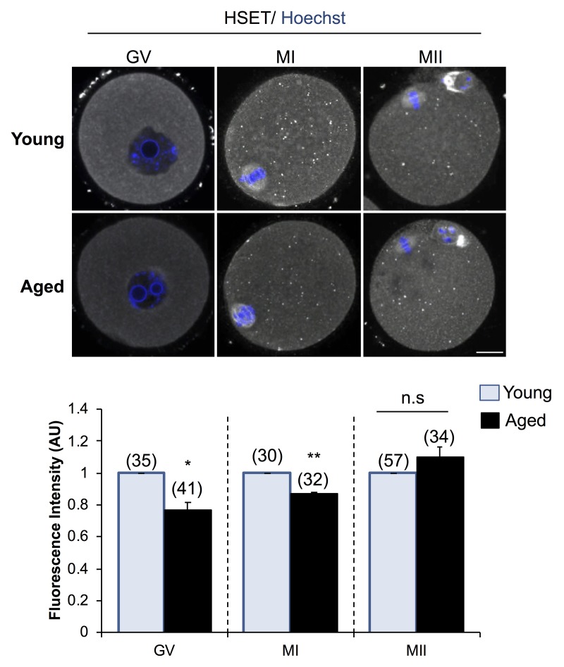
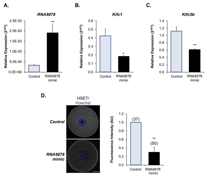
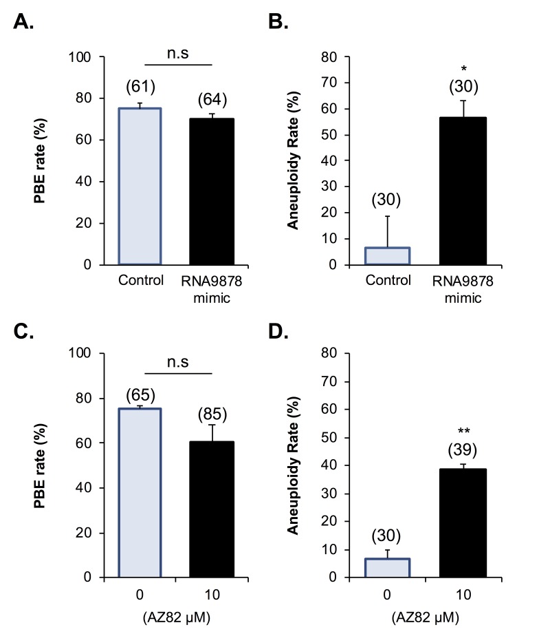
Oocytes are reliant on messenger RNA (mRNA) stores to support their survival and integrity during a protracted period of transcriptional dormancy as they await ovulation. Oocytes are, however, known to experience an age-associated alteration in mRNA transcript abundance, a phenomenon that contributes to reduced developmental potential. Here we have investigated whether the expression profile of small non-protein-coding RNAs (sRNAs) is similarly altered in aged mouse oocytes. The application of high throughput sequencing revealed substantial changes to the global sRNA profile of germinal vesicle stage oocytes from young (4-6 weeks) and aged mice (14-16 months). Among these, 160 endogenous small-interfering RNAs (endo-siRNAs) and 10 microRNAs (miRNAs) were determined to differentially accumulate within young and aged oocytes. Further, we revealed decreased expression of two members of the kinesin protein family, Kifc1 and Kifc5b, in aged oocytes; family members selectively targeted for expression regulation by endo-siRNAs of elevated abundance. The implications of reduced Kifc1 and Kifc5b expression were explored using complementary siRNA-mediated knockdown and pharmacological inhibition strategies, both of which led to increased rates of aneuploidy in otherwise healthy young oocytes. Collectively, our data raise the prospect that altered sRNA abundance, specifically endo-siRNA abundance, could influence the quality of the aged oocyte.

Keywords: aneuploidy; endo-siRNA; kinesin; maternal aging; meiosis; miRNA; oocyte; small non-coding RNA.


Conflict of interest statement

CONFLICTS OF INTEREST: The authors declare that there is no conflict of interest that could be perceived as prejudicing the impartiality of the research reported.

Figures

Figure 1
Figure 1
Endo-siRNA and miRNA signatures of young and aged oocytes. Following filtering and normalization, sRNA reads were mapped to known endo-siRNA and miRNA from RNAcentral sequence database (August 2017, RNAcentral) (https://rnacentral.org/) to explore changes in the endo-siRNA and miRNA landscape between young and aged oocytes. (A) Endo-siRNA and (B) miRNA sequence length distribution between young and aged GV oocytes. (C) Venn diagram illustrating the total number of endo-siRNA and (E) miRNA identified in young and aged oocytes. (D) Graphical representation of the proportion of endo-siRNAs and (F) miRNAs, identified as being expressed at equivalent levels (unchanged) or that were up- or down-regulated (increased or decreased, respectively) between young and aged oocytes. (G) Volcano plots depicting log2-fold changes (x-axis) and false discovery rate (FDR; y-axis) of endo-siRNAs and (H) miRNAs between young and aged oocytes. Solid dots represent sRNAs that were selected for RT-qPCR validation. Counts of ≥ 10 reads aligning to a specific sRNA was used as a threshold for a positive endo-siRNA or miRNA identification in this study. sRNAs experiencing a threshold of ≥ ± 2-fold change and false discovery rate of < 0.05 were considered as being differently expressed between young and aged oocytes.
Figure 2
Figure 2
RT-qPCR validation of miRNA and endo-siRNA abundance in young and aged oocytes. To validate RNA-Seq data, five endo-siRNAs and five miRNAs were selected for quantification using RT-qPCR. Candidate sRNA included four representatives with increased expression in aged oocytes (RNA9878, RNA10696, mmu-miR-486b-5p, and mmu-miR-486b-3p), four with decreased expression in aged oocytes (RNA10927, RNA12022, mmu-miR-143-3p, and mmu-miR-199b-3p), and two that remained at equivalent levels (RNA36 and mmu-miR-6944-3p). cDNA generation and RT-qPCR experiments were performed in technical and biological triplicate, with each biological replicate comprising 10 oocytes randomly sampled from a pool of oocytes isolated from three animals. The U6 small nuclear RNA was employed as an endogenous control to normalize the expression levels of target sRNAs. Values are shown as a mean of all replicates ± SEM. Statistical analyses were performed using Student’s t-test, * p < 0.05, ** p < 0.01, **** p < 0.0001. Log2 fold changes based on RNA-Seq are represented as dark red line graphs while the relative abundance (2-ΔCt) of each sRNA determined by RT-qPCR is represented by columns.
Figure 3
Figure 3
Predicted mRNA targets of differentially expressed endo-siRNA and miRNA. Endo-siRNA and miRNA sequences determined to be differentially expressed between young and aged oocytes (i.e. threshold of ≥ 2-fold change and false discovery rate of < 0.05) were surveyed using NCBI and miRDB target prediction algorithms, respectively to identify putative mRNA targets. The number of mRNA targets of (A) endo-siRNA and (B) miRNA predicted on the basis of this analysis are depicted.
Figure 4
Figure 4
The expression of putative mRNA target genes of endo-siRNAs differentially abundant in young and aged oocytes. To verify that the changes in endo-siRNA abundance led to altered target gene expression in aged oocytes, six mRNAs were selected for RT-qPCR assessment. Candidate mRNAs included three representatives with targeting endo-siRNAs whose abundance was upregulated in aged oocytes (Kifc1, Kifc5b, and Zcchc3), two that were potentially targeted by downregulated endo-siRNAs (Gpr149 and Sp110) and one mRNA potentially targeted by an endo-siRNA with unchanged abundance in mRNA potentially targeted by an endo-siRNA with unchanged abundance in between young and aged oocytes (Oog4). cDNA generation and RT-qPCR experiments were performed in technical and biological triplicate, with each biological replicate comprising 10 oocytes randomly sampled from a pool of oocytes isolated from three animals. Ppia was employed as an endogenous control to normalize the expression levels of target mRNAs. Values are shown as a mean of all replicates ± SEM. Statistical analyses were performed using Student’s t-test, * p < 0.05, *** p < 0.001. Log2 fold changes based on RNA-Seq are represented as pink, red, and orange line graphs while the relative abundance (2-ΔCt) of each sRNA determined by RT-qPCR is represented by columns.
Figure 5
Figure 5
Expression of Kifc1 and Kifc5b targeting endo-siRNA and mRNA in young and aged oocytes throughout meiosis. RT-qPCR of young and aged GV, MI and MII stage oocytes was utilized to determine the impact of increased Kifc1 and Kifc5b targeting endo-siRNAs on Kifc1 and Kifc5b mRNA expression during meiosis. (A) RT-qPCR of RNA9878 in young and aged GV, MI, and MII oocytes (ANOVA; p > 0.0001). RT-qPCR of (B) Kifc1 (ANOVA; p ≥ 0.0266) and (C) Kifc5b (ANOVA; p ≥ 0.0128) in young and aged GV, MI, and MII oocytes. cDNA generation and RT-qPCR experiments were performed in technical and biological triplicate, with each biological replicate comprising 10 oocytes randomly sampled from a pool of oocytes isolated from three animals. The U6 small nuclear RNA and Ppia were employed as endogenous control to normalize the expression levels of target sRNAs and mRNAs, respectively. Values are shown as a mean of all replicates ± SEM. **** p < 0.0001.
Figure 6
Figure 6
HSET expression throughout oocyte meiosis. Immunofluorescence analysis was utilized to track the spatial profile of HSET distribution in GV, MI, anaphase I/telophase I, and MII stage oocytes. Inserts highlight the localization of HSET to the nuclear envelope (white arrowheads), microtubules (yellow arrowheads), between the chromosomes (grey arrowheads), and at the partitioning of the polar body (blue arrowheads). Oocytes were dual labelled with anti-HSET and anti-α-tubulin antibodies followed by either appropriate goat anti-rabbit 633 Alexa Fluor (red) or goat anti-mouse 488 Alexa Fluor-conjugated (green) secondary antibodies, respectively. Oocytes were then counterstained with the nuclear stain Hoechst 33342 (blue) and viewed using confocal microscopy. Scale bar = 20 μm. These experiments were repeated using three independent biological replicates, with each comprising a minimum of 10 oocytes, and representative images are shown.
Figure 7
Figure 7
Expression of HSET protein in young and aged oocytes. Immunofluorescence analysis of HSET in young and aged GV, MI, and MII stage oocytes was utilized to determine whether an age-related decrease in Kifc1 and Kifc5b transcript abundance presages an equivalent decrease in protein abundance. Oocytes were labelled with anti-HSET antibodies followed by goat anti-rabbit 633 Alexa Fluor-conjugated (grey) secondary antibodies. Oocytes were then counterstained with the nuclear stain Hoechst 33342 (blue) and viewed using confocal microscopy. Scale bar = 20 μm. These experiments were repeated using three independent biological replicates, with each comprising a minimum of 10 oocytes, and representative images are shown. The immunofluorescence intensity of the entire cell was calculated for each oocyte as described in the Materials and Methods, and the mean of each biological replicate values ± SEM are shown. Statistical analyses were performed using Student’s t-test, * p < 0.05, ** p < 0.01. AU, arbitrary units.
Figure 8
Figure 8
Examination of endo-siRNA target gene knockdown. To confirm the functional significance of the endo-siRNA, RNA9878, GV oocytes were microinjected with a synthetic RNA9878 small RNA mimic or a non-targeting negative control. (A) To confirm successful microinjection of the RNA9878 mimic, the expression of RNA9878 was assessed via RT-qPCR immediately after injection. At 24 h post-injection, the relative levels of (B) Kifc1 and (C) Kifc5b were assessed in non-targeting and RNA9878 mimic injected oocytes via RT-qPCR. The U6 small nuclear RNA and Ppia were employed as endogenous controls to normalize the expression levels of the target endo-siRNA and mRNAs, respectively. (D) Non-targeting and RNA9878 mimic injected oocytes were then fixed and the expression of HSET was examined. Oocytes were labelled with anti-HSET antibodies followed by goat anti-rabbit 633 Alexa Fluor-conjugated (grey) secondary antibodies. Oocytes were then counterstained with the nuclear stain Hoechst 33342 (blue) and viewed using confocal microscopy. Scale bar = 20 μm. RT-qPCR experiments were performed in technical and biological triplicate, with each biological replicate comprising 10 oocytes randomly sampled from a pool of oocytes isolated from three animals. Similarly, immunofluorescence experiments were repeated on three biological replicates, with each replicate comprising a minimum of 10 oocytes randomly sampled from a pool of oocytes isolated from three animals. Values are shown as a mean of each replicate ± SEM. Statistical analyses were performed using Student’s t-test, * p < 0.05, ** p < 0.01. AU, arbitrary units.
Figure 9
Figure 9
Biological impact of endo-siRNA target gene knockdown and HSET inhibition. To confirm the biological significance of the endo-siRNA-mediated knockdown, RNA9878, GV oocytes were microinjected with a synthetic RNA9878 small RNA mimic, a non-targeting negative control, or subjected to pharmacological HSET inhibition (AZ82; 10 μM). (A) Non-targeting, RNA9878 mimic injected, and HSET inhibited oocytes were then subject to IVM to MII where (A, C) PBE and (B, D) aneuploidy rates were recorded. siRNA-mediated knockdown and HSET inhibition experiments were repeated on three biological replicates, with each replicate comprising a minimum of 20 oocytes randomly sampled from a pool of oocytes isolated from three animals. Values are shown as a mean of each replicate ± SEM. * Statistical analyses were performed using Student’s t-test, p < 0.05, ** p < 0.01. AU, arbitrary units.

Similar articles

Cited by

References

    1. Pan H, Ma P, Zhu W, Schultz RM. Age-associated increase in aneuploidy and changes in gene expression in mouse eggs. Dev Biol. 2008; 11 10 2968 2997: 397–407. 10.1016/j.ydbio.2008.01.048 - DOI - PMC - PubMed
    1. Duncan FE, Gerton JL. Mammalian oogenesis and female reproductive aging. Aging (Albany NY). 2018; 10:162–63. 10.18632/aging.101381 - DOI - PMC - PubMed
    1. Nabti I, Grimes R, Sarna H, Marangos P, Carroll J. Maternal age-dependent APC/C-mediated decrease in securin causes premature sister chromatid separation in meiosis II. Nat Commun. 2017; 8:15346. 10.1038/ncomms15346 - DOI - PMC - PubMed
    1. Abe K, Inoue A, Suzuki MG, Aoki F. Global gene silencing is caused by the dissociation of RNA polymerase II from DNA in mouse oocytes. J Reprod Dev. 2010; 56:502–07. 10.1262/jrd.10-068A - DOI - PubMed
    1. Jukam D, Shariati SA, Skotheim JM. Zygotic Genome Activation in Vertebrates. Dev Cell. 2017; 42:316–32. 10.1016/j.devcel.2017.07.026 - DOI - PMC - PubMed

Publication types