

Improved sensitivity and resolution of in-cell NMR spectra
- PMID: 31128785
- PMCID: PMC6988085
- DOI: 10.1016/bs.mie.2019.02.029
Improved sensitivity and resolution of in-cell NMR spectra
Abstract
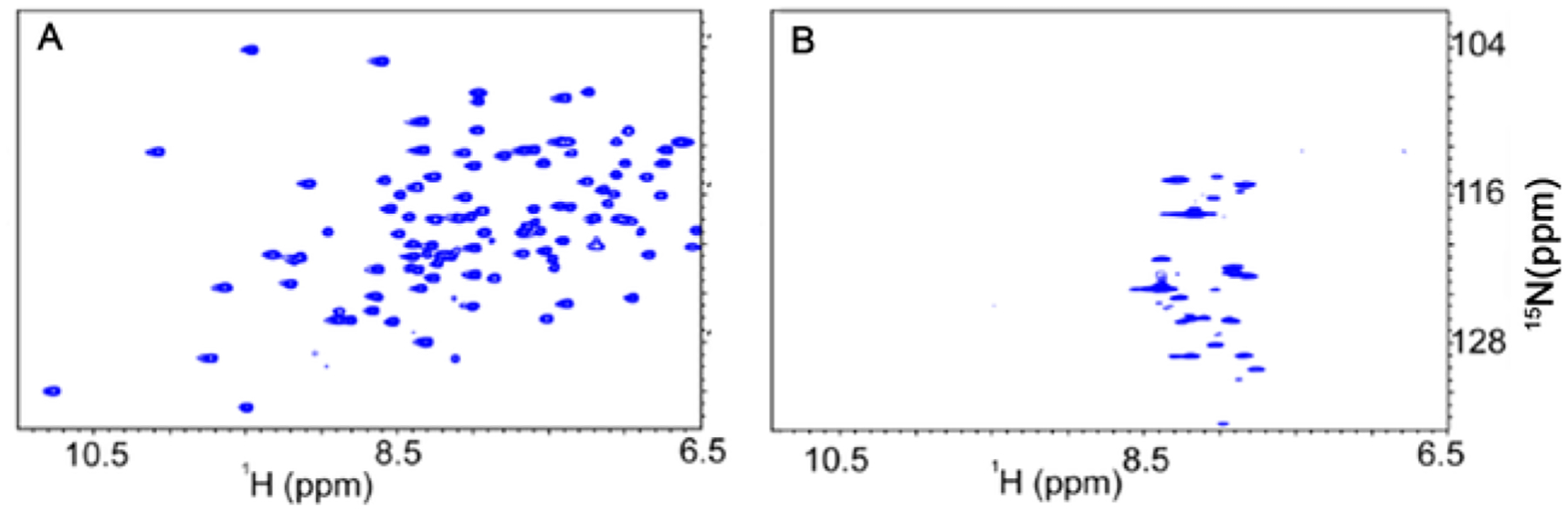
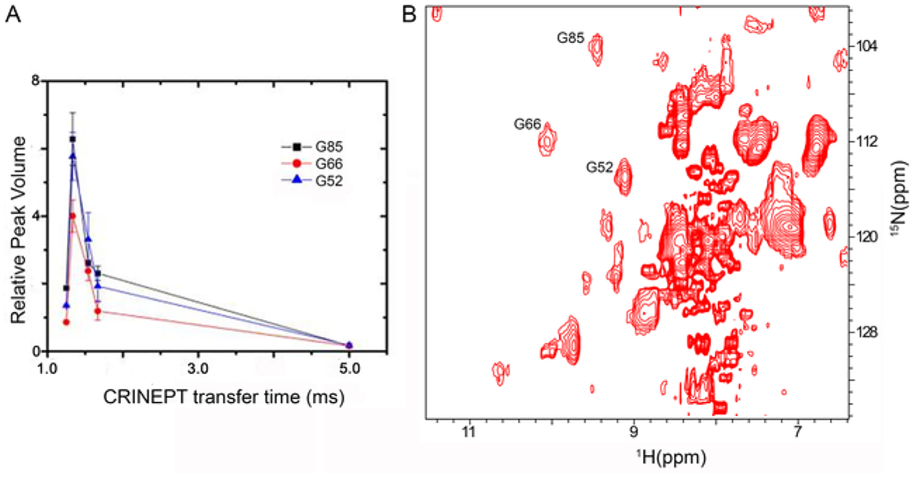
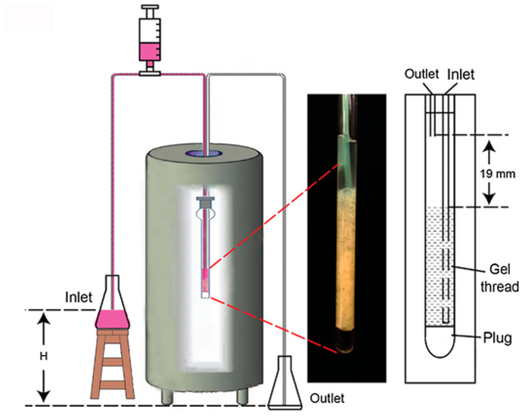
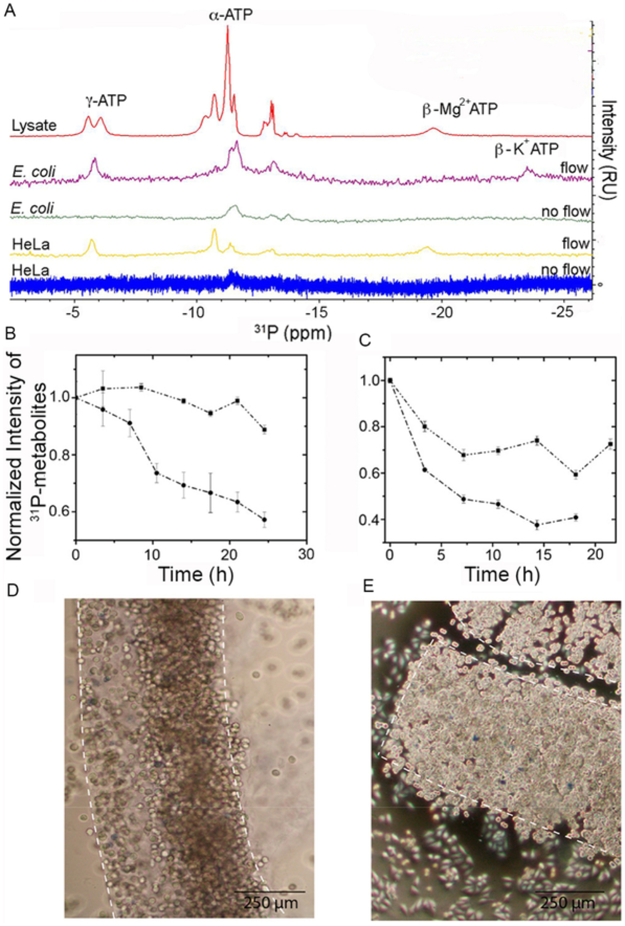
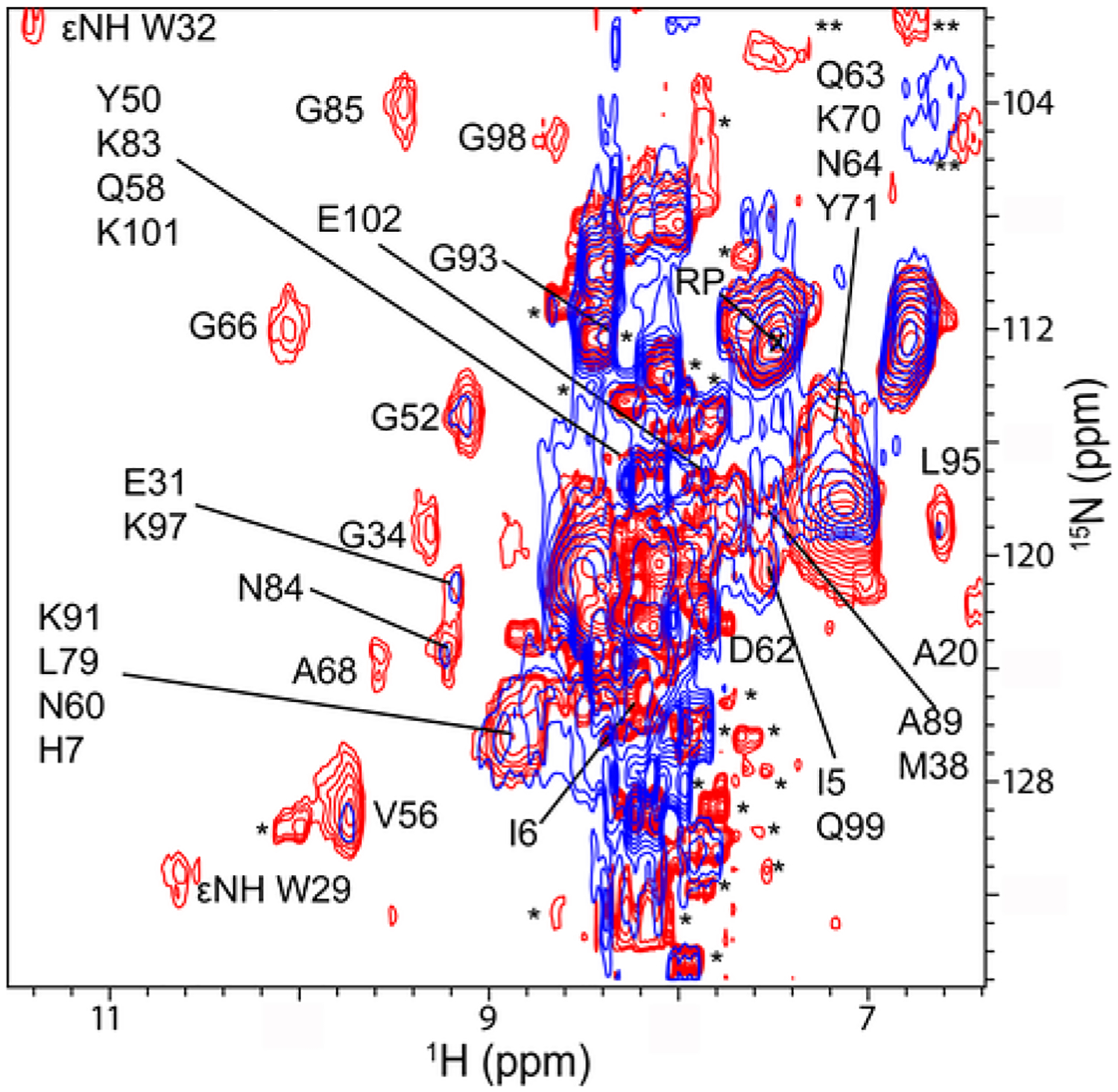
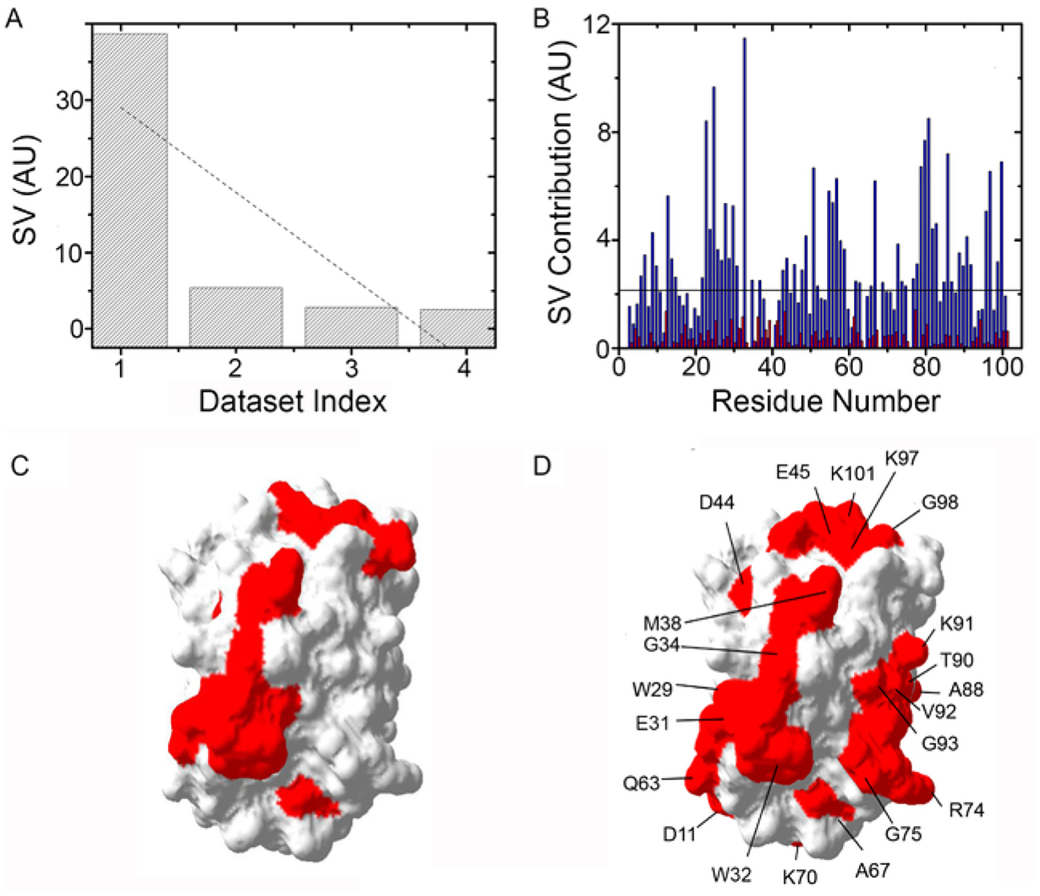
In-cell NMR spectroscopy is a powerful tool to study protein structures and interactions under near physiological conditions in both prokaryotic and eukaryotic living cells. The low sensitivity and resolution of in-cell NMR spectra and limited lifetime of cells over the course of an in-cell experiment have presented major hurdles to wide acceptance of the technique, limiting it to a few select systems. These issues are addressed by introducing the use of the CRINEPT pulse sequence to increase the sensitivity and resolution of in-cell NMR spectra and the use of a bioreactor to maintain cell viability for up to 24h. Application of advanced pulse sequences and bioreactor during in-cell NMR experiments will facilitate the exploration of a wide range of biological processes.
Keywords: Antibiotics; Atomic resolution structure; In vivo biochemistry; In-cell biochemistry; Nucleic acids; Protein interactions; Protein structure; Protein-drug interactions; RNA; Ribosome; Thioredoxin.
© 2019 Elsevier Inc. All rights reserved.
Figures
Similar articles
-
Real-Time In-Cell Nuclear Magnetic Resonance: Ribosome-Targeted Antibiotics Modulate Quinary Protein Interactions.Biochemistry. 2018 Feb 6;57(5):540-546. doi: 10.1021/acs.biochem.7b00938. Epub 2018 Jan 8. Biochemistry. 2018. PMID: 29266932 Free PMC article.
-
Watching protein structure at work in living cells using NMR spectroscopy.Curr Opin Chem Biol. 2012 Dec;16(5-6):609-13. doi: 10.1016/j.cbpa.2012.10.022. Epub 2012 Nov 21. Curr Opin Chem Biol. 2012. PMID: 23176973 Review.
-
In-cell NMR spectroscopy in Escherichia coli.Methods Mol Biol. 2012;831:261-77. doi: 10.1007/978-1-61779-480-3_15. Methods Mol Biol. 2012. PMID: 22167679 Review.
-
Automation of biomolecular NMR screening.Curr Top Med Chem. 2003;3(1):55-67. doi: 10.2174/1568026033392688. Curr Top Med Chem. 2003. PMID: 12570777 Review.
-
NMR protein structure determination in living E. coli cells using nonlinear sampling.Nat Protoc. 2010 Jun;5(6):1051-60. doi: 10.1038/nprot.2010.69. Epub 2010 May 13. Nat Protoc. 2010. PMID: 20539281
Cited by
-
Phenotypic screening of 1,953 FDA-approved drugs reveals 26 hits with potential for repurposing for Peyronie's disease.PLoS One. 2022 Dec 12;17(12):e0277646. doi: 10.1371/journal.pone.0277646. eCollection 2022. PLoS One. 2022. PMID: 36508413 Free PMC article.
-
Towards cost-effective side-chain isotope labelling of proteins expressed in human cells.J Biomol NMR. 2024 Dec;78(4):237-247. doi: 10.1007/s10858-024-00447-6. Epub 2024 Aug 22. J Biomol NMR. 2024. PMID: 39172315 Free PMC article.
-
Determination of intracellular protein-ligand binding affinity by competition binding in-cell NMR.Acta Crystallogr D Struct Biol. 2021 Oct 1;77(Pt 10):1270-1281. doi: 10.1107/S2059798321009037. Epub 2021 Sep 27. Acta Crystallogr D Struct Biol. 2021. PMID: 34605430 Free PMC article.
-
Protein structure determination in human cells by in-cell NMR and a reporter system to optimize protein delivery or transexpression.Commun Biol. 2022 Dec 2;5(1):1322. doi: 10.1038/s42003-022-04251-6. Commun Biol. 2022. PMID: 36460747 Free PMC article.
-
Secretomes from Conventional Insemination and Intra-Cytoplasmic Sperm Injection Derived Embryos Differentially Modulate Endometrial Cells In Vitro.Reprod Sci. 2024 Jul;31(7):2080-2091. doi: 10.1007/s43032-024-01504-z. Epub 2024 Mar 12. Reprod Sci. 2024. PMID: 38472711 Free PMC article.
References
Publication types
MeSH terms
Substances
Grants and funding
LinkOut - more resources
Full Text Sources
