

Alterations of structural and functional connectivity in profound sensorineural hearing loss infants within an early sensitive period: A combined DTI and fMRI study
- PMID: 31129460
- PMCID: PMC6969342
- DOI: 10.1016/j.dcn.2019.100654
Alterations of structural and functional connectivity in profound sensorineural hearing loss infants within an early sensitive period: A combined DTI and fMRI study
Erratum in
-
Corrigendum to "Alterations of structural and functional connectivity in profound sensorineural hearing loss infants within an early sensitive period: A combined DTI and fMRI study" [Dev. Cogn. Neurosci. 38 (2019) 100654].Dev Cogn Neurosci. 2020 Feb;41:100689. doi: 10.1016/j.dcn.2019.100689. Epub 2020 Jan 7. Dev Cogn Neurosci. 2020. PMID: 31923749 Free PMC article. No abstract available.
Abstract
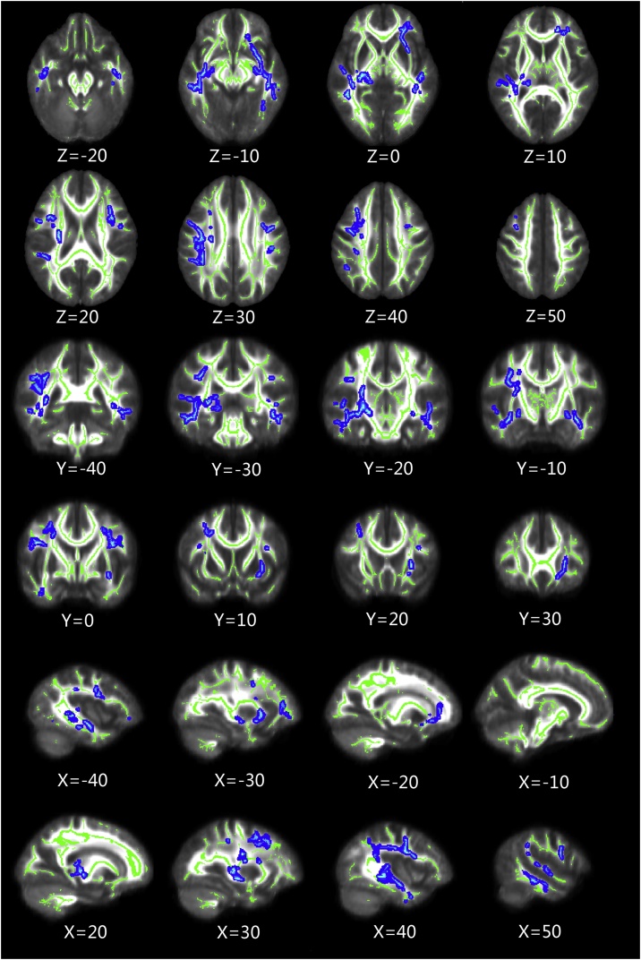
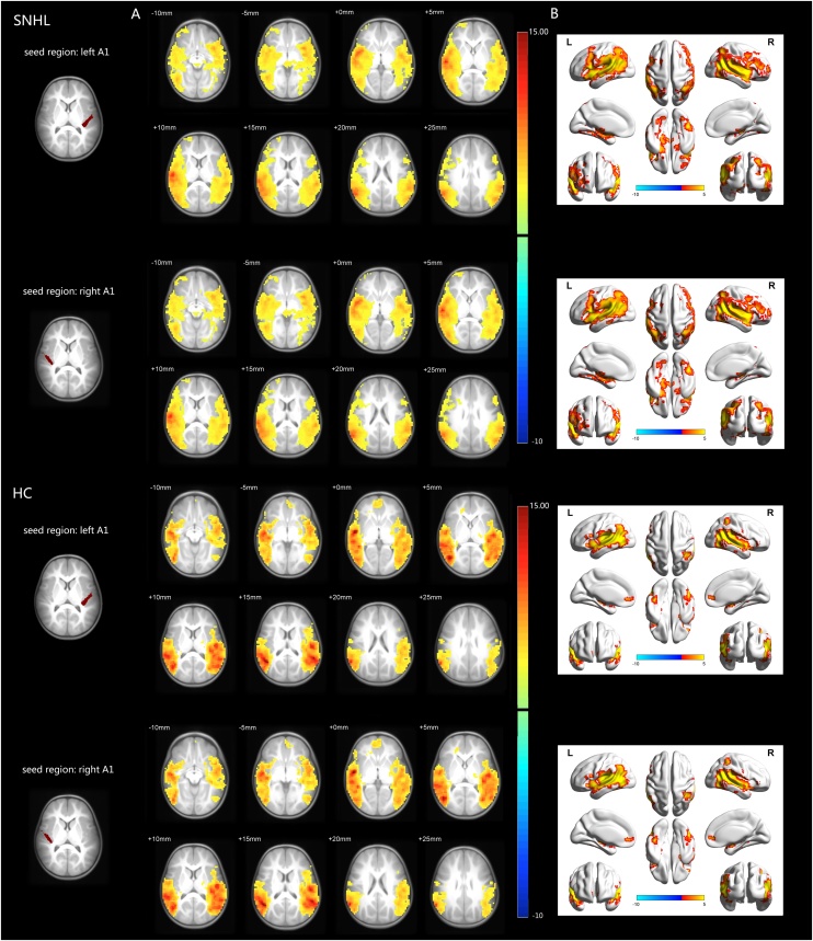
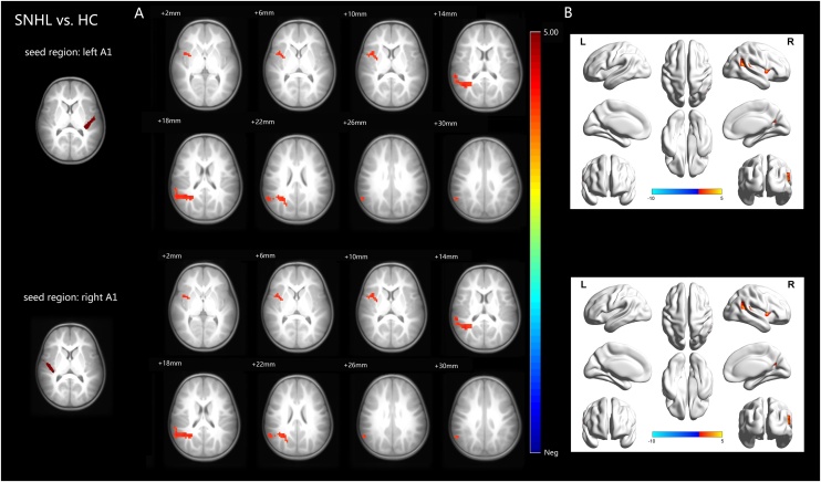
Due to heightened level of neuroplasticity, there is a sensitive period (2-4 years after birth) that exists for optimal central auditory development. Using diffusion tensor imaging combined with resting-state functional connectivity (rsFC) analysis, this study directly investigates the structural connectivity alterations of the whole brain white matter (WM) and the functional reorganization of the auditory network in infants with sensorineural hearing loss (SNHL) during the early sensitive period. 46 bilateral profound SNHL infants prior to cochlear implantation (mean age, 17.59 months) and 33 healthy controls (mean age, 18.55 months) were included in the analysis. Compared with controls, SNHL infants showed widespread WM alterations, including bilateral superior longitudinal fasciculus, inferior fronto-occipital fasciculus, inferior longitudinal fasciculus, right corticospinal tract, posterior thalamic radiation and left uncinate fasciculus. Moreover, SNHL infants demonstrated increased rsFC between left/right primary auditory cortex seeds and right insula and superior temporal gyrus. In conclusion, this study suggests that SNHL in the early sensitive period is associated with diffuse WM alterations that mainly affect the auditory and language pathways. Furthermore, increased rsFC in areas mainly associated with auditory and language networks may potentially reflect reorganization and compensatory activation in response to auditory deprivation during the early sensitive period.
Keywords: Diffusion tensor imaging; Functional connectivity; Resting-state functional magnetic resonance imaging; Sensitive period; Sensorineural hearing loss.
Copyright © 2019 The Authors. Published by Elsevier Ltd.. All rights reserved.
Figures
Similar articles
-
Altered resting-state functional network connectivity in profound sensorineural hearing loss infants within an early sensitive period: A group ICA study.Hum Brain Mapp. 2021 Sep;42(13):4314-4326. doi: 10.1002/hbm.25548. Epub 2021 Jun 1. Hum Brain Mapp. 2021. PMID: 34060682 Free PMC article.
-
Quantitative analyses of high-angular resolution diffusion imaging (HARDI)-derived long association fibers in children with sensorineural hearing loss.Int J Dev Neurosci. 2020 Dec;80(8):717-729. doi: 10.1002/jdn.10071. Epub 2020 Oct 31. Int J Dev Neurosci. 2020. PMID: 33067827 Free PMC article.
-
Diffusion tensor imaging and MR spectroscopy of microstructural alterations and metabolite concentration changes in the auditory neural pathway of pediatric congenital sensorineural hearing loss patients.Brain Res. 2016 May 15;1639:228-34. doi: 10.1016/j.brainres.2014.12.025. Epub 2014 Dec 20. Brain Res. 2016. PMID: 25536303
-
Diffusion Tensor Imaging of Central Auditory Pathways in Patients with Sensorineural Hearing Loss: A Systematic Review.Otolaryngol Head Neck Surg. 2018 Mar;158(3):432-442. doi: 10.1177/0194599817739838. Epub 2017 Nov 7. Otolaryngol Head Neck Surg. 2018. PMID: 29112481 Free PMC article.
-
A systematic review of altered resting-state networks in early deafness and implications for cochlear implantation outcomes.Eur J Neurosci. 2024 May;59(10):2596-2615. doi: 10.1111/ejn.16295. Epub 2024 Mar 5. Eur J Neurosci. 2024. PMID: 38441248
Cited by
-
Impact of inner ear malformation and cochlear nerve deficiency on the development of auditory-language network in children with profound sensorineural hearing loss.Elife. 2023 Sep 12;12:e85983. doi: 10.7554/eLife.85983. Elife. 2023. PMID: 37697742 Free PMC article.
-
Structural Alterations in a Rat Model of Short-Term Conductive Hearing Loss Are Associated With Reduced Resting State Functional Connectivity.Front Syst Neurosci. 2021 Aug 12;15:655172. doi: 10.3389/fnsys.2021.655172. eCollection 2021. Front Syst Neurosci. 2021. PMID: 34456689 Free PMC article.
-
Altered Functional Network in Infants With Profound Bilateral Congenital Sensorineural Hearing Loss: A Graph Theory Analysis.Front Neurosci. 2022 Jan 14;15:810833. doi: 10.3389/fnins.2021.810833. eCollection 2021. Front Neurosci. 2022. PMID: 35095404 Free PMC article.
-
Language networks of normal-hearing infants exhibit topological differences between resting and steady states: An fNIRS functional connectivity study.Hum Brain Mapp. 2024 Sep;45(13):e70021. doi: 10.1002/hbm.70021. Hum Brain Mapp. 2024. PMID: 39258437 Free PMC article.
-
Establishment of Reference Values for Early Auditory Preverbal Skills of Children with Cochlear Implants.Trends Hear. 2022 Jan-Dec;26:23312165221128435. doi: 10.1177/23312165221128435. Trends Hear. 2022. PMID: 36482731 Free PMC article.
References
-
- Altman N.R., Bernal B. Brain activation in sedated children: auditory and visual functional MR imaging. Radiology. 2001;221:56–63. - PubMed
-
- Baldoli C., Scola E., Della Rosa P.A., Pontesilli S., Longaretti R., Poloniato A., Scotti R., Blasi V., Cirillo S., Iadanza A., Rovelli R., Barera G., Scifo P. Maturation of preterm newborn brains: a fMRI-DTI study of auditory processing of linguistic stimuli and white matter development. Brain Struct. Funct. 2015;220:3733–3751. - PubMed
-
- Ball G., Counsell S.M., Merchant N., Arichi T., Doria V., Rutherford M.A., Edwards A.D., Rueckert D., Boardman J.P. An optimised tract-based spatial statistics protocol for neonates: applications to prematurity and chronic lung disease. Neuroimage. 2010;53:94–102. - PubMed
-
- Bamiou D.E., Musiek F.E., Luxon L.M. The insula (Island of reil) and its role in auditory processing. Literature review. Brain Res. Rev. 2003;42:143. - PubMed
MeSH terms
LinkOut - more resources
Full Text Sources
Medical
