. 2019 Jun 4;116(11):2103-2120.
doi: 10.1016/j.bpj.2019.04.030. Epub 2019 May 3.

Modulating Hinge Flexibility in the APP Transmembrane Domain Alters γ-Secretase Cleavage

Affiliations

Modulating Hinge Flexibility in the APP Transmembrane Domain Alters γ-Secretase Cleavage

Alexander Götz et al. Biophys J. .

Abstract

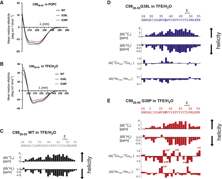
Intramembrane cleavage of the β-amyloid precursor protein C99 substrate by γ-secretase is implicated in Alzheimer's disease pathogenesis. Biophysical data have suggested that the N-terminal part of the C99 transmembrane domain (TMD) is separated from the C-terminal cleavage domain by a di-glycine hinge. Because the flexibility of this hinge might be critical for γ-secretase cleavage, we mutated one of the glycine residues, G38, to a helix-stabilizing leucine and to a helix-distorting proline. Both mutants impaired γ-secretase cleavage and also altered its cleavage specificity. Circular dichroism, NMR, and backbone amide hydrogen/deuterium exchange measurements as well as molecular dynamics simulations showed that the mutations distinctly altered the intrinsic structural and dynamical properties of the substrate TMD. Although helix destabilization and/or unfolding was not observed at the initial ε-cleavage sites of C99, subtle changes in hinge flexibility were identified that substantially affected helix bending and twisting motions in the entire TMD. These resulted in altered orientation of the distal cleavage domain relative to the N-terminal TMD part. Our data suggest that both enhancing and reducing local helix flexibility of the di-glycine hinge may decrease the occurrence of enzyme-substrate complex conformations required for normal catalysis and that hinge mobility can thus be conducive for productive substrate-enzyme interactions.


Figures

Figure 1
Figure 1
C99 G38P and G38L mutants distinctly alter γ-secretase cleavage and processivity. (A) The primary structure of C99 (Aβ numbering) and its major γ-secretase cleavage sites are shown. (B) Levels of AICD were analyzed by immunoblotting after incubation of C100-His6 WT and mutant constructs with CHAPSO-solubilized HEK293 membrane fractions at 37°C. As controls, samples were incubated at 4°C or at 37°C in the presence of the γ-secretase inhibitor L-685,458 (60). (C) Quantification of AICD levels from (B) is shown. Values are shown as a percentage of the WT, which was set to 100%. Data are represented as mean ± standard error of the mean (n = 3, each n represents the mean of three technical replicates). (D and E) Corresponding analysis of Aβ is shown. (F) Representative MALDI-TOF spectra of the different Aβ species generated for WT and the G38 mutants are shown. The intensities of the highest Aβ peaks were set to 100% in the spectra.
Figure 2
Figure 2
Probability of initial contacts of C9926–55 peptides with γ-secretase is not altered for G38 mutants compared to WT, as revealed by in silico modeling of the encounter complex. (A) Kernel densities of the center-of-mass location of the C9926–55 peptide are shown. Darker colors indicate higher contact probabilities. The representation shows the parts of γ-secretase that are embedded in the membrane, pertaining to the subunits nicastrin (green), PS1 NTF (blue), PS1 CTF (cyan), APH-1a (purple), and PEN-2 (yellow). Black arrows highlight TMD2, TMD3, and TMD6 of PS1, and the active-site aspartate residues in PS1 TMD6 and TMD7 are indicated by red spheres. (B) Normalized proximities between residues of γ-secretase subunits and the C9926–55 peptide are shown. Gray areas indicate residues that are part of the indicated TMDs of PS1. (C) Normalized proximities between C9926–55 residues and TMD2 of PS1 are shown.
Figure 3
Figure 3
Helicity of C9926–55 TMD peptides is increased by the G38L and distorted by the G38P mutation. (A) CD spectra of C9926–55 WT and G38L and G38P mutant peptides reconstituted in POPC model membranes and (B) dissolved in TFE/H2O are shown. (C) Chemical shift indices (Δδ) for 13Cα and 1Hα atoms of each residue of C9926–55 WT obtained from solution NMR in TFE/H2O are shown. (D and E) Results of solution NMR measurements as in (C) of the G38L and G38P mutants, respectively, are shown, in which the differences between Δδ values of mutants and WT are also depicted.
Figure 4
Figure 4
DHX rates along the TMD of C9926–55 reveal an impact of the G38 mutations on H-bond stability around the mutation sites but not at the ε-sites. (A) Overall DHX kinetics of C9926–55 WT and G38L and G38P mutant peptides measured with MS-DHX is shown. Complete deuteration was followed by back-exchange in TFE/H2O (pH 5.0), T = 20°C. Exchange kinetics during the first 60 min (inset) and 72 h were measured (n = 3, error bars showing SD are smaller than the size of the symbols). Note that the lower deuterium content in G38P mainly resulted from the lack of one amide deuteron at the cyclic side chain of proline. (B) Site-specific DHX rate constants (kexp,DHX (min−1)) of C9926–55 WT and G38L and G38P mutants dissolved in TFE/H2O are shown, as determined by MS-ETD (error bars show 95% CI). (C) Site-specific HDX rate constants (kexp,HDX (min−1)) determined by NMR are shown (n = 3, error bars show SD). (D) Site-specific kDHX (min−1) computed from MD simulations are shown (error bars show 95% CI). (E) Backbone H-bond occupancy of the individual residues of C9926–55 WT and its G38L and G38P mutants in POPC and (F) in TFE/H2O is shown, calculated by MD simulations (error bars show 95% CI). An amide H-bond is counted as closed if either the α- or 310-H-bond is formed. Note that G38P cannot form H-bonds at residue 38 because of the chemical nature of proline.
Figure 5
Figure 5
G38 mutations of the C9926–55 peptide do not significantly alter its global membrane orientation but change the relative orientation of the ε-sites region. (A) Heat maps of tilt (τ) and azimuthal rotation (ρ) angle combinations of C9926–55 WT and G38L and G38P mutant peptides in a POPC bilayer are given as determined by ssNMR. The colors represent the RMSDNorm of the given (τ,ρ) pair. Maxima (dark areas) represent possible orientations. The circles represent the likeliest (red), second likeliest (blue), and third likeliest (green) solutions. (B) Probability distributions P (τ,ρ) of τ and ρ angle combinations of C9926–55 WT and G38L and G38P mutants in a POPC bilayer are shown, calculated from MD simulations. Dark areas represent high probabilities. (C) Probability distributions of bending (θ) and swivel (ϕ) angle combinations characterizing the orientation of ε-sites in C9926–55 WT and G38L and G38P mutants in POPC and in TFE/H2O are shown, calculated from MD simulations. (D and E) Representative conformations for WT and G38 mutants in (D) POPC and (E) TFE/H2O determined by K-means clustering of (θ,ϕ) combinations in cos-sin space are shown. Domains colored in red (domain A) represent the TMD-N segment I31-M35 and were also used to overlay the structures. Domains colored in blue (domain B) indicate the TMD-C segment I47-M51 carrying the ε-sites. The G37G38 hinge is colored in green. For the G38 mutants, the L and P residues are depicted in orange. Green spheres represent the Cα atom of G33 used as reference for the determination of swivel angles. (F) Distribution of conformations according to their bending angles θ are shown. The last class summarizes all conformations with θ > 80°.
Figure 6
Figure 6
G38 mutations alter global bending and twisting motions. (A) The fundamental motions of helices exemplified for the C9926–55 WT peptide are shown. Motion types are bending (B) and twisting (T) coordinated by a single hinge, as well as combinations of bending and twisting (types BB, BT, TB, and TT) coordinated by a pair of hinges. Helical segments moving as quasi-rigid domains are colored in blue and red. Residues that act as flexible hinges are colored in green. Spheres represent Cα atoms of G37 and G38 and are colored according to the domain in which they are located. Screw axes passing the hinge regions are shown in gray. A screw axis perpendicular to the helix axis indicates a bending-like motion, whereas a screw axis parallel to the helix axis indicates a twisting-like motion. For mixed bending and twisting motions, a larger projection of the screw axis with respect to the helix axis indicates a higher percentage of twisting. (B) Probability of all six types of hinge bending and twisting motions in POPC and (C) TFE/H2O is shown. (D and E) Probability of each residue to act as a hinge site in the single-hinge (B + T) and double-hinge motions (BB + BT + TB + TT) for peptides in (D) POPC and (E) TFE/H2O is shown.

References

    1. Wolfe M.S. Intramembrane-cleaving proteases. J. Biol. Chem. 2009;284:13969–13973. - PMC - PubMed
    2. Wolfe, M. S. 2009. Intramembrane-cleaving proteases. J. Biol. Chem. 284:13969-13973. - PMC - PubMed
    1. Sun L., Li X., Shi Y. Structural biology of intramembrane proteases: mechanistic insights from rhomboid and S2P to γ-secretase. Curr. Opin. Struct. Biol. 2016;37:97–107. - PubMed
    2. Sun, L., X. Li, and Y. Shi. 2016. Structural biology of intramembrane proteases: mechanistic insights from rhomboid and S2P to γ-secretase. Curr. Opin. Struct. Biol. 37:97-107. - PubMed
    1. Madala P.K., Tyndall J.D., Fairlie D.P. Update 1 of: proteases universally recognize β strands in their active sites. Chem. Rev. 2010;110:PR1–PR31. - PubMed
    2. Madala, P. K., J. D. Tyndall, …, D. P. Fairlie. 2010. Update 1 of: proteases universally recognize β strands in their active sites. Chem. Rev. 110:PR1-PR31. - PubMed
    1. Timmer J.C., Zhu W., Salvesen G.S. Structural and kinetic determinants of protease substrates. Nat. Struct. Mol. Biol. 2009;16:1101–1108. - PMC - PubMed
    2. Timmer, J. C., W. Zhu, …, G. S. Salvesen. 2009. Structural and kinetic determinants of protease substrates. Nat. Struct. Mol. Biol. 16:1101-1108. - PMC - PubMed
    1. Belushkin A.A., Vinogradov D.V., Kazanov M.D. Sequence-derived structural features driving proteolytic processing. Proteomics. 2014;14:42–50. - PMC - PubMed
    2. Belushkin, A. A., D. V. Vinogradov, …, M. D. Kazanov. 2014. Sequence-derived structural features driving proteolytic processing. Proteomics. 14:42-50. - PMC - PubMed

Publication types

MeSH terms

Substances

LinkOut - more resources