. 2019 Jun 3;29(11):1885-1894.e4.
doi: 10.1016/j.cub.2019.04.065. Epub 2019 May 23.

Artificially Enhancing and Suppressing Hippocampus-Mediated Memories

Affiliations

Artificially Enhancing and Suppressing Hippocampus-Mediated Memories

Briana K Chen et al. Curr Biol. .

Abstract

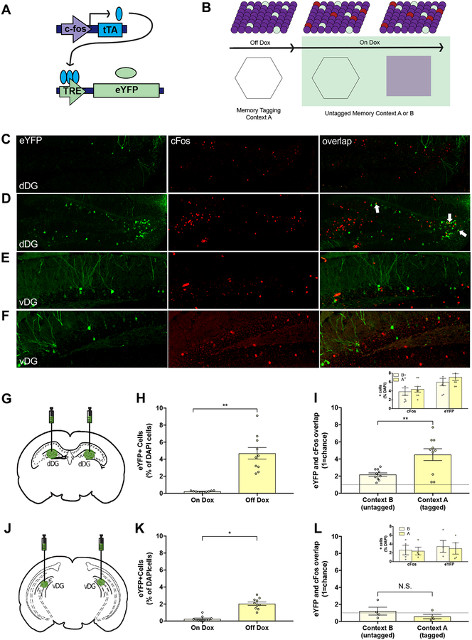
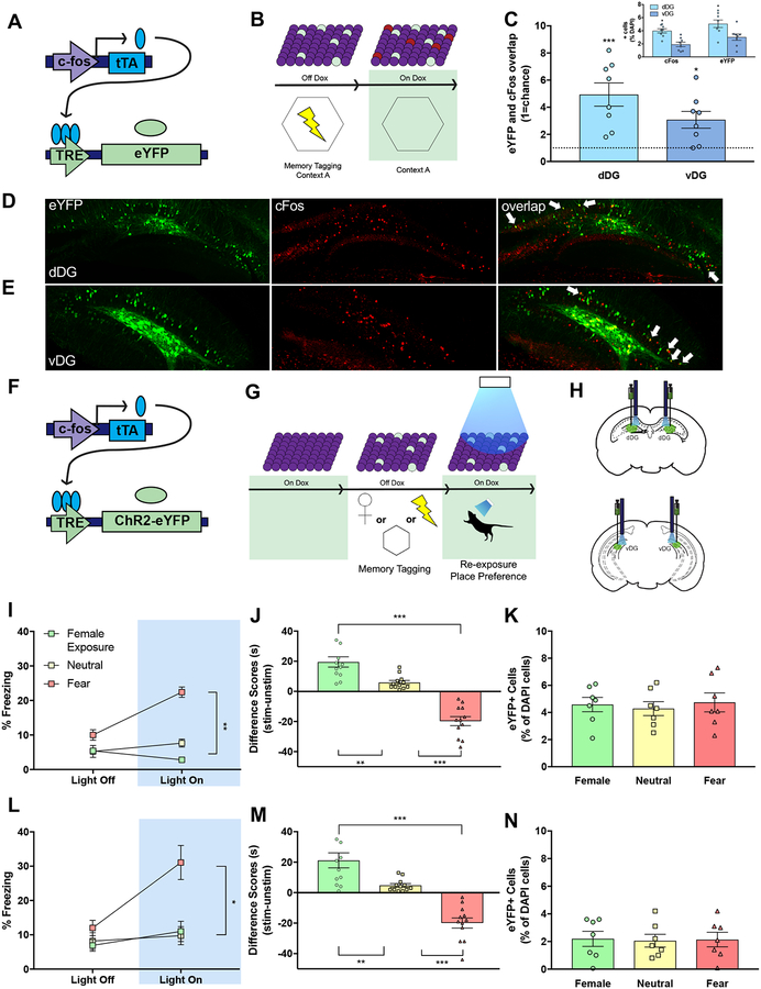
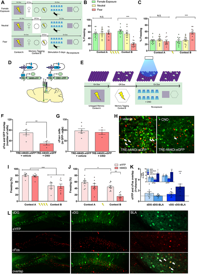
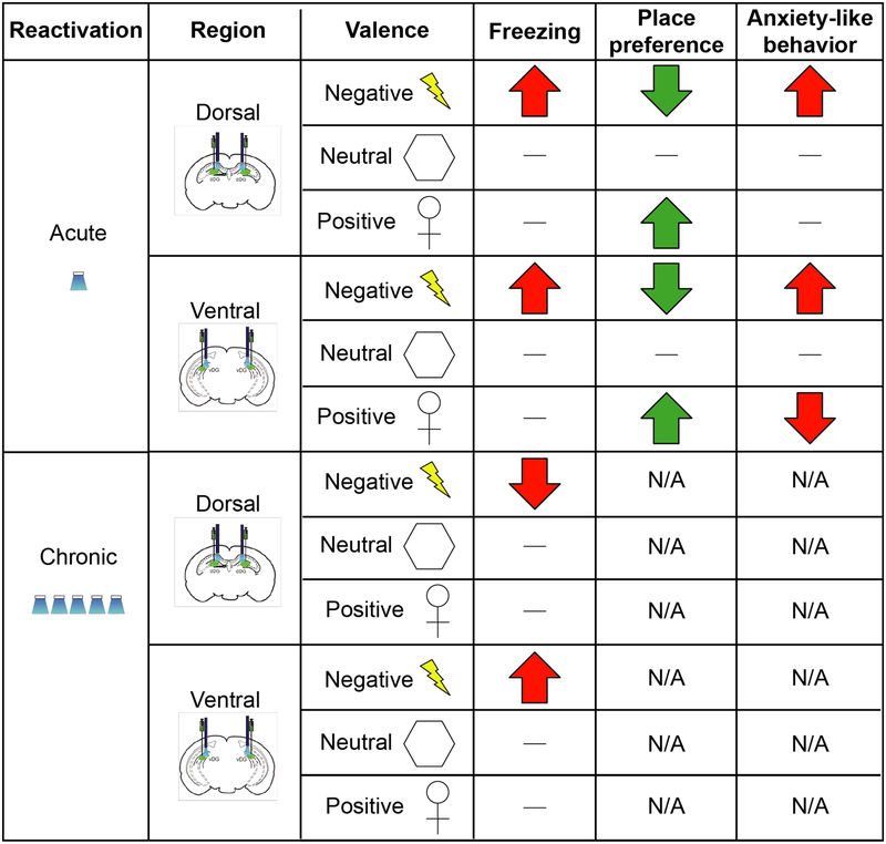
Emerging evidence indicates that distinct hippocampal domains differentially drive cognition and emotion [1, 2]; dorsal regions encode spatial, temporal, and contextual information [3-5], whereas ventral regions regulate stress responses [6], anxiety-related behaviors [7, 8], and emotional states [8-10]. Although previous studies demonstrate that optically manipulating cells in the dorsal hippocampus can drive the behavioral expression of positive and negative memories, it is unknown whether changes in cellular activity in the ventral hippocampus can drive such behaviors [11-14]. Investigating the extent to which distinct hippocampal memories across the longitudinal axis modulate behavior could aid in the understanding of stress-related psychiatric disorders known to affect emotion, memory, and cognition [15]. Here, we asked whether tagging and stimulating cells along the dorsoventral axis of the hippocampus could acutely, chronically, and differentially promote context-specific behaviors. Acute reactivation of both dorsal and ventral hippocampus cells that were previously active during memory formation drove freezing behavior, place avoidance, and place preference. Moreover, chronic stimulation of dorsal or ventral hippocampal fear memories produced a context-specific reduction or enhancement of fear responses, respectively, thus demonstrating bi-directional and context-specific modulation of memories along the longitudinal axis of the hippocampus. Fear memory suppression was associated with a reduction in hippocampal cells active during retrieval, while fear memory enhancement was associated with an increase in basolateral amygdala activity. Together, our data demonstrate that discrete sets of cells throughout the hippocampus provide key nodes sufficient to bi-directionally reprogram both the neural and behavioral expression of memory.

Keywords: emotion; engram; hippocampus; longitudinal axis; memory; optogenetics.


Conflict of interest statement

DECLARATION OF INTERESTS

The authors declare no competing interests.

Figures

Figure 1.
Figure 1.. Activity-dependent and inducible expression of enhanced yellow fluorescent protein (eYFP) in dorsal and ventral dentate gyrus (DG).
(A) A virus cocktail of AAV9-c-Fos-tTA and AAV9-TRE-eYFP was infused into the dorsal or ventral DG in mice on a doxycycline diet (Dox). (B) Dox was removed prior to placement in Context A and returned to diet following exposure to label active cells with eYFP. The following day, mice were returned to the same or different context and expression of c-Fox was visualized. (C–F) Representative images (20X) of eYFP+ (green), c-Fos+ (red), and overlap of cells in the dorsal (dDG) or ventral (vDG) dentate gyrus. eYFP+, c-Fos+, and overlap in mice exposed to a different (C & E) or the same (D & F) context. (G & H) In the dorsal DG, 4.7% (+/−0.7%) of DAPI+ cells were labeled with eYFP while Off Dox compared to <0.3% of On Dox (t(18)=6.58, p<0.01). (I) Mice exposed to the same context (Context A; tagged) had significantly more overlap between eYFP+ and c-Fos+ granule cells than those exposed to a different context (Context B; untagged) relative to chance (dashed line; n=10/group) (t(18)=3.24, p<0.01). Both Contexts A and B recruited similar amounts of cFos+ and eYFP+ cells (inset). (J & K) In the ventral DG, 2.1% (+/−0.2) of ventral DG granule cells were labeled while Off Dox compared to < 0.3% while On Dox (t(18)=7.96, p<0.01) (top; n=10/group). (L) Re-exposure to the same or different context did not result in significant overlap relative to chance (ns) (bottom; n=4/group). The number of cFos+ and eYFP+ cells was comparable between both Contexts A and B (inset). White arrows indicate cells expressing eYFP and c-Fos.
Figure 2.
Figure 2.. Acute activation of distinct memories drives discrete behavior across the longitudinal axis of the hippocampus.
(A) A virus cocktail of AAV9-c-Fos-tTA and AAV9-TRE-eYFP was infused into the dorsal or ventral DG. (B) While on Dox, mice were fear conditioned in Context A. Dox was then removed prior to exposure to a novel Context B, and overlap of cells in the dDG or vDG was visualized. (C) Overlap between eYFP+ and c-Fos+ neurons was comparable between the dDG and vDG and, in both regions, significantly above chance (dashed line). Inset represents %positive cfos and % positive eYFP labeled cells. There were significantly more cFos+ and eYFP+ in the dDG than in the vDG. (D–E) Representative images (20X) of eYFP+ (green), c-Fos+ (red), and overlap of cells in the dDG or vDG. (F) A virus cocktail of AAV9-c-Fos-tTA and AAV9-TRE-ChR2-eYFP was infused into the dorsal or ventral DG. (G) While off Dox, mice were exposed to a female, neutral, or fear memory to label active cells with ChR2, returned to Dox, and administered light stimulation during re-exposure to the training context and an OPP/OPA paradigm. (H) Representative schematic of viral injection and optogenetic stimulation in the dorsal (top) and ventral (bottom) DG. (I, L) Activation of cells (Light On) processing a fear, but not a neutral or a female memory, in both the dorsal (I; female group: n=11; neutral group: n=12; fear group: n=12) and ventral (L; n=12/group) hippocampus drove freezing behavior in a neutral context relative to Light Off epochs. Repeated-measures ANOVA, significant Group × Epoch interaction, dorsal DG [F(2, 32)=41.23, p<0.01] and ventral DG [F(2, 33)=6.48, p<0.01]. (J, M) Light-activation of fear memories drove place avoidance while light-activation of female exposure memories drove place preference in both dorsal (one-way ANOVA, J, dDG [F(2,33)=53.44, p<0.01] and M, vDG [F(2,33)=34.75, p<0.01]). (K, N) In the absence of acute stimulation, fear, neutral, and female memories labeled a comparable amount of eYFP+ neurons in both the dDG (K) and vDG (N). White arrows indicate cells expressing eYFP and c-Fos.
Figure 3.
Figure 3.. Chronic activation of memories induces long-lasting and bidirectional changes in behavior.
(A) A virus cocktail of AAV9-c-Fos-tTA and AAV9-TRE-ChR2-eYFP was infused into the dorsal and ventral DG. All groups were first fear conditioned in context A, followed by fear conditioning in context B a day later, during which the fear group was off Dox. A separate group was exposed to a neutral context to label active cells with ChR2 and returned to being on Dox. Memories were reactivated twice a day for 5 days in a novel context. (B) Upon re-exposure to Contexts A and B, chronic reactivation of cells processing a four-shock fear memory in the dorsal DG resulted in reduced freezing in the tagged context (e.g. Context B) compared to reactivation of cells processing female and neutral memories (n=10/group. Repeated measures ANOVA, Group × Context, [F(2, 27)=12.67, p<0.01]). Newman-Keules posthoc test revealed significantly lower levels of freezing in Context B compared to Context A only in fear group (p<0.001). (C) When returned to contexts A and B, chronic reactivation of cells processing a single-shock fear memory in the ventral DG resulted in increased freezing in the tagged context (e.g. Context B) compared to reactivation of cells processing female and neutral memories (n=10/group: Repeated measures ANOVA, Group × Context, [F(2, 27)=10.88, p<0.01]). Posthoc revealed significantly higher levels of freezing in Context B compared to Context A only in fear group (p<0.001). (D,E) A virus cocktail of AAV9-c-Fos-tTA and AAV9-TRE-ChR2-eYFP (ChR2) was infused into the dorsal or ventral hippocampus and AAV9-c-Fos-tTA and AAV9-TRE-hM4Di-eYFP (hM4Di) or AAV9-TRE-eYFP (eYFP) was infused into the basolateral amygdala (BLA). Mice were fear conditioned in Context A while on Dox and in Context B while off Dox to label DG and BLA cells associated with Context B fear memory with ChR2 and hM4Di or eYFP, respectively. Following tagging, mice underwent a total of 10 sessions over 5 days of optical stimulation while in a novel environment; all mice received 1 mg/kg CNO (i.p.) 30 minutes prior to each session. Finally, mice were placed back into Context A and Context B and their levels of freezing were assessed (in the absence of either light activation or CNO injection). (F) Treatment with CNO significantly reduced cFos and GFP overlap when compared to treatment with a vehicle control (Student’s t-test, p = 0.006). (G) cFos expression in GFP cells was comparable between CNO- and vehicle-treated brains following CNO infusion. (H) Representative images of cFos (red) and eGFP (green) overlap in the BLA following CNO treatment. (I) Chronic reactivation of cells processing a strong four-shock fear conditioning memory in the dorsal DG led to a context-specific reduction of freezing behavior, an effect not affected by hM4Di-expression in the BLA. (eYFP: n=6; hM4Di: n=8; Repeated-Measures ANOVA, Main effect of Context [F(1,12)=41.93, p<0.001] without a main effect of Group or Context × Group interaction (ns). (J) Chronic reactivation of cells processing a weak single-shock fear conditioning memory in the ventral DG led to a context-specific increase in freezing behavior. This effect was ablated by chemogenetic inactivation of the BLA (eYFP: n = 10; hM4Di: n = 7; Repeated-Measures ANOVA, Main effect of Context [F(1,15) = 23.92, p < 0.001], Main effect of Group [F(1,15) = 15.47, p < 0.01]). Posthoc revealed significantly lower levels of freezing between the eYFP and hM4Di group in Context A (p < 0.001). (K) Following chronic activation of dDG fear memories, there was no significant change in overlap between eYFP+ and cFos+ cells in either the dDG or BLA (denoted here as dDG-BLA). However, after chronic activation of vDG fear memories, there was a significant amount of overlap between eYFP+ and c-Fos+ cells above chance (dashed line) in the BLA (denoted by vDG-BLA), but not in the vDG. Inset represents % positive cFos+ and eYFP+ cells. cFos+ and eYFP+ cells were significantly greater in the BLA after chronic vDG activation compared to all other groups. (L) Representative images of eYFP (green), c-Fos (red), and overlap in the dDG, vDG, and BLA. White arrows indicate cells expressing eYFP and c-Fos. See also Figures S1 and S2.
Figure 4.
Figure 4.. Graphical summary of behavioral results.
Dash indicates no change. “N/A” indicates not measured.

References

    1. Poppenk J, Evensmoen HR, Moscovitch M, and Nadel L (2013). Long-axis specialization of the human hippocampus. Trends in cognitive sciences 17, 230–240. - PubMed
    1. Fanselow MS, and Dong HW (2010). Are The Dorsal and Ventral Hippocampus functionally distinct structures? Neuron 65, 7. - PMC - PubMed
    1. Moser MB, Moser EI, Forrest E, Andersen P, and Morris RG (1995). Spatial learning with a minislab in the dorsal hippocampus. Proceedings of the National Academy of Sciences of the United States of America 92, 9697–9701. - PMC - PubMed
    1. Jung MW, Wiener SI, and McNaughton BL (1994). Comparison of spatial firing characteristics of units in dorsal and ventral hippocampus of the rat. The Journal of neuroscience : the official journal of the Society for Neuroscience 14, 7347–7356. - PMC - PubMed
    1. Komorowski RW, Garcia CG, Wilson A, Hattori S, Howard MW, and Eichenbaum H (2013). Ventral hippocampal neurons are shaped by experience to represent behaviorally relevant contexts. The Journal of neuroscience : the official journal of the Society for Neuroscience 33, 8079–8087. - PMC - PubMed

Publication types