. 2019 May 3:10:985.
doi: 10.3389/fimmu.2019.00985. eCollection 2019.

Functional Characterization of Dark Sleeper (Odontobutis obscura) TBK1 on IFN Regulation

Affiliations

Functional Characterization of Dark Sleeper (Odontobutis obscura) TBK1 on IFN Regulation

Jian Chen et al. Front Immunol. .

Abstract

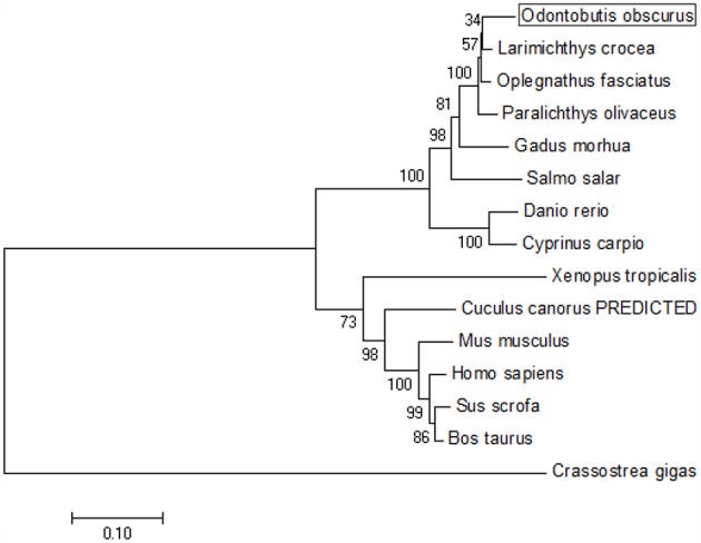
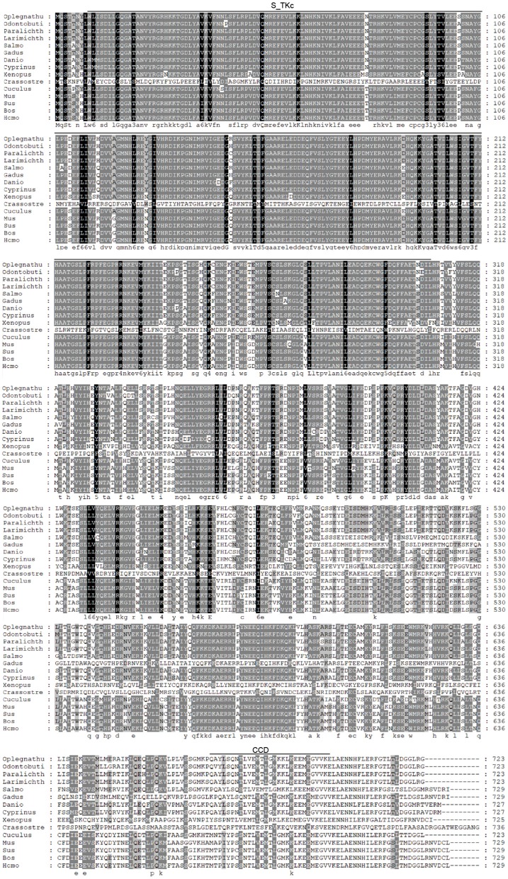
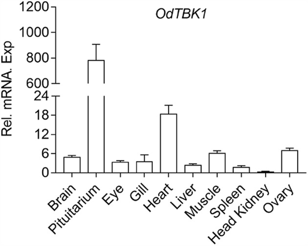
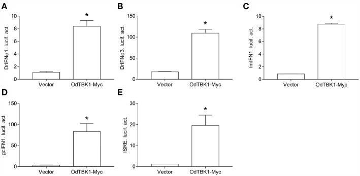
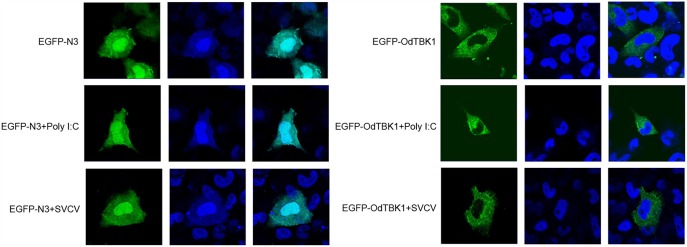
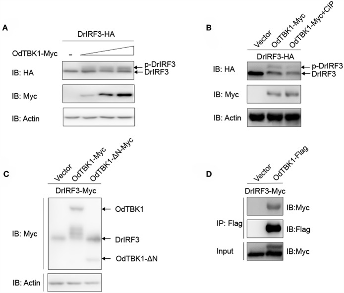
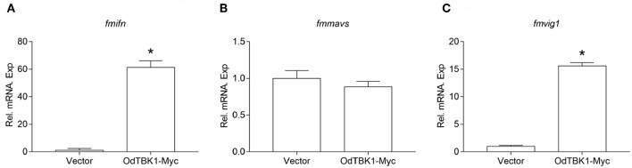
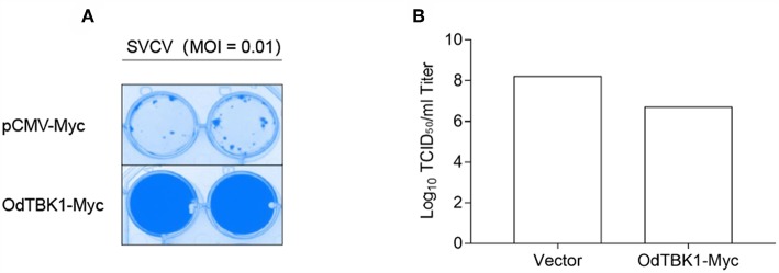
In East Asia, the dark sleeper, Odontobutis obscura (O. obscura) is a crucial commercial species of freshwater fish; however, its molecular biology research is still undeveloped, including its innate immune system, which is pivotal to antiviral responses. In this study, we cloned and identified the characterization and kinase function of dark sleeper TANK-binding kinase 1 (TBK1), supplementing the evidence of the conservation of this classical factor in fish. First, the ORF of Odontobutis obscurus (O. obscura) TBK1 (OdTBK1) was cloned from liver tissue by RACE-PCR. Subsequent nucleic acid and amino acid sequence analysis suggested that OdTBK1 is homologous with other fish TBK1, and the N-terminal Serine/Threonine protein kinases catalytic domain (S_TKc) and C-terminal coiled coil domain (CCD) are conserved. Subsequently, the cellular distribution demonstrated that OdTBK1 was located in the cytoplasm region. With regard to the identification of functions, OdTBK1 activated several interferon (IFN) promoters' activity and induced downstream IFN-stimulated genes (ISGs) expression. In a canonical manner, wild-type OdTBK1 significantly phosphorylated interferon regulatory factor 3 (IRF3) but failed when the N-terminal region was truncated. Furthermore, overexpression of OdTBK1 decreased viral proliferation remarkably. Collectively, these data systematically analyzed the characterization and function of OdTBK1, initiating the study of the innate antiviral response of dark sleeper.

Keywords: IFN; Odontobutis obscura; RLRs; TBK1; antiviral.


Figures

Figure 1
Figure 1
Phylogenetic tree of various TBK1 members. The tree was depicted on the overall sequences by neighbor-joining method. The 0.1 scale indicates the genetic distance. Accession numbers of TBK1 amino acid sequences are as follows: Odontobutis obscura (dark sleeper), MK637832; Oplegnathus fasciatus (Stone bream), AHX37216.1; Paralichthys olivaceus (bastard halibut), AGL54166.1; Larimichthys crocea (large yellow croaker), AKM77645.1; Salmo salar (Atlantic salmon), AEA42006.1; Gadus morhua (Atlantic cod), ADL60136.1; Danio rerio (zebra fish), NP_001038213.2; Cyprinus carpio (koi), ADZ55455.1; Xenopus tropicalis (Western clawed frog), NP_001135652.1; Crassostrea gigas (Thunberg), APX43288.1; Cuculus canorus (Cuckoo), XP_009554581.1; Mus musculus (Mouse), AAF05990.1; Sus scrofa (Pig), NP_001098762.1; Bos taurus (Bovine), NP_001179684.1; Homo sapiens (Human), AAF05989.1.
Figure 2
Figure 2
Sequence alignment of the amino acid sequence of TBK1 from several species was carried out by Gene Doc. Conserved N-terminal Serine/Threonine protein kinases catalytic domain (S_TKc) was showed in the black box, C-terminal coiled coil domain (CCD) was showed in the gray box.
Figure 3
Figure 3
Expression analysis of OdTBK1 in different tissues. Total RNAs of various tissues in dark sleeper were extracted to examine the transcripts of OdTBK1, β-actin was used as an internal control for normalization to give relative expression. The expression levels in the other tissues are shown as fold induction compared with the head kidney (for which their relative expressions were set to 1). Error bars represents the means ± SEM (n = 3), and the experiments were repeated three time with similar results.
Figure 4
Figure 4
(A–E) Activation of DrIFNϕ1, DrIFNϕ3 and ISRE promoters by overexpression of OdTBK1. EPC cells were seeded on 24-well-plates overnight and co-transfected with pCMV-Myc or OdTBK1-Myc and DrIFNϕ1pro-Luc (A), DrIFNϕ3pro-Luc (B), fmIFNpro-Luc (C). GCO cells were treated at the same way and co-transfected with pCMV-Myc or OdTBK1-Myc and gcIFN1pro-Luc (D), ISRE-Luc (E). The luciferase assays were performed 24 h after transfection. The promoter activity is presented as relative light units normalized to Renilla luciferase activity. Error bars represents the means ± SEM (n = 3), and the experiments were repeated three time with similar results. Asterisks indicate significant differences from control (*p < 0.05).
Figure 5
Figure 5
Subcellular localization of OdTBK1. EPC cells seeded onto microscopy cover glass in 6-well plates were transfected with 2 μg EGFP-OdTBK1 or the empty vector. After 24 h, the cells were untreated (null) or transfected with 2 μg poly (I:C) or infected with 4 μl SVCV for 12 h, then the cells were fixed and subjected to confocal microscopy analysis. Green staining represents the OdTBK1 protein signal, and blue staining indicates the nucleus region (original magnification, ×63; oil immersion objective). Bar, 10 μm. All experiments were repeated at least three times, with similar results.
Figure 6
Figure 6
IRF3 was phosphorylated by OdTBK1. (A) Overexpression of OdTBK1 induces the phosphorylation of DrIRF3 in a dose-dependent manner. HEK 293T cells were seeded in six-well plates overnight and co-transfected with 1 μg DrIRF3-HA (attachment of a HA tag to the N terminus of DrIRF3) and 1 μg empty vector, or OdTBK1-Myc (0.25, 0.5 and 1 μg, respectively) for 24 h. The cell lysates were subjected to IB with the anti-HA, anti-Myc, and anti-β-actin Abs. (B) OdTBK1 mediates the phosphorylation of DrIRF3. HEK 293T cells were seeded in six-well plates overnight and co-transfected with 1 μg DrIRF3-HA and 1 μg empty vector or OdTBK1-Myc for 24 h. The cell lysates (100 μg) were treated with or without CIP (10 U) for 40 min at 37°C. The lysates were then subjected to IB with the anti-HA, anti-Myc, and anti-β-actin Abs. (C) IRF3 was phosphorylated by the N terminus of OdTBK1. HEK 293T cells were seeded in six-well plates overnight and transfected with the indicated plasmids (1 μg each) for 24 h. The cell lysates were subjected to IB with the anti-Myc and anti-β-actin Abs. (D) OdTBK1 associates with DrIRF3. HEK 293T cells seeded into 10-cm2 dishes were transfected with the indicated plasmids (10 μg each). After 24 h, cell lysates were immunoprecipitated (IP) with anti-Flag affinity gel. The immunoprecipitates and cell lysates were then analyzed by IB with anti-Myc and anti-Flag Abs, respectively. All experiments were repeated at least three times with similar results.
Figure 7
Figure 7
Overexpression of OdTBK1 induces the expressions of ISGs in EPC cells. (A–C) qPCR detection of the transcript levels of ISGs in OdTBK1-overexpressing cells. EPC cells seeded in 6-well plates overnight were transfected with 1.5μg OdIRF3-Myc or empty vector for 24 h. Then total RNAs were extracted to examine the transcripts of ifn (A), mavs (B), and vig1 (C) by qPCR. β-actin was used as an internal control for normalization and the relative expression is represented as fold induction relative to the expression level in control cells (set to 1). Error bars represents the means ± SEM (n = 3), and the experiments were repeated three time with similar results. Asterisks indicate significant differences from control (*p < 0.05).
Figure 8
Figure 8
Antiviral assay on OdTBK1. (A,B) EPC cells seeded in 24-well plates overnight were transfected with 0.5 μg OdTBK1-Myc or pCMV-Myc vector. At 24 h after transfection, the EPC cells were infected with SVCV (MOI = 0.01) per well and incubated at 28°C. The culture media was then harvested to detect the virus titers at 48 h post infection. (A) The cell monolayers were fixed with 4% PFA for 1 h and stained with 1% crystal violet overnight. (B) The viral titers of the supernatants were determined by TCID50 assays on EPC cells. The experiments were performed for three times with similar results.

Similar articles

Cited by

References

    1. Loo YM, Gale M, Jr. Immune signaling by RIG-I-like receptors. Immunity. (2011) 34:680–92. 10.1016/j.immuni.2011.05.003 - DOI - PMC - PubMed
    1. He TS, Chen T, Wang DD, Xu LG. HAUS8 regulates RLR-VISA antiviral signaling positively by targeting VISA. Mol Med Rep. (2018) 18:2458–66. 10.3892/mmr.2018.9171 - DOI - PubMed
    1. Zhang P, Li Y, Xia J, He J, Pu J, Xie J, et al. . IPS-1 plays an essential role in dsRNA-induced stress granule formation by interacting with PKR and promoting its activation. J Cell Sci. (2014) 127 (Pt. 11):2471–82. 10.1242/jcs.139626 - DOI - PubMed
    1. Haynes LD, Verma S, McDonald B, Wu R, Tacke R, Nowyhed HN, et al. . Cardif (MAVS) Regulates the Maturation of NK Cells. J Immunol. (2015) 195:2157–67. 10.4049/jimmunol.1402060 - DOI - PMC - PubMed
    1. Liu S, Cai X, Wu J, Cong Q, Chen X, Li T, et al. . Phosphorylation of innate immune adaptor proteins MAVS, STING, and TRIF induces IRF3 activation. Science. (2015) 347:aaa2630. 10.1126/science.aaa2630 - DOI - PubMed

Publication types