

Dose-dependent effects of NMDA on retinal and optic nerve morphology in rats
- PMID: 31131232
- PMCID: PMC6520264
- DOI: 10.18240/ijo.2019.05.08
Dose-dependent effects of NMDA on retinal and optic nerve morphology in rats
Abstract
Aim: To investigate dose-dependent effects of N-methyl-D-aspartate (NMDA) on retinal and optic nerve morphology in rats.
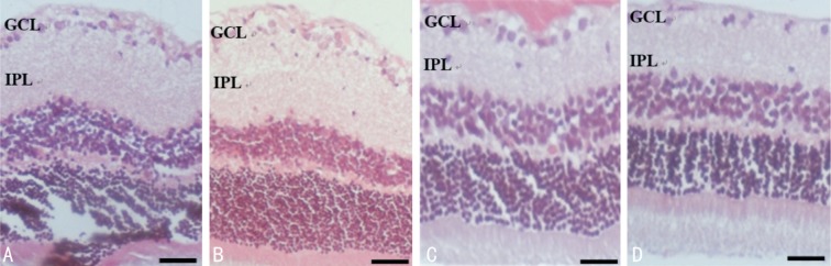
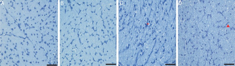
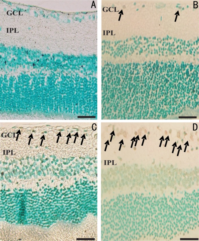
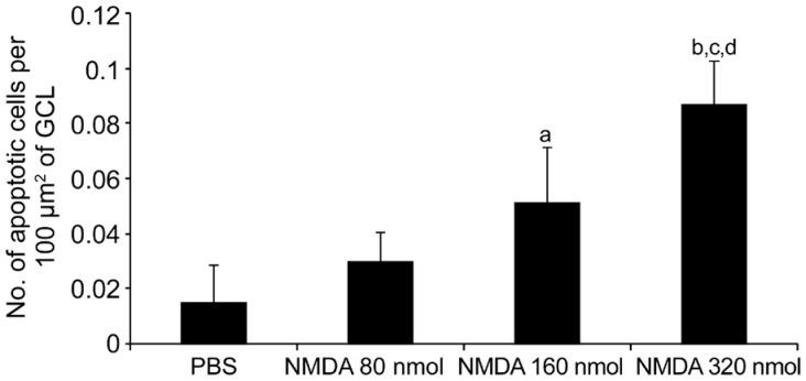
Methods: Sprague Dawley rats, 180-250 g in weight were divided into four groups. Groups 1, 2, 3 and 4 were intravitreally administered with vehicle and NMDA at the doses 80, 160 and 320 nmol respectively. Seven days after injection, rats were euthanized, and their eyes were taken for optic nerve toluidine blue and retinal hematoxylin and eosin stainings. The TUNEL assay was done for detecting apoptotic cells.
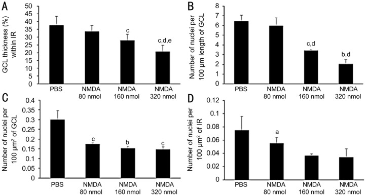
Results: All groups treated with NMDA showed significantly reduced ganglion cell layer (GCL) thickness within inner retina, as compared to control group. Group NMDA 160 nmol showed a significantly greater GCL thickness than the group NMDA 320 nmol. Administration of NMDA also resulted in a dose-dependent decrease in the number of nuclei both per 100 µm GCL length and per 100 µm2 of GCL. Intravitreal NMDA injection caused dose-dependent damage to the optic nerve. The degeneration of nerve fibres with increased clearing of cytoplasm was observed more prominently as the NMDA dose increased. In accordance with the results of retinal morphometry analysis and optic nerve grading, TUNEL staining demonstrated NMDA-induced excitotoxic retinal injury in a dose-dependent manner.
Conclusion: Our results demonstrate dose-dependent effects of NMDA on retinal and optic nerve morphology in rats that may be attributed to differences in the severity of excitotoxicity and oxidative stress. Our results also suggest that care should be taken while making dose selections experimentally so that the choice might best uphold study objectives.
Keywords: N-methyl-D-aspartate; dose-dependent effects; excitotoxicity; glaucoma; optic nerve; retina.
Figures

Similar articles
-
Intravitreal Trans-Resveratrol Ameliorates NMDA-Induced Optic Nerve and Retinal Injury.Curr Eye Res. 2022 Jun;47(6):866-873. doi: 10.1080/02713683.2022.2033270. Epub 2022 Mar 22. Curr Eye Res. 2022. PMID: 35188034
-
Neuroprotective Effect of Magnesium Acetyltaurate Against NMDA-Induced Excitotoxicity in Rat Retina.Neurotox Res. 2017 Jan;31(1):31-45. doi: 10.1007/s12640-016-9658-9. Epub 2016 Aug 27. Neurotox Res. 2017. PMID: 27568334
-
Antiapoptotic effect of taurine against NMDA-induced retinal excitotoxicity in rats.Neurotoxicology. 2019 Jan;70:62-71. doi: 10.1016/j.neuro.2018.10.009. Epub 2018 Oct 30. Neurotoxicology. 2019. PMID: 30385388
-
Philanthotoxin-343 attenuates retinal and optic nerve injury, and protects visual function in rats with N-methyl-D-aspartate-induced excitotoxicity.PLoS One. 2020 Jul 24;15(7):e0236450. doi: 10.1371/journal.pone.0236450. eCollection 2020. PLoS One. 2020. PMID: 32706792 Free PMC article.
-
[Morphological changes of retina after N-methyl-D-aspartate induced damage in rats].Zhong Nan Da Xue Xue Bao Yi Xue Ban. 2004 Jun;29(3):287-91. Zhong Nan Da Xue Xue Bao Yi Xue Ban. 2004. PMID: 16136962 Chinese.
Cited by
-
Neuroprotective effects of MK-801 against cerebral ischemia reperfusion.Heliyon. 2024 Jun 28;10(13):e33821. doi: 10.1016/j.heliyon.2024.e33821. eCollection 2024 Jul 15. Heliyon. 2024. PMID: 39040387 Free PMC article.
-
Early inner plexiform layer thinning and retinal nerve fiber layer thickening in excitotoxic retinal injury using deep learning-assisted optical coherence tomography.Acta Neuropathol Commun. 2024 Feb 1;12(1):19. doi: 10.1186/s40478-024-01732-z. Acta Neuropathol Commun. 2024. PMID: 38303097 Free PMC article.
-
Heat Shock Protein Upregulation Supplemental to Complex mRNA Alterations in Autoimmune Glaucoma.Biomolecules. 2022 Oct 21;12(10):1538. doi: 10.3390/biom12101538. Biomolecules. 2022. PMID: 36291747 Free PMC article.
-
YAP activation in Müller cells protects against NMDA-induced retinal ganglion cell injury by regulating Bcl-xL expression.Front Pharmacol. 2024 Aug 6;15:1446521. doi: 10.3389/fphar.2024.1446521. eCollection 2024. Front Pharmacol. 2024. PMID: 39166115 Free PMC article.
-
Retinal Glutamate Neurotransmission: From Physiology to Pathophysiological Mechanisms of Retinal Ganglion Cell Degeneration.Life (Basel). 2022 Apr 25;12(5):638. doi: 10.3390/life12050638. Life (Basel). 2022. PMID: 35629305 Free PMC article. Review.
References
-
- Tham YC, Li X, Wong TY, Quigley HA, Aung T, Cheng CY. Global prevalence of glaucoma and projections of glaucoma burden through 2040. Ophthalmology. 2014;121(11):2081–2090. - PubMed
-
- Chiu SL, Chu CL, Muo CH, Chen CL, Lan SJ. The prevalence and the incidence of diagnosed open-angle glaucoma and diagnosed angle-closure glaucoma: changes from 2001 to 2010. J Glaucoma. 2016;25(5):e514–e519. - PubMed
-
- Agarwal R. Recent advances in pathophysiology and pharmacotherapy of glaucoma. Med Heal Rev. 2009;1(2):75–94.
-
- Ebneter A, Chidlow G, Wood JP, Casson RJ. Protection of retinal ganglion cells and the optic nerve during short-term hyperglycemia in experimental glaucoma. Arch Ophthalmol. 2011;129(10):1337–1344. - PubMed
-
- Casson RJ, Chidlow G, Wood JP, Crowston JG, Goldberg I. Definition of glaucoma: clinical and experimental concepts. Clin Exp Ophthalmol. 2012;40(4):341–349. - PubMed
LinkOut - more resources
Full Text Sources