

doi: 10.18240/ijo.2019.05.29.
eCollection 2019.
A presumed iridocyclitis developed to panophthalmitis caused by a non-metallic intraocular foreign body
Affiliations
- PMID: 31131253
- PMCID: PMC6520279
- DOI: 10.18240/ijo.2019.05.29
Item in Clipboard
A presumed iridocyclitis developed to panophthalmitis caused by a non-metallic intraocular foreign body
Int J Ophthalmol.
.
No abstract available
Figures

A: The fundus image of the patient one month after the hit by the branch. The arrow indicated the foreign body in the vitreous cavity. B: The foreign body during the surgery. The arrow indicated the foreign body in the vitreous cavity. C: The foreign body was about 1.2 mm length.
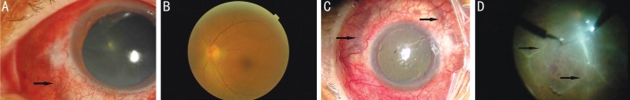
A: The arrow showed the sclera necrosis before the surgery; B: The fundus image before surgery; C: The arrow showed that sclera necrosis was seen around the eyeball during the surgery; D: The arrow showed the retinal vessels were occluded during the surgery.
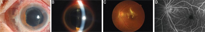
A: Anterior segment image of the patient under diffused light. The necrotizing of the sclera ceased. The choroidal pigment was still seen from the sclera. B: Anterior segment image of the patient under slit light. The posterior capsule opacification was found clearly. C: The fundus image of the patient. All the vessels were infused with blood flow. D: The fluorescein angiography image of the fundus. Reperfusion of the retinal vessels was seen.
Similar articles
-
An intraocular foreign body masquerading as idiopathic chronic iridocyclitis.Ophthalmic Surg Lasers. 1998 Apr;29(4):336-7. Ophthalmic Surg Lasers. 1998. PMID: 9571669
-
Bacillus cereus panophthalmitis associated with intraocular gas bubble.Br J Ophthalmol. 1989 Jan;73(1):25-8. doi: 10.1136/bjo.73.1.25. Br J Ophthalmol. 1989. PMID: 2493262 Free PMC article.
-
Gas gangrene panophthalmitis is a rare condition that can occur following penetrating injury with retained intraocular foreign body.Retina. 1992;12(1):74. Retina. 1992. PMID: 1565874 No abstract available.
-
Acute aseptic panophthalmitis caused by a copper foreign body.Fortschr Ophthalmol. 1990;87(4):362-3. Fortschr Ophthalmol. 1990. PMID: 2210563
-
Clostridium perfringens panophthalmitis and orbital cellulitis: a case report.BMC Ophthalmol. 2018 Apr 10;18(1):88. doi: 10.1186/s12886-018-0751-0. BMC Ophthalmol. 2018. PMID: 29631556 Free PMC article.
Cited by
-
Establishment of a prediction tool for ocular trauma patients with machine learning algorithm.Int J Ophthalmol. 2021 Dec 18;14(12):1941-1949. doi: 10.18240/ijo.2021.12.20. eCollection 2021. Int J Ophthalmol. 2021. PMID: 34926212 Free PMC article.
-
Development, comparison, and internal validation of prediction models to determine the visual prognosis of patients with open globe injuries using machine learning approaches.BMC Med Inform Decis Mak. 2024 May 21;24(1):131. doi: 10.1186/s12911-024-02520-4. BMC Med Inform Decis Mak. 2024. PMID: 38773484 Free PMC article.
-
Development of a nomogram for predicting early visual acuity outcomes and reoperation rate in patients with open globe injury.BMC Ophthalmol. 2025 Jan 13;25(1):16. doi: 10.1186/s12886-025-03845-y. BMC Ophthalmol. 2025. PMID: 39806301 Free PMC article.
References
-
- Obuchowska I, Sidorowicz A, Napora KJ, Mariak Z. Clinical characteristics of penetrating ocular injuries with intraocular foreign body. Part II. Diagnostics and treatment. Klin Oczna. 2010;112(1-3):77–81. - PubMed
-
- Loporchio D, Mukkamala L, Gorukanti K, Zarbin M, Langer P, Bhagat N. Intraocular foreign bodies: a review. Surv Ophthalmol. 2016;61(5):582–596. - PubMed
-
- Mester V, Kuhn F. Intraocular foreign bodies. Ophthalmol Clin North Am. 2002;15(2):235–242. - PubMed
LinkOut - more resources
Full Text Sources